Review
. 2019 Apr 9;14(1):29.
doi: 10.1186/s13000-019-0802-8.

Applications of molecular neuro-oncology - a review of diffuse glioma integrated diagnosis and emerging molecular entities

Affiliations
Review

Applications of molecular neuro-oncology - a review of diffuse glioma integrated diagnosis and emerging molecular entities

Matthew D Wood et al. Diagn Pathol. .

Abstract

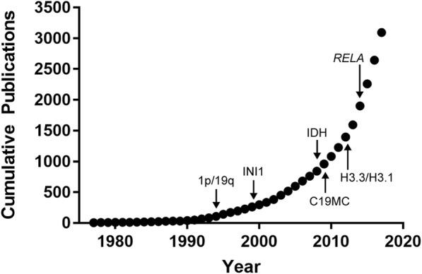
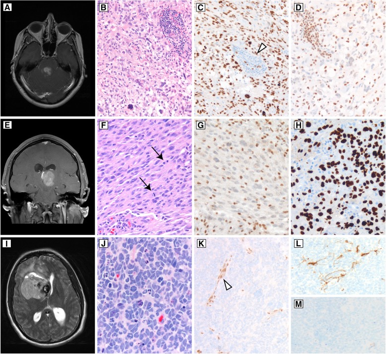
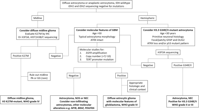
Insights into the molecular underpinnings of primary central nervous system tumors have radically changed the approach to tumor diagnosis and classification. Diagnostic emphasis has shifted from the morphology of a tumor under the microscope to an integrated approach based on morphologic and molecular features, including gene mutations, chromosomal copy number alterations, and gene rearrangements. In 2016, the World Health Organization provided guidelines for making an integrated diagnosis that incorporates both morphologic and molecular features in a subset of brain tumors. The integrated diagnosis now applies to infiltrating gliomas, a category that includes diffusely infiltrating astrocytoma grades II, III, and IV, and oligodendroglioma, grades II and III, thereby encompassing the most common primary intra-axial central nervous system tumors. Other neoplasms such as medulloblastoma, embryonal tumor with multilayered rosettes, certain supratentorial ependymomas, and atypical teratoid/rhabdoid tumor are also eligible for integrated diagnosis, which can sometimes be aided by characteristic immunohistochemical markers. Since 2016, advances in molecular neuro-oncology have resulted in periodic updates and clarifications to the integrated diagnostic approach. These advances reflect expanding knowledge on the molecular pathology of brain tumors, but raise a challenge in rapidly incorporating new molecular findings into diagnostic practice. This review provides a background on the molecular characteristics of primary brain tumors, emphasizing the molecular basis for classification of infiltrating gliomas, the most common entities that are eligible for an integrated diagnosis. We then discuss entities within the diffuse gliomas that do not receive an integrated diagnosis by WHO 2016 criteria, but have distinctive molecular features that are important to recognize because their clinical behavior can influence clinical management and prognosis. Particular attention is given to the histone H3 G34R/G34V mutant astrocytomas, an entity to consider when faced with an infiltrating glioma in the cerebral hemisphere of children and young adults, and to the group of histologically lower grade diffuse astrocytic gliomas with molecular features of glioblastoma, an important category of tumors to recognize due to their aggressive clinical behavior.

Keywords: Brain tumors; Diffuse glioma; Integrated diagnosis; Molecular pathology; WHO 2016.


Conflict of interest statement

Ethics approval and consent to participate

Clinical data and photomicrographs are gathered under a Neurosurgery/Neuropathology Data Repository, which has been approved by the Oregon Health & Science University Institutional Review Board, with a waiver of patient consent (STUDY0018895).

Consent for publication

Not applicable.

Competing interests

The authors declare that they have no competing interests.

Publisher’s Note

Springer Nature remains neutral with regard to jurisdictional claims in published maps and institutional affiliations.

Figures

Fig. 1
Fig. 1
Exponential growth in brain tumor molecular information. Pubmed search results for the combined terms “brain tumor” and “molecular” from 1979 to 2017. Annotations show the year of identification for key molecular features that were incorporated into the WHO 2016 diagnostic recommendations, including 1p/19q-codeletion for oligodendroglioma (1p/19q), loss of INI1 protein for atypical teratoid/rhabdoid tumor (INI1), isocitrate dehydrogenase alterations for lower grade infiltrating gliomas (IDH), histone H3 mutations for diffuse midline and other pediatric high-grade gliomas (H3.3/H3.1), RELA fusions for supratentorial ependymoma (RELA), and chromosome 19 microRNA cluster alterations for embryonal tumor with multilayered rosettes (C19MC)
Fig. 2
Fig. 2
Typical histologic features of infiltrating gliomas. In diffuse astrocytomas (a), cellularity is increased due to infiltrating neoplastic astrocytes with irregular, hyperchromatic nuclei and scant associated cytoplasm. Immunohistochemistry for IDH1 R132H mutant protein (A, inset) can be helpful when infiltrating cells are sparse or rare. b Anaplastic astrocytoma is distinguished by mitotic activity (black arrow). Note the infiltrating tumor cells around a non-neoplastic neuron (white arrowhead). c Palisading necrosis (left) and endothelial proliferation (upper right) are histologic features of glioblastoma, though neither feature is absolutely specific. d WHO grade II oligodendroglioma with uniform, rounded nuclei and perinuclear clearing, the latter feature being an artifact of formalin fixation. Infiltrative growth is demonstrated by entrapped non-neoplastic neurons (white arrowheads). e Oligodendrogliomas with high cellularity, cytologic atypia, significant mitotic activity (black arrows) generally defined as 6 or more mitoses per 10 high-power fields, and vascular proliferation qualify for anaplastic oligodendroglioma, WHO grade III. Necrosis often accompanies vascular proliferation and mitotic activity, but is not required. f Gemistocytic astrocytoma appears as cells with abundant, glassy, eosinophilic cytoplasm, and frequently is associated with perivascular inflammatory infiltrates. This astrocytoma subtype has a propensity toward rapid malignant progression
Fig. 3
Fig. 3
Assessment for whole-arm 1p and 19q loss. Examples of single nucleotide polymorphism chromosomal arrays (SNP microarrays) for (a) IDH-mutant oligodendroglioma and (b) IDH-wildtype glioblastoma. Red arrows mark a neutral copy number, and blue arrows accompanied by red shading indicate areas of copy number loss. Note that in oligodendroglioma, the entire 1p and 19q arms show copy number loss. In contrast, for the glioblastoma, there is only focal loss of distal 1p, and a focal interstitial loss on 19q, with these areas encompassing the region that corresponds to FISH probes for 1p (orange shading) and 19q (green shading). The probe annealing regions are not to scale
Fig. 4
Fig. 4
Immunohistochemical features in typical lower grade infiltrating gliomas. Oligodendrogliomas (a) are immunoreactive for IDH1 R132H (b) unless they have a less common non-canonical mutation, in which case sequencing of IDH1 and IDH2 is required; they lack ATRX mutations and therefore retain nuclear reactivity of ATRX protein in tumor cell nuclei (c), and p53 immunoreactivity is generally low (d), except in anaplastic cases. Astrocytomas with IDH mutation (e-f) and ATRX mutations almost always show loss of ATRX staining in tumor nuclei, while endothelial cells (black arrow) and entrapped normal cells are positive, providing an internal positive control (g). TP53 mutations are associated with increased staining in tumor cell nuclei (h)
Fig. 5
Fig. 5
Examples of histone H3 mutant gliomas. a Post-contrast T1-weighted imaging of an enhancing pontine mass occurring in a 33-year-old woman. Biopsy showed a markedly pleomorphic astrocytoma with frequent mitotic figures and prominent perivascular inflammation (b). Tumor cell nuclei were positive for histone H3 K27M mutant protein (c), while inflammatory cells were negative for this marker (white arrowhead). Immunohistochemistry for trimethylated histone H3 is negative in tumor nuclei (d), and appropriately stains inflammatory cells. e Post-contrast T1-weighted imaging of a left thalamic tumor occurring in a 31-year-old man who presented with headaches. Sections showed an astrocytic neoplasm with mitotic activity (black arrows) (f). ATRX was absent in tumor cell nuclei, and stained a background population of macrophages and microglial cells (g). An immunostain for H3 K27M was positive in tumor nuclei (h), supporting the diagnosis of diffuse midline glioma, H3 K27M-mutant, WHO grade IV. (i) T2-weighted imaging of a right frontal cerebral hemispheric tumor in a 35-year-old man who presented with headaches. Sections showed a tumor with brisk mitotic activity, frequent apoptotic cells, high nuclear to cytoplasmic ratios, and nuclear molding (j). ATRX was negative in tumor cell nuclei (k), with preserved staining in the endothelial cells (white arrowhead). GFAP showed only focal staining (l), and OLIG2 was completely negative (m). p53 (not pictured) was strongly positive. Sequencing of this tumor showed wildtype IDH1 and IDH2, and a further sequencing study revealed an H3F3A G34R mutation
Fig. 6
Fig. 6
An approach to IDH-wildtype diffuse astrocytoma and anaplastic astrocytoma. See text for details. Abbreviations: IDH – isocitrate dehydrogenase; IHC – immunohistochemistry; ATRX – ataxia telangiectasia and mental retardation syndrome, X-linked; K27M – histone H3 K27M; G34R/V – histone H3.3 G34R or G34V; PA – pilocytic astrocytoma; GG – ganglioglioma; GBM – glioblastoma; NOS – not otherwise specified; NEC – not elsewhere classified

References

    1. Ostrom QT, Gittleman H, Truitt G, Boscia A, Kruchko C, Barnholtz-Sloan JS. CBTRUS Statistical Report: Primary Brain and Other Central Nervous System Tumors Diagnosed in the United States in 2011–2015. Neuro-Oncology. 2018;20(suppl_4):iv1–iv86. doi: 10.1093/neuonc/noy131. - DOI - PMC - PubMed
    1. Phillips J, Tihan T, Fuller G. Practical molecular pathology and histopathology of embryonal tumors. Surg Pathol Clin. 2015;8(1):73–88. doi: 10.1016/j.path.2014.10.003. - DOI - PubMed
    1. Stone TJ, Rowell R, Jayasekera BAP, Cunningham MO, Jacques TS. Review: molecular characteristics of long-term epilepsy-associated tumours (LEATs) and mechanisms for tumour-related epilepsy (TRE) Neuropathol Appl Neurobiol. 2018;44(1):56–69. doi: 10.1111/nan.12459. - DOI - PubMed
    1. Blumcke I, Aronica E, Becker A, Capper D, Coras R, Honavar M, et al. Low-grade epilepsy-associated neuroepithelial tumours - the 2016 WHO classification. Nat Rev Neurol. 2016;12(12):732–740. doi: 10.1038/nrneurol.2016.173. - DOI - PubMed
    1. Bailey OT. Genesis of the Percival Bailey-Cushing classification of gliomas. Pediatr Neurosci. 1985;12(4–5):261–265. doi: 10.1159/000120262. - DOI - PubMed

LinkOut - more resources