. 2019 Apr 9;51(4):1-13.
doi: 10.1038/s12276-019-0227-1.

LFA-1 (CD11a/CD18) and Mac-1 (CD11b/CD18) distinctly regulate neutrophil extravasation through hotspots I and II

Affiliations

LFA-1 (CD11a/CD18) and Mac-1 (CD11b/CD18) distinctly regulate neutrophil extravasation through hotspots I and II

Young-Min Hyun et al. Exp Mol Med. .

Abstract

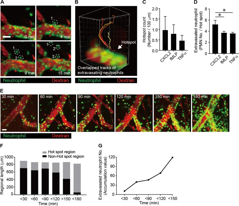
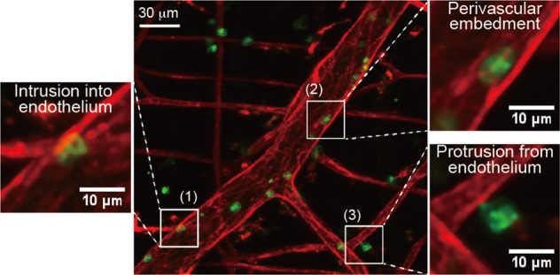
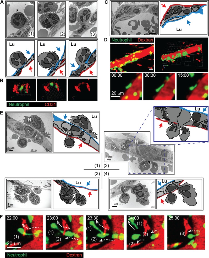
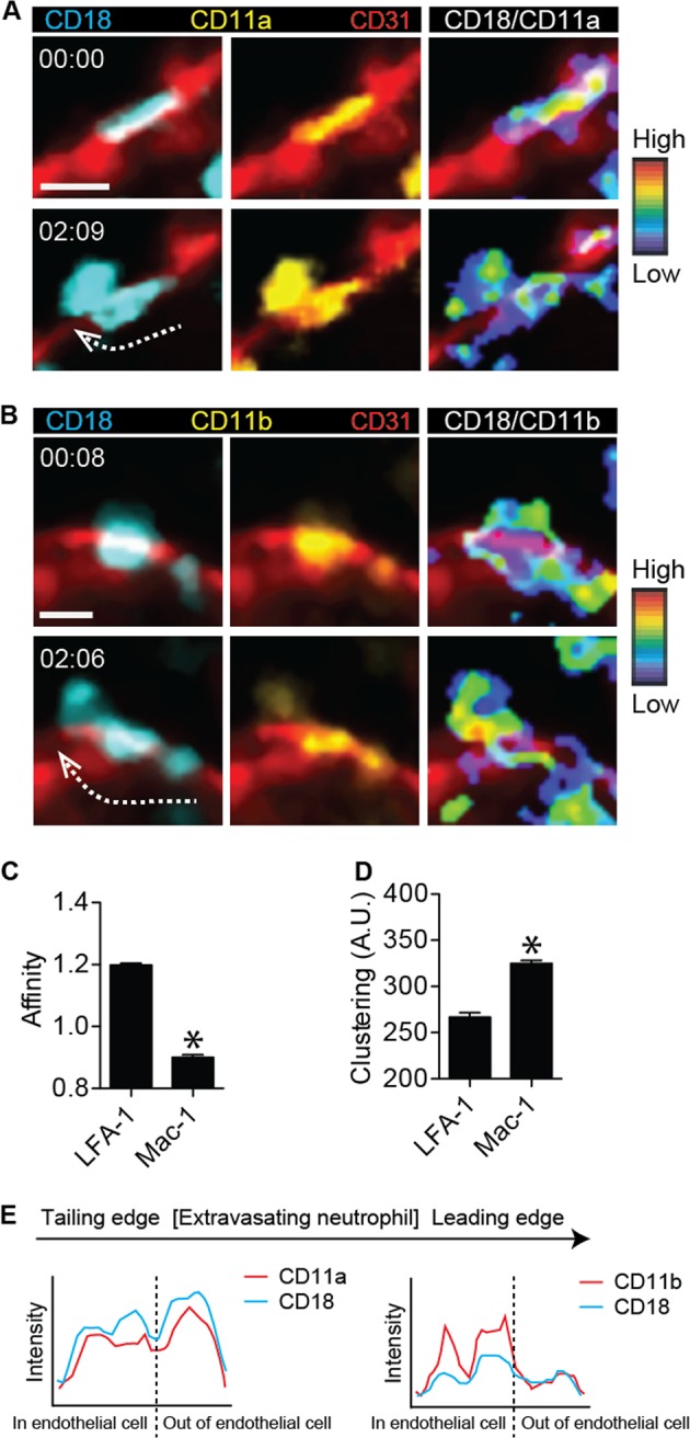
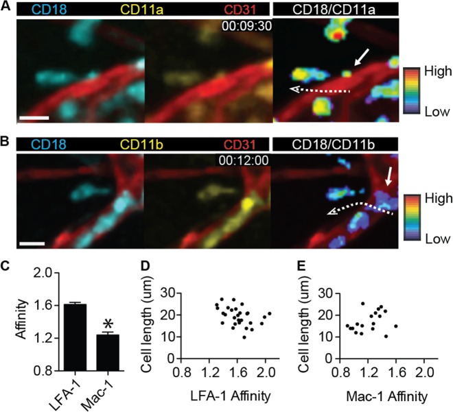
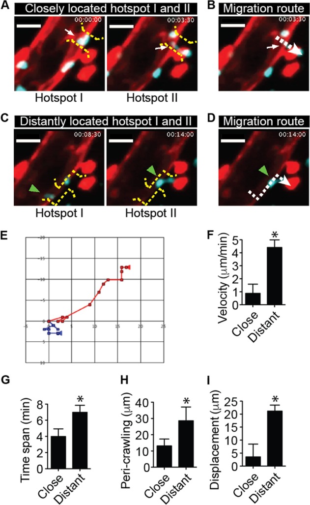
Precise spatiotemporal regulation of leukocyte extravasation is key for generating an efficient immune response to injury or infection. The integrins LFA-1(CD11a/CD18) and Mac-1(CD11b/CD18) play overlapping roles in neutrophil migration because they bind the same as well as different ligands in response to extracellular signaling. Using two-photon intravital imaging and transmission electron microscopy, we observed the existence of preferred sites for neutrophil entrance into the endothelial cell monolayer and exit from the basement membrane and pericyte sheath during neutrophil extravasation, namely, hotspots I and II, by elucidating distinctive roles of LFA-1 and Mac-1. To penetrate the vascular endothelium, neutrophils must first penetrate the endothelial cell layer through hotspot I (i.e., the point of entry into the endothelium). Neutrophils frequently remain in the space between the endothelial cell layer and the basement membrane for a prolonged period (>20 min). Subsequently, neutrophils penetrate the basement membrane and pericyte sheath at hotspot II, which is the final stage of exiting the vascular endothelium. To further investigate the roles of LFA-1 and Mac-1, we newly generated LFA-1 FRET (CD11a-YFP/CD18-CFP) mice and Mac-1 FRET (CD11b-YFP/CD18-CFP) mice. Using both FRET mice, we were able to determine that LFA-1 and Mac-1 distinctly regulate the neutrophil extravasation cascade. Our data suggest that the vascular endothelium functions as a double-layered barrier in the steps of neutrophil extravasation. We propose that the harmonized regulation of neutrophil penetration through the endothelium via hotspots I and II may be critical for vascular homeostasis during inflammation.


Conflict of interest statement

The authors declare that they have no conflict of interest.

Figures

Fig. 1
Fig. 1. Neutrophils penetrate the endothelium at hotspots during the early stage of inflammation.
a Analysis of neutrophil infiltration at a hotspot from the cremaster muscle blood vessel stimulated with N-formylmethionyl-leucyl-phenylalanine (fMLP). Dotted circles indicate neutrophils from the same hotspot. Scale bar, 50 μm. b The tracks of six extravasating neutrophils overlapped, indicating that neutrophils penetrated through a hotspot. a, b were analyzed from Video 1. c Hotspots were counted per 100 μm of blood vessels upon stimulation with C-X-C chemokine motif ligand 2 (CXCL2), fMLP, and tumor necrosis factor α (TNFα). d The numbers of neutrophils undergoing extravasation per hotspot were determined upon stimulation with CXCL2, fMLP, and TNFα. *P < 0.005. e Neutrophils penetrated the endothelial cell layer at a hotspot in the early stage of inflammation. Two-photon intravital imaging of LysM-GFP mice was performed upon stimulation with fMLP. Scale bar, 30 μm. See Video 2. f Analysis of neutrophil infiltration at a hotspot region vs. non-hotspot region at each time point upon stimulation with fMLP. g The accumulated number of neutrophils undergoing extravasation with increasing time after stimulation. f, g were analyzed from Video 2. Representative data from five independently repeated experiments are shown
Fig. 2
Fig. 2. Visualization of the substeps of neutrophil extravasation by two-photon microscopy.
Analysis of neutrophil infiltration visualized from three-dimensional two-photon images of anti-CD31 antibody-labeled blood vessels in mouse cremaster upon stimulation with N-formylmethionyl-leucyl-phenylalanine: (1) intrusion into the endothelium, in which the neutrophil cell body started to penetrate the endothelial cell layer; (2) most of the neutrophil cell body then was colocalized within the CD31-labeled endothelial cell layer; (3) as the final step, the tailing edge of the extravasating neutrophil was attached. Please see Video 3
Fig. 3
Fig. 3. The vascular endothelium is a double-layered barrier for neutrophil extravasation.
a Transmission electron microscopy revealed a gradual intrusion of neutrophils into the endothelial cell layer (upper panel). The cartoons of the transmission electron micrographs from the upper panel indicate the endothelial cell layer in red and pericytes in blue (lower panel). b Two-photon three-dimensional microscopy revealed the presence of transendothelial migratory neutrophils. See Video 4. c Transmission electron microscopy revealed neutrophil embedment in the endothelial basement between the endothelial cell layer and pericytes (upper panel). The cartoons of the transmission electron micrographs from the upper panel indicate the endothelial cell layer shown in red and pericytes in blue (lower panel). d Two-photon intravital microscopy revealed that neutrophils were embedded in the endothelial basement membrane. See Videos 5 and 6. e Transmission electron microscopy revealed that neutrophils penetrated the pericyte sheath. (1) The leading edge of an embodied neutrophil protruded from the endothelial basement membrane between the endothelial cell layer and the pericyte sheath. (2) Two neutrophils were undergoing penetration at the same site of the pericyte sheath in the panel. (3) Three neutrophils were undergoing penetration at the same site of the pericyte sheath in the panel. (4) The head and tail regions of a neutrophil are localized outside the pericyte sheath in the panel. In all of the corresponding cartoons, the endothelial cells are shown in blue, and the pericytes are in red. Blue and red arrows indicate endothelial cells and pericytes, respectively. N neutrophil, Lu lumen. f Two-photon intravital microscopy revealed three neutrophils that were continually penetrating the vascular endothelium at the same spot within 5 min of each other. The dotted arrows indicate the trajectories of the extravasating neutrophils. See Video 7. For all experiments, the data are representative of five independently repeated experiments with N-formylmethionyl-leucyl-phenylalanine stimulation
Fig. 4
Fig. 4. High-affinity LFA-1 and Mac-1 clustering were distinctly localized on neutrophils during transendothelial migration upon stimulation with N-formylmethionyl-leucyl-phenylalanine.
a CD18 expression (left panel), CD11a expression (middle panel), and the CD18-to-CD11a ratio (right panel) were visualized at two different time points during transendothelial migration of a neutrophil in LFA-1 FRET (CD11a-mYFP/CD18-mCFP) mice. Red, endothelial cellular border imaged with an Alexa 594-stained anti-CD31 antibody. Two-photon intravital two-dimensional imaging of LFA-1 FRET mice was performed. The cyan fluorescent protein/yellow fluorescent protein (CFP/YFP) ratio is visualized using the rainbow scale. The dotted arrow shows the direction of transendothelial migration of the neutrophil. Scale bar, 10 μm. See Video 8. The images are representative of at least five independent videos. b CD18 expression (left panel), CD11b expression (middle panel), and the CD18-to-CD11b ratio (right panel) were visualized at two different time points during transendothelial migration of a neutrophil in Mac-1 FRET (CD11b-mYFP/CD18-mCFP) mice. Red, endothelial cellular border imaged with an Alexa-594 stained anti-CD31 antibody. Two-photon intravital two-dimensional imaging of Mac-1 FRET mice was performed. The CFP/YFP ratio is visualized using the rainbow scale. The dotted arrow shows the direction of transendothelial migration of the neutrophil. Scale bar, 10 μm. See Video 9. The images are representative of at least five independent videos. c, d The affinity and clustering of LFA-1 and Mac-1 were quantified based on CFP, YFP, and the CFP-to-YFP ratio during two-photon intravital imaging of both LFA-1 FRET and Mac-1 FRET mice. *P < 0.0001. A.U. arbitrary units. e The intensities of CD18 and CD11a in LFA-1 mice and CD18 and CD11b in Mac-1 FRET mice were, respectively, quantified along a line from the trailing edge to the leading edge of the transendothelial migratory neutrophils. The images are representative of analyses of at least five independent neutrophils
Fig. 5
Fig. 5. High-affinity LFA-1 is localized at the trailing edge of the neutrophil during the elongation step of extravasation upon stimulation with N-formylmethionyl-leucyl-phenylalanine.
a CD18 expression (upper left panel), CD11a expression (upper right panel), Alexa 594-anti-CD31 antibody staining of an endothelial cellular border, and the CD18-to-CD11a ratio (lower right panel), visualized in the step of uropod elongation of a neutrophil in LFA-1 FRET (CD11a-mYFP/CD18-mCFP) mice. Two-photon intravital imaging of LFA-1 FRET mice was performed. The cyan fluorescent protein/yellow fluorescent protein (CFP/YFP) ratio is visualized using the rainbow scale. The arrow indicates LFA-1 affinity status at the trailing edge of an elongated neutrophil. The dotted arrow indicates the elongation of an extravasating neutrophil. Scale bar, 10 μm. See Video 10. The images are representative of at least five independent videos. b CD18 expression (upper left panel), CD11b expression (upper right panel), Alexa 594-anti-CD31 antibody staining of an endothelial cellular border, and the CD18-to-CD11a ratio (lower right panel), visualized in the step of uropod elongation of a neutrophil in Mac-1 FRET (CD11b-mYFP/CD18-mCFP) mice. Two-photon intravital imaging of Mac-1 FRET mice was performed. The CFP/YFP ratio was visualized using the rainbow scale. The arrow indicates the Mac-1 affinity status at the trailing edge of an elongated neutrophil. The dotted arrow indicates the elongation of an extravasating neutrophil. Scale bar, 10 μm. See Video 11. The images are representative of at least five independent videos. c The affinities of LFA-1 and Mac-1 were compared at the trailing edges of neutrophils during uropod elongation. *P < 0.0001. d Plot of the correlation of cell length with LFA-1 affinity. Data from two-photon intravital imaging of neutrophils in LFA-1 FRET mice during uropod elongation, as the final step of neutrophil extravasation, were analyzed. A dot indicates the correlation value of an elongated neutrophil. e Plot of the correlation of cell length with Mac-1 affinity. Data from two-photon intravital imaging of neutrophils in Mac-1 FRET mice during uropod elongation, as the final step of neutrophil extravasation, were analyzed. A dot indicates the correlation value of an elongated neutrophil
Fig. 6
Fig. 6. The distance between hotspots I and II determines the perivascular crawling pattern upon stimulation with N-formylmethionyl-leucyl-phenylalanine.
a The neutrophil quickly penetrated the vascular endothelium through closely located hotspots I and II. The white arrow indicates a hotpot, which is located between the yellow dotted brackets. b The migration route of the neutrophil through the closely located hotspots I and II is indicated by the white dotted arrow. c The neutrophil spent considerable time in the endothelial basement membrane when using the route of distantly located hotspots I and II. The green arrowhead indicates a hotpot between the yellow dotted brackets. d The migration route of the neutrophil through the distantly located hotspots I and II is indicated by the green arrowhead. ad were generated from Video 12. Scale bar, 20 μm. e The migratory patterns of neutrophils using the quick route and round route during extravasation were compared in rectangular coordinates. Unit; μm. Velocity (f), time span (g), perivascular crawling distance (h), and displacement (i) were compared between fast penetrating and round penetrating neutrophils. *P < 0.005. A representative dataset from >10 extravasating neutrophils from 3 independent intravital imaging experiments is shown

Similar articles

Cited by

References

    1. Auffray C, et al. Monitoring of blood vessels and tissues by a population of monocytes with patrolling behavior. Science. 2007;317:666–670. doi: 10.1126/science.1142883. - DOI - PubMed
    1. Muller WA. Leukocyte-endothelial-cell interactions in leukocyte transmigration and the inflammatory response. Trends Immunol. 2003;24:327–334. - PubMed
    1. Phillipson M, et al. Intraluminal crawling of neutrophils to emigration sites: a molecularly distinct process from adhesion in the recruitment cascade. J. Exp. Med. 2006;203:2569–2575. doi: 10.1084/jem.20060925. - DOI - PMC - PubMed
    1. Schenkel AR, Mamdouh Z, Muller WA. Locomotion of monocytes on endothelium is a critical step during extravasation. Nat. Immunol. 2004;5:393–400. doi: 10.1038/ni1051. - DOI - PubMed
    1. Vestweber D. Adhesion and signaling molecules controlling the transmigration of leukocytes through endothelium. Immunol. Rev. 2007;218:178–196. doi: 10.1111/j.1600-065X.2007.00533.x. - DOI - PubMed

Publication types

MeSH terms