. 2019 Nov;40(11):1424-1435.
doi: 10.1038/s41401-019-0221-0. Epub 2019 Apr 9.

Sevoflurane promotes migration, invasion, and colony-forming ability of human glioblastoma cells possibly via increasing the expression of cell surface protein 44

Affiliations

Sevoflurane promotes migration, invasion, and colony-forming ability of human glioblastoma cells possibly via increasing the expression of cell surface protein 44

Ren-Chun Lai et al. Acta Pharmacol Sin. 2019 Nov.

Abstract

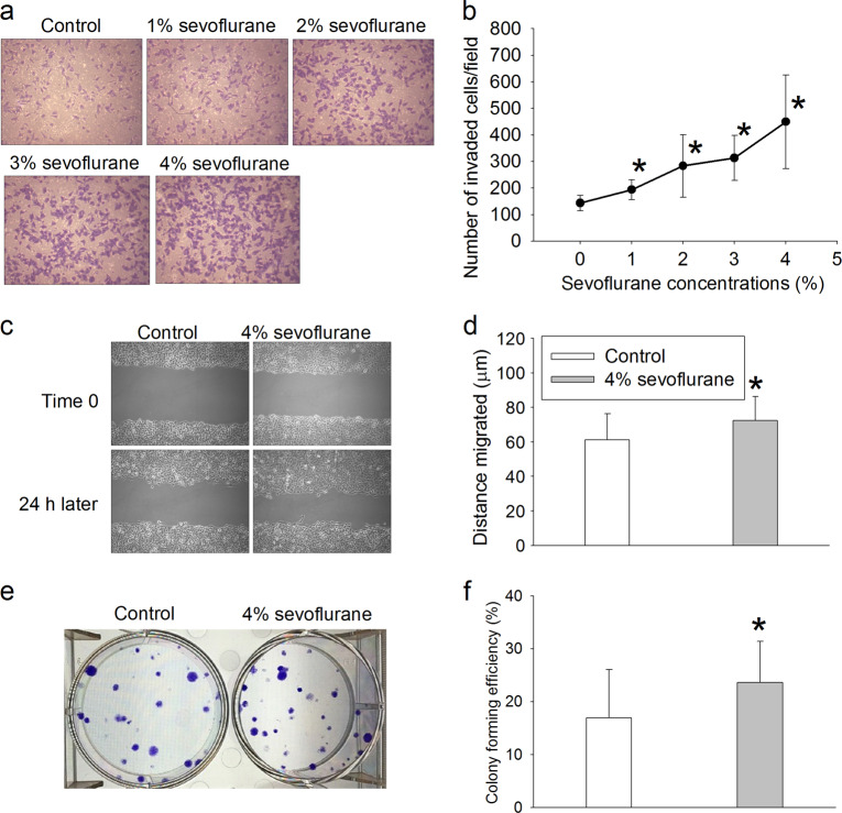
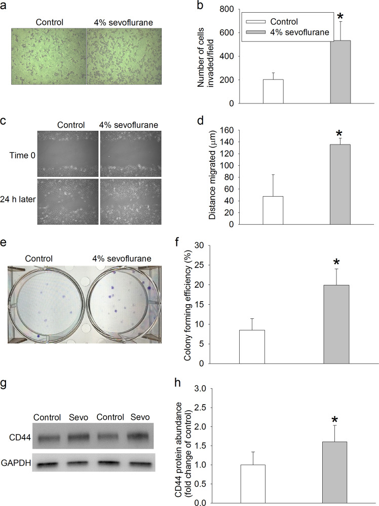
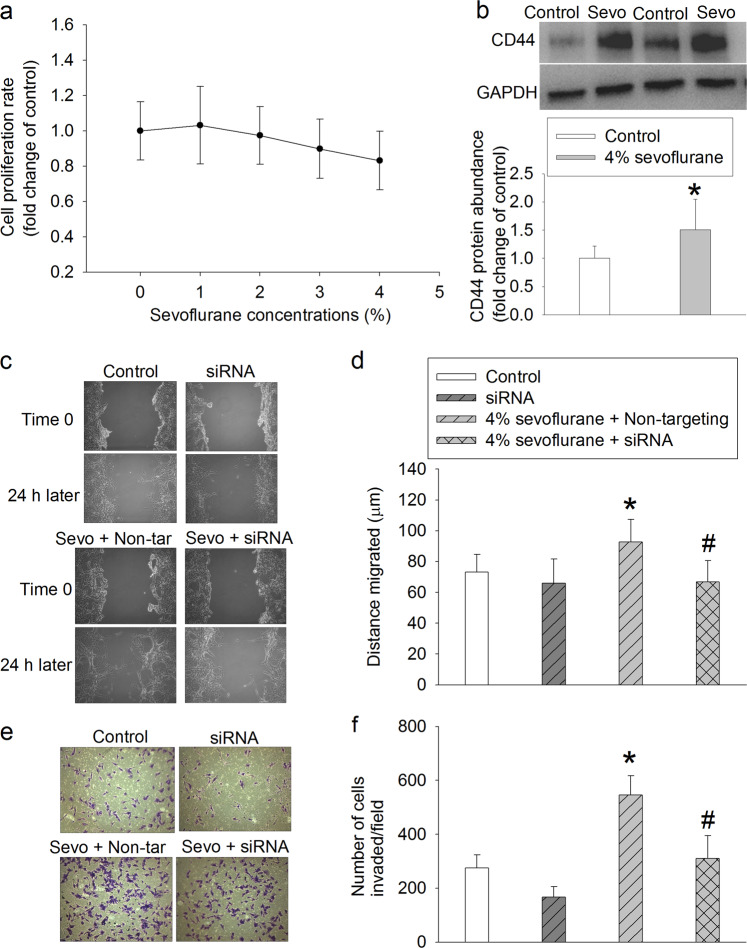
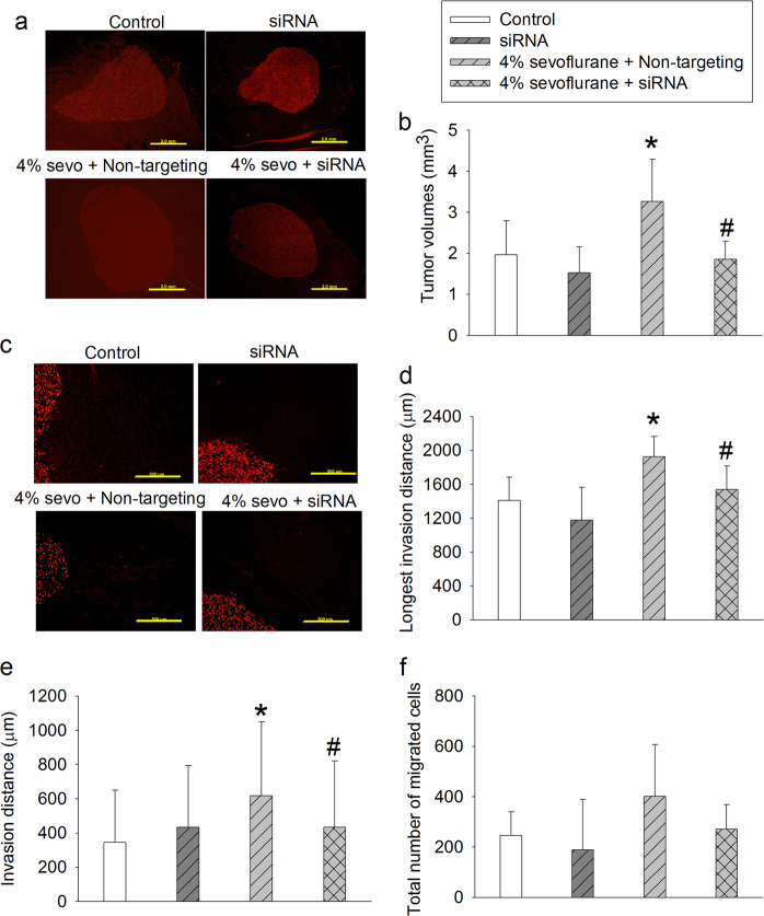
Surgical resection of primary solid tumor under anesthesia remains a common practice. It has been concerned whether general anesthetics, especially volatile anesthetics, may promote the growth, migration, and invasion of cancer cells. In this study, we examined the effects of sevoflurane on human glioblastoma cells and determined the role of cluster of differentiation (CD) 44, a cell surface protein involved in cell growth, migration, and invasion, in sevoflurane's effects. We showed that exposure to 1%-4% sevoflurane did not change the cell proliferation, but concentration-dependently increased the invasion of human glioblastoma U251 cells. Furthermore, 4% sevoflurane significantly increased the migration and colony-forming ability of U251 cells. Similar results were observed in human glioblastoma A172 cells. Exposure to sevoflurane concentration-dependently increased the activity of calpains, a group of cysteine proteinases, and CD44 protein in U251 and A172 cells. Knockdown of CD44 with siRNA abolished sevoflurane-induced increases in calpain activity, migration, invasion, and colony-forming ability of U251 cells. Inhalation of 4% sevoflurane significantly increased the tumor volume and invasion/migration distance of U87 cells from the tumor mass in the nude mice bearing human glioblastoma U87 xenograft in the brain. The aggravation by sevoflurane was attenuated by CD44 silencing. In conclusion, sevoflurane increases the migration, invasion, and colony-forming ability of human glioblastoma cells in vitro, and their tumor volume and invasion/migration in vivo. Sevoflurane enhances these cancer cell biology features via increasing the expression of CD44.

Keywords: CD44; anesthetics; calpains; colony forming; human glioblastoma; invasion; migration; sevoflurane.


Conflict of interest statement

The authors declare no competing interests.

Figures

Fig. 1
Fig. 1
No effect of sevoflurane on proliferation and cell death in U251 cells. U251 cells were exposed to various concentrations of sevoflurane for 4 h. Cells were used for experiments 24 h later. a MTT results, b trypan blue exclusion results, and c total number of cells. The results are shown as the mean ± S.D. (n = 8 for a, and n = 12 for b and c)
Fig. 2
Fig. 2
Sevoflurane-induced increase in invasion, migration, and colony-formation in U251 cells. U251 cells were exposed to various concentrations of sevoflurane for 4 h. Cells were used for experiments 24 h later. a Representative images of invaded cells, b quantitative results of cell invasion assay, c representative images of migration assay, d quantitative results of migration assay, e representative images of cell colonies, f quantitative results of colony-formation assay. The results are shown as the mean ± S.D. (n = 6 for b, n = 17 for d and n = 14 for f). *P < 0.05 compared with control
Fig. 3
Fig. 3
Sevoflurane-induced increase in A172 cell invasion, migration, and colony formation. A172 cells were exposed to 4% sevoflurane for 4 h. Cells were used for experiments 24 h later. a Representative images of invaded cells, b quantitative results of cell invasion assays, c representative images of migration assays, d quantitative results of migration assays, e representative images of cell colonies, f quantitative results of colony formation assays, g representative images of Western blotting analysis of CD44, with two representative results from control and 4% sevoflurane exposure conditions, and h quantitative results of CD44 expression analysis. The results are shown as the mean ± S.D. (n = 6 for b, n = 18 for d, n = 12 for f, and n = 6 for h). *P < 0.05 compared with control
Fig. 4
Fig. 4
Sevoflurane-induced increase in calpain activity and CD44 protein expression in U251 cells. U251 cells were exposed to various concentrations of sevoflurane for 4 h. Cells were used for experiments 24 h later. a Representative zymogram images of MMP-2 and MMP-9, b quantitative assessment of MMP-2 activity, c quantitative assessment of MMP-9 activity, d quantitative assessment of calpain activity, e representative images of Western blotting analysis of CD44, and f quantitative assessment of CD44 expression. The results are shown as the mean ± S.D. (n = 8 for b and c, n = 6 for d, and n = 7 for f). *P < 0.05 compared with control
Fig. 5
Fig. 5
Blocking of the sevoflurane-induced increase in calpain activity and invasion of U251 cells via CD44 downregulation. U251 cells were transfected with CD44 siRNA or non-targeting siRNA for 48 h and harvested for analysis to generate the results shown in a and b or exposed to 4% sevoflurane for 4 h and then used in experiments to generate the results shown in cg. a Representative Western blotting images of CD44 expression in cells without sevoflurane exposure, b quantitative results of CD44 expression in cells without sevoflurane exposure, c representative images of Western blotting of CD44 in cells exposed to sevoflurane, d quantitative results of CD44 expression in cells exposed to sevoflurane, e quantitative assessment of calpain activity, f representative images of invaded cells, g quantitative results of invasion assays. The results are shown as the mean ± S.D. (b, e) or in a box plot in which the box represents the 25% to 75% interval and the median of the data is shown as a line inside the box (d, g) (n = 6). *P < 0.05 compared with control. &P < 0.05 compared with sevoflurane alone. #P < 0.05 compared with sevoflurane plus non-targeting siRNA. Sevo: 4% sevoflurane, non-tar: non-targeting RNA
Fig. 6
Fig. 6
Blocking of the sevoflurane-induced increase in migration and colony formation of U251 cells via CD44 downregulation. U251 cells were transfected with CD44 siRNA or non-targeting siRNA for 48 h, exposed to 4% sevoflurane for 4 h and then used in experiments to generate the results shown in ad. a Representative images of migration assays, b quantitative results of migration assays, c representative images of cell colonies, d quantitative results of colony-formation assays. The results are shown as the mean ± S.D. (n = 9–42). #P < 0.05 compared with sevoflurane plus non-targeting siRNA. Sevo: sevoflurane, non-tar: non-targeting RNA
Fig. 7
Fig. 7
Role of CD44 in sevoflurane-induced increase in U87 cell migration and invasion. U87 cells were exposed to various concentrations of sevoflurane for 4 h. Cells were used for MTT assays 24 h later to generate the results shown in a. U87 cells were exposed to 4% sevoflurane for 4 h and used 24 h later for Western blotting to generate the results shown in b. In another experiment, U87 cells were transfected with CD44 siRNA or non-targeting siRNA for 48 h, exposed to 4% sevoflurane for 4 h and then used in experiments to generate the results shown in cf. a MTT results, b CD44 expression (top panel: representative Western blotting images of CD44 expression, with two representative blots from control and 4% sevoflurane exposure conditions; bottom panel: quantitative assessment of CD44 expression), c representative images of migration assays, d quantitative results of migration assays, e representative images of invaded cells, f quantitative results of invasion assays. The results are shown as the mean ± S.D. (n = 17–32). *P < 0.05 compared with control. #P < 0.05 compared with sevoflurane plus non-targeting siRNA. Sevo: 4% sevoflurane, non-tar: non-targeting RNA
Fig. 8
Fig. 8
Role of CD44 in xenograft growth and cell invasion in mouse brains. U87 cells were transfected with CD44 siRNA or non-targeting siRNA for 48 h and then implanted into mouse striatum. Mice were then exposed to sevoflurane on days 4 and 8 for 2 h and brains were harvested on day 14 after siRNA injection. a Representative images of brain sections after immunofluorescence staining to show tumor mass (scale bar = 2 mm), b tumor volumes, c representative images of brain sections after immunofluorescence staining to show cell invasion (scale bar = 0.5 mm), d longest invasion distance of cells in each mouse, e invasion distances of all visible U87 cells in mice, f number of all visible U87 cells outside of the main mass in each mouse. The results are shown as the mean ± S.D. (n = 6–9 for b, d, and f, and n = 1124–2410 cells for e). *P < 0.05 compared with control. #P < 0.05 compared with sevoflurane plus non-targeting siRNA. Sevo: 4% sevoflurane, non-targeting: non-targeting RNA

References

    1. Heaney A, Buggy DJ. Can anaesthetic and analgesic techniques affect cancer recurrence or metastasis? Br J Anaesth. 2012;109(Suppl 1):i17–i28. doi: 10.1093/bja/aes421. - DOI - PubMed
    1. Benzonana LL, Perry NJ, Watts HR, Yang B, Perry IA, Coombes C, et al. Isoflurane, a commonly used volatile anesthetic, enhances renal cancer growth and malignant potential via the hypoxia-inducible factor cellular signaling pathway in vitro. Anesthesiology. 2013;119:593–605. doi: 10.1097/ALN.0b013e31829e47fd. - DOI - PubMed
    1. Muller-Edenborn B, Roth-Z’graggen B, Bartnicka K, Borgeat A, Hoos A, Borsig L, et al. Volatile anesthetics reduce invasion of colorectal cancer cells through down-regulation of matrix metalloproteinase-9. Anesthesiology. 2012;117:293–301. doi: 10.1097/ALN.0b013e3182605df1. - DOI - PubMed
    1. Kalhori V, Tornquist K. MMP2 and MMP9 participate in S1P-induced invasion of follicular ML-1 thyroid cancer cells. Mol Cell Endocrinol. 2015;404:113–22. doi: 10.1016/j.mce.2015.01.037. - DOI - PubMed
    1. Hoskin V, Szeto A, Ghaffari A, Greer PA, Cote GP, Elliott BE. Ezrin regulates focal adhesion and invadopodia dynamics by altering calpain activity to promote breast cancer cell invasion. Mol Biol Cell. 2015;26:3464–79. doi: 10.1091/mbc.E14-12-1584. - DOI - PMC - PubMed