Review
. 2019 Sep;16(9):549-562.
doi: 10.1038/s41571-019-0204-6.

Cell-state dynamics and therapeutic resistance in melanoma from the perspective of MITF and IFNγ pathways

Affiliations
Review

Cell-state dynamics and therapeutic resistance in melanoma from the perspective of MITF and IFNγ pathways

Xue Bai et al. Nat Rev Clin Oncol. 2019 Sep.

Abstract

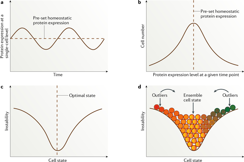
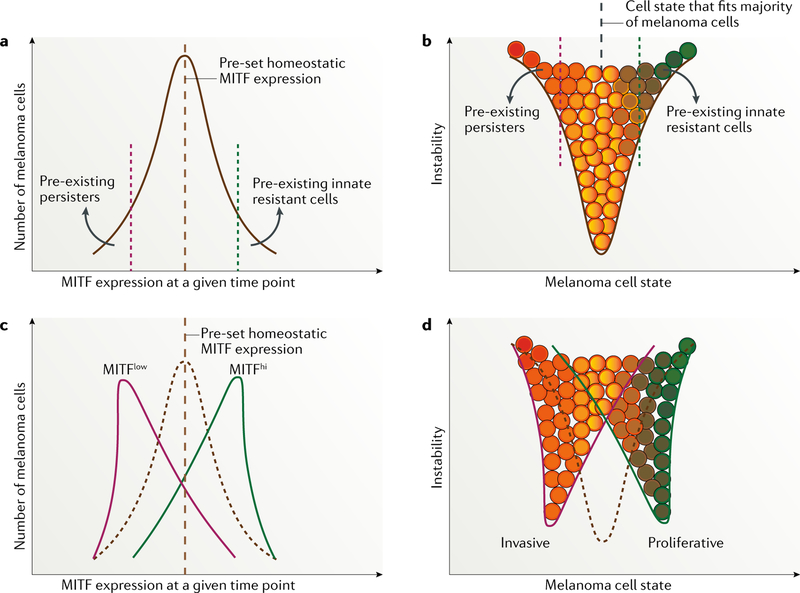
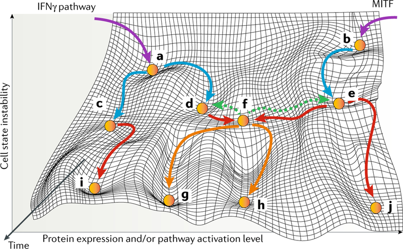
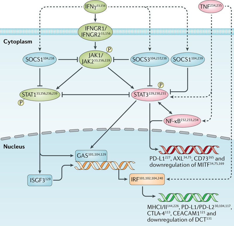
Targeted therapy and immunotherapy have greatly improved the prognosis of patients with metastatic melanoma, but resistance to these therapeutic modalities limits the percentage of patients with long-lasting responses. Accumulating evidence indicates that a persisting subpopulation of melanoma cells contributes to resistance to targeted therapy or immunotherapy, even in patients who initially have a therapeutic response; however, the root mechanism of resistance remains elusive. To address this problem, we propose a new model, in which dynamic fluctuations of protein expression at the single-cell level and longitudinal reshaping of the cellular state at the cell-population level explain the whole process of therapeutic resistance development. Conceptually, we focused on two different pivotal signalling pathways (mediated by microphthalmia-associated transcription factor (MITF) and IFNγ) to construct the evolving trajectories of melanoma and described each of the cell states. Accordingly, the development of therapeutic resistance could be divided into three main phases: early survival of cell populations, reversal of senescence, and the establishment of new homeostatic states and development of irreversible resistance. On the basis of existing data, we propose future directions in both translational research and the design of therapeutic strategies that incorporate this emerging understanding of resistance.


Figures

Fig. 1 |
Fig. 1 |. Dynamic fluctuation model.
a | Dynamically fluctuating expression pattern of a given protein longitudinally at the single-cell level. b | Cell distribution based on expression levels of a particular protein, with the vast majority of cells located around the predefined homeostatic protein expression level. c | Cell state defined by a particular protein or a group of correlated proteins entangled within a regulatory pathway, with the majority of cells located at the optimal ensemble state. The more distant from this pre-set state a cell is, the more unstable it is likely to be, with a strong tendency to switch back to the point of homeostatic balance. d | Distribution of cells of different states (depicted in different colours). Continuous colour changes indicate that cell-state transitions occur across a spectrum. Unlike the perfectly symmetric distribution shown, the cell-state curve varies for proteins that define different cell states. Immortalized melanoma cells are probably skewed towards one end of the spectrum in a context-dependent way. Importantly, unstable outliers are in a cell state that differs substantially from the majority of the cell population and are therefore expected to behave differently in a given situation, such as stress.
Fig. 2 |
Fig. 2 |. MITF expression and phenotype switching in melanoma before and after treatment with targeted agents.
a,b | Microphthalmia-associated transcription factor (MITF) expression and melanoma cell-state pattern before exposure to targeted therapy, representing a unimodal distribution of MITF expression in the same population of melanoma cells. Of note, variations in predefined points exist between melanoma cell lines, mouse models and patient samples , owing to differences in melanoma initiation and in microenvironment selective pressure. c,d | Alterations in MITF expression and variation in cell state after starting therapy.
Fig. 3 |
Fig. 3 |. Reshaping of cell states at different stages in melanoma.
The initiation, progression and development of initial and secondary therapy resistance are designated with purple, blue, red and orange arrows, respectively. Green dashed arrows demonstrate the complete reversibility of a cellular state soon after therapy initiation. Melanoma initiation can occur through an IFNγ-dependent and immunogenicity-dependent mechanism (part a) or a microphthalmia-associated transcription factor (MITF)-related mechanism (part b). Through immune editing, melanoma cells bifurcate into different states characterized by IFNγ hypoactivation and low immunogenicity (part c) and IFNγ hyperactivation and high immunogenicity (part d). MITF-related cell state is maintained, with inter-individual variations, during disease progression (part e). A persister cell state is characterized by IFNγ hyperactivation, high immunogenicity, low expression of MITF and an invasive phenotype that contribute to resistance to both targeted agents and immunotherapy (part f). This persister state is not stable and will reverse back to that seen in part d or part e if therapy is withdrawn or, otherwise, through the accumulation of both epigenetic and genetic aberrations, will evolve into more stable states (parts g,h). Epigenetic and genetic alterations can lead to a cell state of IFNγ hypoactivation and low immunogenicity (part i) that contributes to resistance to immunotherapy and to a state with extremely high MITF expression and a senescent phenotype (part j) that contributes to resistance to targeted agents.
Fig. 4 |
Fig. 4 |. IFNγ–JAK1/JAK2–STAT1/STAT3 pathway in melanoma.
Dashed lines represent positive regulation with no specific regulatory mechanism elucidated yet. Examples of regulated downstream molecules are provided. CEACAM1, carcinoembryonic antigen-related cell adhesion molecule 1; CTLA-4, cytotoxic T lymphocyte antigen 4; GAS, growth arrest-specific protein; IFNGR, IFNγ receptor; IRF, interferon regulatory factor; ISGF, interferon-stimulated transcription factor; MITF, microphthalmia-associated transcription factor; P, phosphorylated molecule; PD-L, programmed cell death 1 ligand; SOCS, suppressor of cytokine signalling; STAT, signal transducer and activator of transcription.

References

    1. Siegel RL, Miller KD & Jemal A. Cancer statistics, 2015. CA Cancer J. Clin. 65, 5–29 (2015). - PubMed
    1. Siegel RL, Miller KD & Jemal A. Cancer statistics, 2016. CA Cancer J. Clin. 66, 7–30 (2016). - PubMed
    1. Siegel RL, Miller KD & Jemal A. Cancer Statistics, 2017. CA Cancer J. Clin. 67, 7–30 (2017). - PubMed
    1. Siegel RL, Miller KD & Jemal A. Cancer statistics, 2018. CA Cancer J. Clin. 68, 7–30 (2018). - PubMed
    1. Flaherty KT et al. Inhibition of mutated, activated BRAF in metastatic melanoma. N. Engl. J. Med. 363, 809–819 (2010). - PMC - PubMed

Publication types

MeSH terms

Substances