. 2019 Mar 22:10:222.
doi: 10.3389/fphar.2019.00222. eCollection 2019.

Repeated Sigma-1 Receptor Antagonist MR309 Administration Modulates Central Neuropathic Pain Development After Spinal Cord Injury in Mice

Affiliations

Repeated Sigma-1 Receptor Antagonist MR309 Administration Modulates Central Neuropathic Pain Development After Spinal Cord Injury in Mice

Sílvia Castany et al. Front Pharmacol. .

Abstract

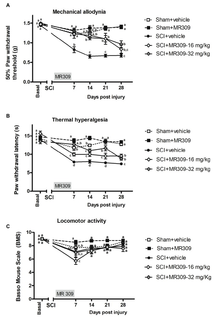
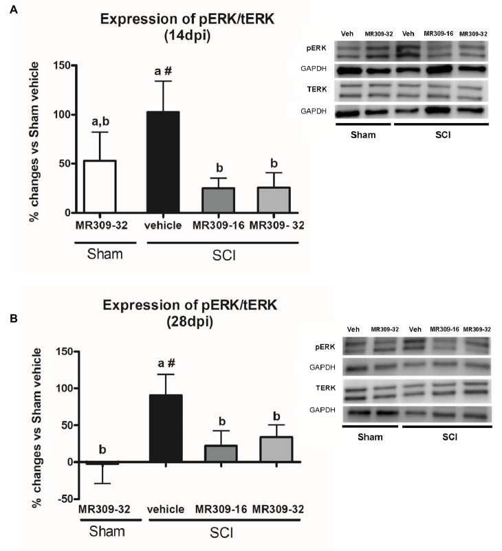
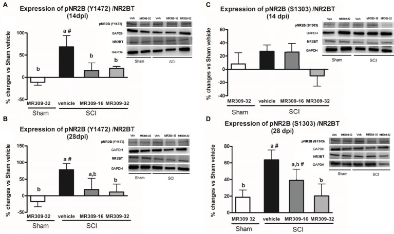
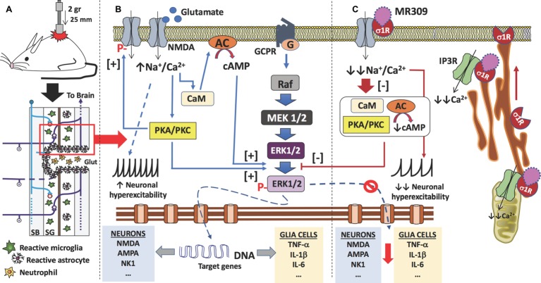
Up to two-thirds of patients affected by spinal cord injury (SCI) develop central neuropathic pain (CNP), which has a high impact on their quality of life. Most of the patients are largely refractory to current treatments, and new pharmacological strategies are needed. Recently, it has been shown that the acute administration of the σ1R antagonist MR309 (previously developed as E-52862) at 28 days after spinal cord contusion results in a dose-dependent suppression of both mechanical allodynia and thermal hyperalgesia in wild-type CD-1 Swiss female mice. The present work was addressed to determine whether MR309 might exert preventive effects on CNP development by repeated administration during the first week after SCI in mice. To this end, the MR309 (16 or 32 mg/kg i.p.) modulation on both thermal hyperalgesia and mechanical allodynia development were evaluated weekly up to 28 days post-injury. In addition, changes in pro-inflammatory cytokine (TNF-α, IL-1β) expression and both the expression and activation (phosphorylation) of the N-methyl-D-aspartate receptor subunit 2B (NR2B-NMDA) and extracellular signal-regulated kinases (ERK1/2) were analyzed. The repeated treatment of SCI-mice with MR309 resulted in significant pain behavior attenuation beyond the end of the administration period, accompanied by reduced expression of central sensitization-related mechanistic correlates, including extracellular mediators (TNF-α and IL-1β), membrane receptors/channels (NR2B-NMDA) and intracellular signaling cascades (ERK/pERK). These findings suggest that repeated MR309 treatment after SCI may be a suitable pharmacologic strategy to modulate SCI-induced CNP development.

Keywords: MR309; central neuropathic pain; central sensitization-related biomarkers; pro-inflammatory cytokines; spinal cord injury.


Figures

Figure 1
Figure 1
Time-course assessment of mechanical allodynia, thermal hyperalgesia, and locomotor activity after preventive sigma-1 receptor antagonist MR309 treatment. Each point and vertical line represent the mean ± SEM. Experimental groups: Sham-Veh (n = 17), Sham-MR309 (n = 13), SCI-Veh (n = 12), SCI-MR309-16 mg/kg (n = 7), and SCI-MR309-32 mg/kg (n = 13). a–c: groups not sharing a letter are significantly different, p < 0.05, by Duncan’s test (A) The MANOVA analysis of mechanical allodynia indicated significant effects on day (F(4,54) = 66.08, p < 0.001), treatment (F(4,57) = 46.59, p < 0.001), and interaction for day × treatment factors (F(16,165) = 8.53, p < 0.001). Significant group differences were found on post-injury days 7, 14, 21, and 28 (all p’s < 0.001) by ANOVA analysis. On the whole, mechanical allodynia is prevented in treated SCI-animals up to 14 dpi and attenuated until the end of experimental period. (B) The MANOVA analysis of thermal hyperalgesia indicated significant effects on day (F(4,58) = 55.59, p < 0.001), treatment (F(4,61) = 47.01, p < 0.001), and interaction for day × treatment factors (F(16,177) = 7.66, p < 0.001). Significant group differences were found on post-injury days 7, 14, 21, and 28 (all p’s < 0.001) by ANOVA analysis. Note that thermal hyperalgesia is prevented in MR309-32 mg/kg animals up to 7 dpi. Both doses significantly attenuate thermal hyperalgesia development up to 28 dpi. (C) The MANOVA analysis of BMS indicated significant effects on day (F(4,54) = 23.57, p < 0.001), treatment (F(4,57) = 5.01, p = 0.002) factors, and a lack of significant interaction for day × treatment (F(16,168) = 1.63, p = 0.089). Significant group differences were identified only at 7 dpi by ANOVA analysis. Considering BMS scale parameters, mild BMS alterations referring to altered paw position but not to altered horizontal locomotion, at 7 dpi. No significant differences between groups from 14 to 28 dpi.
Figure 2
Figure 2
Spinal ERK1/2 phosphorylation (pERK) expression after preventive sigma-1 receptor antagonist MR309 treatment. Quantification and representative immunoblots of total ERK (tERK), pERK, and glyceraldehyde 3-phosphate dehydrogenase (GAPDH). Experimental groups: Sham-Veh (n = 5), Sham-MR309-32 (n = 5), SCI-Veh (n = 4), SCI-MR309-16 (n = 5), and SCI-MR309-32 (n = 5). Protein expressions were normalized to GAPDH, and data are presented as a percentage respect to SCI-Veh mice. ANOVA analysis revealed significant differences at both 14 (F(4,23) = 3.54, p = 0.025) and 28 (F(4,24) = 3.65, p = 0.022) days post-injury. a, b: groups not sharing a letter are significantly different, p < 0.05, by Duncan’s test; #: significant differences vs. SCI-Veh (p < 0.05, Duncan’s test). MR309 treatments prevent pERK upregulation observed in mild spinal cord injured mice at both 14 (A) and 28 (B) days post-injury. Control images were reused either for illustrative purposes or methodological purposes when several protein levels were assessed in one blot. Full-length blots are presented in Supplementary Figure S1.
Figure 3
Figure 3
Spinal Tyr1472 and Ser1303 phosphorylation of N-methyl-D-aspartate (NMDA) receptor NR2B subunit after preventive sigma-1 receptor antagonist MR309 treatment. ANOVA analyses revealed significant group differences in pNR2B-Tyr1472 at both 14 (F(4,23) = 4.39, p = 0.011) and 28 (F(4,24) = 3.52, p = 0.025) dpi, and in pNR2B-Ser1303 at 28 dpi (F(4,19) = 4.83, p = 0.01), but not at 14 dpi (F(4,19) = 1.68, p = 0.206). (A,B) Quantification and representative immunoblots of total NR2B, pY1472NR2B, and glyceraldehyde 3-phosphate dehydrogenase (GAPDH) at 14 and 28 days post-injury. (C,D) Quantification and representative immunoblots of total NR2B, pS1303NR2B, and GAPDH at 14 and 28 days post-injury. Experimental groups: Sham-Veh (n = 5), Sham-MR309 (n = 5), SCI-Veh (n = 5), SCI-MR309-16 (n = 5), and SCI-MR309-32 (n = 5). Protein expressions were normalized to GAPDH, and data are presented as a percentage respect to SCI-Veh mice. a, b: groups not sharing a letter are significantly different, p < 0.05, by Duncan’s test; #: significant differences vs. SCI-Veh (p < 0.05, Duncan’s test). pY1472NR2B upregulation after SCI was prevented by both doses of MR309 (16 and 32 mg/kg), whereas pS1303NR2B was decreased by both doses of MR309 (16 and 32 mg/kg) at 28 days post-injury. Control images were reused either for illustrative purposes or methodological purposes when several protein levels were assessed in one blot. Full-length blots are presented in Supplementary Figure S2.
Figure 4
Figure 4
Spinal inflammatory cytokines [tumor necrosis factor-α (TNF-α) and interleukin-1β (IL-1β)] expression after preventive sigma-1 receptor antagonist MR309 treatment. The ANOVA analysis revealed significant differences between groups in TNF-α and IL-1β expression at 14 (TNF-α F(4,23) = 2.91, p = 0.049; IL-1β F(4,22) = 5.25, p = 0.006) and 28 (TNF-α F(4,18) = 3.43, p = 0.037; IL-1β F(4,24) = 6.26, p = 0.002) dpi (A,B) Quantification and representative immunoblots of TNF-α and glyceraldehyde 3-phosphate dehydrogenase (GAPDH) at 14 and 28 days post-injury. (C,D) Quantification and representative immunoblots of IL-1β and GAPDH at 14 and 28 days post-injury. Experimental groups: Sham-Veh (n = 5), Sham-MR309 (n = 5), SCI-Veh (n = 5), SCI-MR309-16 (n = 5), and SCI-MR309-32 (n = 5). Protein expressions were normalized to GAPDH, and data are presented as a percentage respect to SCI-Veh mice. a, b: groups not sharing a letter are significantly different, p < 0.05, by Duncan’s test; #: significant differences vs. SCI-Veh (p < 0.05, Duncan’s test). Both MR309 doses prevented TNF-α and IL-1β upregulation at 14 days post-injury. However, MR309 dose of 32 mg/kg, but not MR309 dose of 16 mg/kg, exerted this effect at 28 days post-injury. Control images were reused either for illustrative purposes or methodological purposes when several protein levels were assessed in one blot. Full-length blots are presented in Supplementary Figure S3.
Figure 5
Figure 5
Proposed mechanism of action of MR309 to decrease the expression of inflammatory cytokines and the hyperexcitability of spinal neurons after contusion of the spinal cord. (A) Contusion of the spinal cord of the mouse causes a spinal cord injury which results in glutamate (Glut) release by necrotic death of neurons and glial cells, medullary parenchyma destructuring, progressive reactivation of microglia and astrocytes, and the formation of the glial scar that surrounds the area of injury. In this injured area, there is also an infiltration of blood-forming elements, such as neutrophils, lymphocytes, and monocytes, is evidenced. In the neurons and glial cells surviving in the spinal cord injury (red box), the excess of glutamate in the medullary parenchyma causes a set of molecular changes. (B) The glutamate can interact with the NMDA receptors present in neurons and glial cells, causing an increase in the influx of sodium and calcium ions. In both cell types, the increase in intracellular calcium ions causes the activation of calmodulin (CaM), so that the calcium-calmodulin complex in turn activates adenylate-cyclase (AC) with an increase in the synthesis of cAMP, the second messenger that causes the activation (phosphorylation) of ERK1/2. Also, calcium ions activate protein kinases (PKA and PKC), which in turn also facilitate the phosphorylation of ERK1/2. Phosphorylated ERK1/2 translocates to the nucleus and transcription of target genes begins. In the case of spinal nociceptive neurons, genes encoding neurotransmitter receptors (e.g., AMPA, NMDA, and NK1) as well as voltage-gated ion channels are transcribed. While in the case of glial cells (astrocytes and microglia), genes encoding pro-inflammatory cytokines are transcribed (e.g., TNF-α, IL-1β, and IL-6). On the other hand, the influx of sodium ions causes an increase in the neuronal excitability of the spinal nociceptive neurons, with an increase in action potentials by the spinothalamic tract toward the brain. Gene transcription of ion channels and receptors for neurotransmitters also facilitates neuronal hyperexcitability. Finally, activated protein kinases also cause phosphorylation of NMDA-receptors that will further contribute to central sensitization. (C) σ₁ receptor classified as a ligand-regulated molecular chaperone is activated under stress or pathological conditions and interacts with several neurotransmitter receptors and ion channels to modulate their function and its activity can be regulated by endogenous and/or synthetic compounds in an agonist-antagonist manner (Díaz et al., 2009). Upon σ₁R activation under stress or pathological conditions (SCI in this case), σ1R in the ER binds to IP3R to enhance Ca2+ influx into mitochondria and efflux into the cytosol. There is also redistribution of σ1R from mitochondria-associated endoplasmic reticulum membrane to peripheral endoplasmic membranes to bind ion channels, receptors (NMDA) or protein kinases which, in turn will produce a further increase in intracellular Ca2+. Therefore, σ1R antagonism by MR309 would result in a reduced Ca2+ cytosolic mobilization from ER stores (via PLC and IP3R) and in a reduced extracellular entry through NMDAR, favoring the access of negative regulators of the receptor such as Ca2+-CaM, finally resulting in an inhibition of Ca2+ dependent intracellular effectors such as PKC/PKA and CaMKII. This downstream modulation would modulate the ERK pathway by preventing the phosphorylation of ERK1/2, and consequently, the levels of gene expression of NMDAR and proinflammatory cytokines, together with a decrease in the overactivity of NMDAR (mediated by phosphorylations at S1303 and Y1472). All of these changes would decrease neuronal and glial hyperexcitability and thus produce an attenuation in central neuropathic pain development.

References

    1. Abe T., Matsumura S., Katano T., Mabuchi T., Takagi K., Xu L., et al. . (2005). Fyn kinase-mediated phosphorylation of NMDA receptor NR2B subunit at Tyr1472 is essential for maintenance of neuropathic pain. Eur. J. Neurosci. 22, 1445–1454. 10.1111/j.1460-9568.2005.04340.x, PMID: - DOI - PubMed
    1. Ahn S. H., Park H. W., Lee B. S., Moon H. W., Jang S. H., Sakong J., et al. . (2003). Gabapentin effect on neuropathic pain compared among patients with spinal cord injury and different durations of dymptoms. Spine 28, 341–346. 10.1097/01.BRS.0000048464.57011.00, PMID: - DOI - PubMed
    1. Álvarez-Pérez B., Homs J., Bosch-Mola M., Puig T., Reina F., Verdú E., et al. . (2016). Epigallocatechin-3-gallate treatment reduces thermal hyperalgesia after spinal cord injury by down-regulating RhoA expression in mice. Eur. J. Pain 20, 341–352. 10.1002/ejp.722, PMID: - DOI - PubMed
    1. Anwar M. A., Al Shehabi T. S., Eid A. H. (2016). Inflammogenesis of secondary spinal cord injury. Front. Cell. Neurosci. 13:98. 10.3389/fncel.2016.00098 - DOI - PMC - PubMed
    1. Ataoğlu E., Tiftik T., Kara M., Tunç H., Ersöz M., Akkuş S. (2013). Effects of chronic pain on quality of life and depression in patients with spinal cord injury. Spinal Cord 51, 23–26. 10.1038/sc.2012.51, PMID: - DOI - PubMed