. 2019 Mar 22:9:63.
doi: 10.3389/fcimb.2019.00063. eCollection 2019.

Burkholderia pseudomallei BimC Is Required for Actin-Based Motility, Intracellular Survival, and Virulence

Affiliations

Burkholderia pseudomallei BimC Is Required for Actin-Based Motility, Intracellular Survival, and Virulence

Varintip Srinon et al. Front Cell Infect Microbiol. .

Abstract

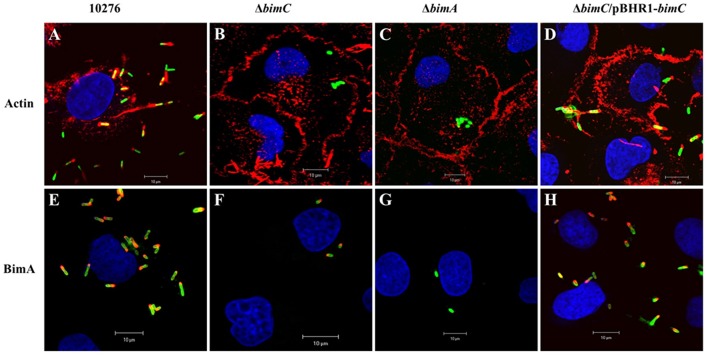
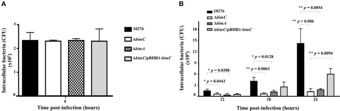
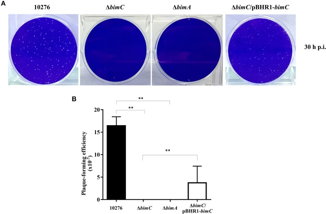
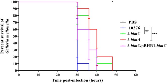
The intracellular pathogen Burkholderia pseudomallei, the etiological agent of melioidosis in humans and various animals, is capable of survival and movement within the cytoplasm of host cells by a process known as actin-based motility. The bacterial factor BimA is required for actin-based motility through its direct interaction with actin, and by mediating actin polymerization at a single pole of the bacterium to promote movement both within and between cells. However, little is known about the other bacterial proteins required for this process. Here, we have investigated the role of the bimC gene (bpss1491) which lies immediately upstream of the bimA gene (bpss1492) on the B. pseudomallei chromosome 2. Conserved amongst all B. pseudomallei, B. mallei and B. thailandensis strains sequenced to date, this gene encodes an iron-binding protein with homology to a group of proteins known as the bacterial autotransporter heptosyltransferase (BAHT) family. We have constructed a B. pseudomallei bimC deletion mutant and demonstrate that it is defective in intracellular survival in HeLa cells, but not in J774.1 macrophage-like cells. The bimC mutant is defective in cell to cell spread as demonstrated by ablation of plaque formation in HeLa cells, and by the inability to form multi-nucleated giant cells in J774.1 cells. These phenotypes in intracellular survival and cell to cell spread are not due to the loss of expression and polar localization of the BimA protein on the surface of intracellular bacteria, however they do correlate with an inability of the bacteria to recruit and polymerize actin. Furthermore, we also establish a role for bimC in virulence of B. pseudomallei using a Galleria mellonella larvae model of infection. Taken together, our findings indicate that B. pseudomallei BimC plays an important role in intracellular behavior and virulence of this emerging pathogen.

Keywords: BimA; BimC; Burkholderia pseudomallei; actin-based motility; intracellular survival; multi-nucleated giant cell; virulence.


Figures

Figure 1
Figure 1
Actin-based motility of B. pseudomallei strains in HeLa cells. B. pseudomallei wild-type strain 10276 (A,E), ΔbimC mutant (B,F), ΔbimA mutant (C,G), and ΔbimC/pBHR1-bimC (D,H) strains were used to infect HeLa cells. At 12 h post infection, the infected epithelial cells and bacteria were stained for bacteria and actin-tails (A–D), or bacteria and BimA protein expression (E–H). Bacteria (green) were stained with anti-B. pseudomallei lipopolysaccharide antibody and detected with anti-rabbit antibody-AlexaFluor488. F-actin (red) was stained with phalloidin-AlexaFluor568 and nuclei (blue) were stained with DAPI. BimA protein (red) was stained with a panel of three monoclonal antibodies detected with anti-mouse antibody-AlexaFluor568. Scale bar = 10 μm.
Figure 2
Figure 2
Net intracellular replication and survival of B. pseudomallei strains within HeLa cells. HeLa cells were infected with B. pseudomallei strains at an MOI of 50. At the indicated time point, the numbers of viable intracellular bacteria (CFU) were determined. Data from 6 h post-infection is shown in A and data from 12, 18, and 24 h post-infection in B. The graphs show data for the wild-type (black bars), ΔbimC mutant (white bars), ΔbimA mutant (dotted bars), and ΔbimC/pBHR1-bimC (gray bars) strains. Error bars represent standard errors of the means from three independent experiments (n = 3 biological replicates). Asterisks indicate significant differences (P < 0.01, t-test).
Figure 3
Figure 3
Intercellular spreading of B. pseudomallei strains in HeLa cells. (A) Representative micrographs of cell monolayers and (B) plaque-forming efficiency of HeLa cells infected with B. pseudomallei wild-type (10276), ΔbimC mutant, ΔbimA mutant, or ΔbimC/pBHR1-bimC strains. Plaque-forming efficiency was calculated as the number of plaques at 30 h post infection divided by bacterial CFU added per well. Error bars represent standard errors of the means from three independent experiments (n = 3 biological replicates). Asterisks indicate significant differences (P < 0.01, t-test).
Figure 4
Figure 4
MNGC formation in J774A.1 cells. B. pseudomallei wild-type 10276 (A), ΔbimC mutant (B), ΔbimA mutant (C), and ΔbimC/pBHR1-bimC (D) strains were used to infect J774A.1 cells. At 24 h post infection, the bacteria were stained with anti-B. pseudomallei lipopolysaccharide antibody and anti-rabbit-AlexaFluor488. F-actin (red) was stained with phalloidin-AlexaFluor568 and nuclei (blue) were stained with DAPI. Scale bar = 10 μm.
Figure 5
Figure 5
Virulence of B. pseudomallei strains in Galleria mellonella larvae. Representative data from an experiment where groups of 10 insect larvae were challenged with 100 CFU of either B. pseudomallei wild-type 10276, ΔbimC mutant, ΔbimA mutant, or the ΔbimC/pBHR1-bimC strain. The numbers of dead larvae were scored at 24, 30, 36, 40, and 48 h post infection. GraphPad Prism software was used to graph and analyze the data using a Log-rank (Mantel-Cox) test. Asterisks indicate significant differences (P < 0.05) in mean time to death (MTTD) between larvae infected with B. pseudomallei wild-type 10276 and the mutant strains. Data is representative of that obtained in three independent experiments (n = 3 biological replicates).

References

    1. Benanti E. L., Nguyen C. M., Welch M. D. (2015). Virulent Burkholderia species mimic host actin polymerases to drive actin-based motility. Cell 161, 348–360. 10.1016/j.cell.2015.02.044 - DOI - PMC - PubMed
    1. Benz I., Schmidt M. A. (2011). Structures and functions of autotransporter proteins in microbial pathogens. Int. J. Med. Microbiol. 301, 461–468. 10.1016/j.ijmm.2011.03.003 - DOI - PubMed
    1. Boddey J. A., Day C. J., Flegg C. P., Ulrich R. L., Stephens S. R., Beacham I. R., et al. (2007). The bacteria‘l gene lfpA influences the potent induction of calcitonin receptor and osteoclast-related genes in Burkholderia pseudomallei-induced TRAP-positive multinucleated giant cells. Cell. Microbiol. 9, 514–531. 10.1111/j.1462-5822.2006.00807.x - DOI - PubMed
    1. Burtnick M. N., Brett P. J. (2013). Burkholderia mallei and Burkholderia pseudomallei cluster 1 type VI secretion system gene expression is negatively regulated by iron and zinc. PLoS ONE 8:e76767. 10.1371/journal.pone.0076767 - DOI - PMC - PubMed
    1. Burtnick M. N., Brett P. J., Harding S. V., Ngugi S. A., Ribot W. J., Chantratita N., et al. (2011). The cluster 1 type VI secretion system is a major virulence determinant in Burkholderia pseudomallei. Infect. Immun. 79, 1512–1525. 10.1128/IAI.01218-10 - DOI - PMC - PubMed

Publication types

LinkOut - more resources