

Review
doi: 10.3389/fmolb.2019.00014.
eCollection 2019.
Highlighting the Proteasome: Using Fluorescence to Visualize Proteasome Activity and Distribution
Affiliations
- PMID: 30968028
- PMCID: PMC6438883
- DOI: 10.3389/fmolb.2019.00014
Item in Clipboard
Review
Highlighting the Proteasome: Using Fluorescence to Visualize Proteasome Activity and Distribution
Front Mol Biosci.
.
Abstract
Proteasomes are critical proteases in the cell responsible for the turnover of many cytoplasmic and nuclear proteins. They are essential for many cellular processes and various diseases are associated with their malfunctioning. Proteasome activity depends on the nature of the catalytic subunits, as well as the interaction with associated proteasome regulators. Here we describe various fluorescence-based methods to study proteasome function, highlighting the use of activity-based probes to study proteasome localization, dynamics, and activity in living cells.
Keywords: activity; distribution; fluorescence; probe; proteasome.
Figures
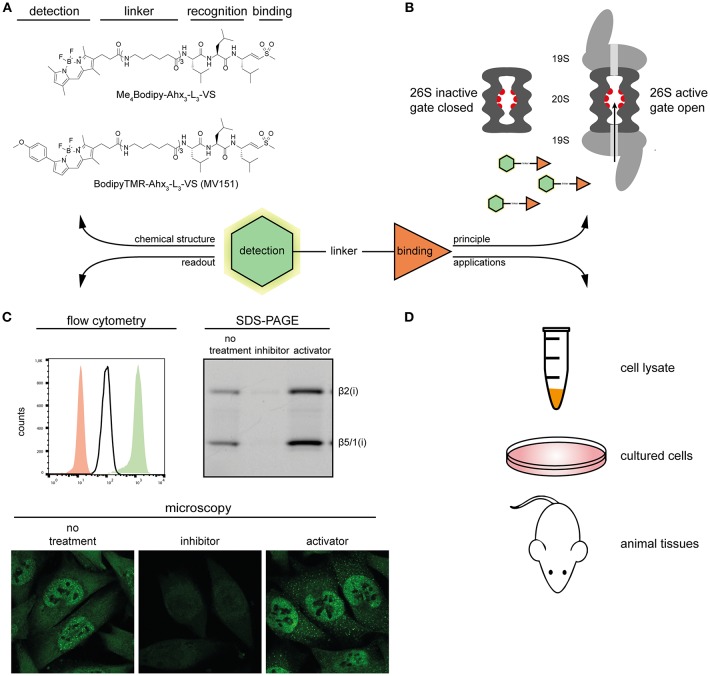
Overview of proteasome ABPs. (A) Molecular structures of two proteasome ABPs. (B) The principle of how probes target the active proteasome: proteasome ABPs enter through the 20S proteasome gate, and covalently target the catalytic sites. (C) Typical examples of the detection methods of proteasome ABPs. Left, overlay of the ABP signal in proteasome inhibitor treated (red), untreated (white), and proteasome activator treated (green) MelJuSo cells; Right, In-gel fluorescence scan showing representative proteasome activity profiles of proteasome inhibitor treated, untreated, and proteasome activator treated MelJuSo cells; Below, confocal microscopy images of the ABP signal in proteasome inhibitor treated, untreated and proteasome activator treated MelJuSo cells. (D) Applications of proteasome ABPs.
References
Publication types
LinkOut - more resources
Full Text Sources
Other Literature Sources