. 2019 Jan-Dec:33:2058738419839592.
doi: 10.1177/2058738419839592.

Immunomodulatory effects of chemotherapy on blood lymphocytes and survival of patients with advanced non-small cell lung cancer

Affiliations

Immunomodulatory effects of chemotherapy on blood lymphocytes and survival of patients with advanced non-small cell lung cancer

Mohanad Aldarouish et al. Int J Immunopathol Pharmacol. 2019 Jan-Dec.

Abstract

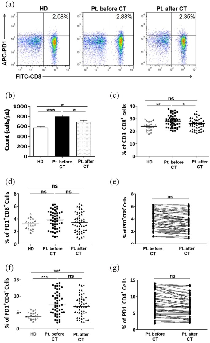
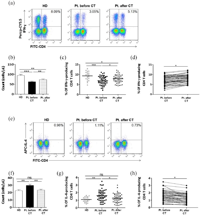
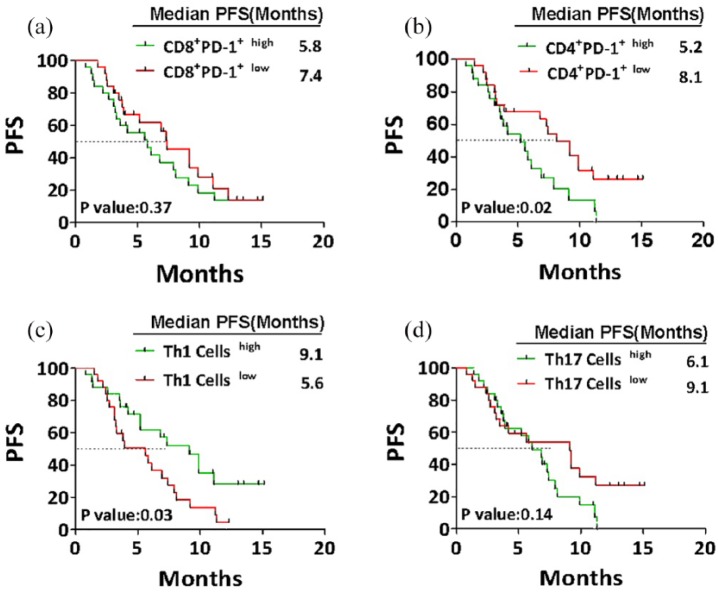
A better understanding of the immune profile of non-small cell lung cancer (NSCLC) and the immunomodulatory impact of chemotherapy is essential to develop current therapeutic approaches. Herein, we collected peripheral blood from 20 healthy donors and 50 patients with advanced NSCLC, before and after chemotherapy, followed by phenotypic analysis of lymphocyte subsets and assessment of the correlation between their post-chemotherapy levels and progression-free survival (PFS). Results showed that, before chemotherapy, the levels of CD8+ lymphocytes, PD-1+CD4+, Th2, and Th17 cells were elevated in patients' peripheral blood, in contrast to natural killer (NK) cells and Th1 cells. Besides, there was no remarkable difference in the frequency of PD-1+CD8+ cells between patients and healthy controls. After chemotherapy, the levels of CD8+ lymphocytes, NK, Th2, Th17, and Treg were declined, in contrast to the level of Th1 cells which was markedly increased. Importantly, chemotherapy had no impact on the frequencies of PD-1+CD8+ and PD-1+CD4+ cells. PFS was significantly better in patients with low percentage of PD-1+CD4+ T cells than those with high percentage. Patients with high content of Th1 cells showed longer PFS than those with low content. The low percentages of Th17 and Treg cells were correlated with longer PFS, even though the difference did not reach statistical significance. In conclusion, the imbalance of lymphocyte subsets is a hallmark of NSCLC. Furthermore, the high level of PD-1+CD4+ cells plays a crucial role in the progression of NSCLC and could be used as a prognostic marker; and the high level of Th1 could predict better clinical outcomes of chemotherapy.

Keywords: NSCLC; PFS; chemotherapy; immunomodulatory; immunophenotyping; lymphocytes.


Conflict of interest statement

Declaration of conflicting interests: The author(s) declared no potential conflicts of interest with respect to the research, authorship, and/or publication of this article.

Figures

Figure 1.
Figure 1.
CD8+ T cells, but not PD-1 expressing CD4+ and CD8+ T cells, were significantly decreased in the peripheral blood from patients with NSCLC after two cycles of chemotherapy. PBMCs were collected from peripheral blood samples of 20 healthy donors and from 50 patients with NSCLC before and after chemotherapy: (a) Representative staining patterns of PD-1+ on CD8+ T cells (CD3+ gated) of one healthy donor and one patient before and after two cycles of chemotherapy. (b, c) The absolute number (b) and frequency (c) of CD3+CD8+ T cells in the peripheral blood of healthy donors and patients with NSCLC before and after chemotherapy. (d, f) Frequencies of PD-1-expressing CD3+CD8+ (d) and CD3+CD4+ (f) T cells in peripheral blood of healthy donors and patients with NSCLC before and after chemotherapy. (e, g) Representing the impact of chemotherapy on the frequencies of PD-1+CD8+ (e) and PD-1+CD4+ (g) T cells in peripheral blood of patients with NSCLC. HD: healthy donor; CT: chemotherapy.
Figure 2.
Figure 2.
Chemotherapy had positive and negative impacts on the number and frequency of Th1 cells and Th2 cells, respectively. PBMCs were collected from peripheral blood samples of 20 healthy donors and from 50 patients with NSCLC patients before and after chemotherapy. (a, e) Representative staining patterns of IFN-γ (a) and IL-4 (e) production by CD4+ T cells (CD3+ gated) of one healthy donor and one patient before and after two cycles of chemotherapy. (b, c) The absolute number (b) and frequency (c) of Th1 cells in the peripheral blood of healthy donors and patients with NSCLC before and after chemotherapy. (f, g) The absolute number (f) and frequency (g) of Th2 cells in the peripheral blood of healthy donors and patients with NSCLC before and after chemotherapy. (d, h) Representing the impact of chemotherapy on the frequencies of Th1 cells (d) and Th2 cells (h) in peripheral blood of patients with NSCLC. HD: healthy donor; CT: chemotherapy.
Figure 3.
Figure 3.
The correlation between post-chemotherapy levels of lymphocyte subsets and patients’ PFS. After two cycles of chemotherapy, 50 patients with NSCLC were divided into two groups each with 25 patients according to the median frequency of CD8+PD-1+, CD4+PD-1+, Th1 cells, and Treg cells. The median value was assessed as cutoff point to define “high” and “low” contents. Kaplan–Meier curves representing the correlation between progression-free survival (PFS) of patients and the relative contents of CD8+PD-1+ (a), CD4+PD-1+ (b), Th1 cells (c), and Treg cells (d). Statistical significance was determined by a log-rank (Mantel–Cox) test.

References

    1. Aldarouish M, Wang C. (2016) Trends and advances in tumor immunology and lung cancer immunotherapy. Journal of Experimental and Clinical Cancer Research: CR 35(1): 157. - PMC - PubMed
    1. Markman JL, Shiao SL. (2015) Impact of the immune system and immunotherapy in colorectal cancer. Journal of Gastrointestinal Oncology 6(2): 208–223. - PMC - PubMed
    1. Matsushita H, Vesely MD, Koboldt DC, et al. (2012) Cancer exome analysis reveals a T-cell-dependent mechanism of cancer immunoediting. Nature 482(7385): 400–404. - PMC - PubMed
    1. Kida H, Ihara S, Kumanogoh A. (2013) Involvement of STAT3 in immune evasion during lung tumorigenesis. Oncoimmunology 2(1): e22653. - PMC - PubMed
    1. Salmon H, Donnadieu E. (2012) Within tumors, interactions between T cells and tumor cells are impeded by the extracellular matrix. Oncoimmunology 1(6): 992–994. - PMC - PubMed

MeSH terms

Substances