Review
. 2019 May 1;34(3):198-215.
doi: 10.1152/physiol.00048.2018.

Hepatic Insulin Clearance: Mechanism and Physiology

Affiliations
Review

Hepatic Insulin Clearance: Mechanism and Physiology

Sonia M Najjar et al. Physiology (Bethesda). .

Abstract

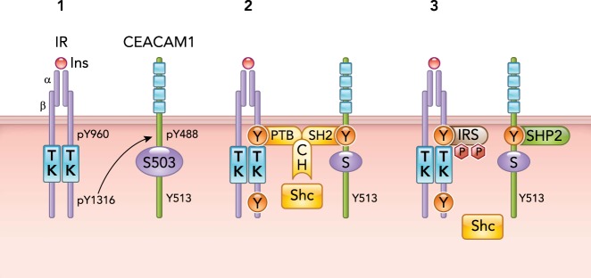
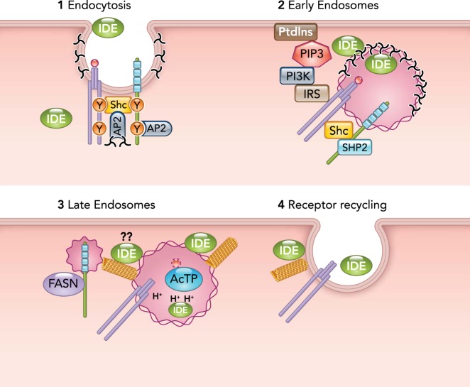
Upon its secretion from pancreatic β-cells, insulin reaches the liver through the portal circulation to exert its action and eventually undergo clearance in the hepatocytes. In addition to insulin secretion, hepatic insulin clearance regulates the homeostatic level of insulin that is required to reach peripheral insulin target tissues to elicit proper insulin action. Receptor-mediated insulin uptake followed by its degradation constitutes the basic mechanism of insulin clearance. Upon its phosphorylation by the insulin receptor tyrosine kinase, carcinoembryonic antigen-related cell adhesion molecule 1 (CEACAM1) takes part in the insulin-insulin receptor complex to increase the rate of its endocytosis and targeting to the degradation pathways. This review summarizes how this process is regulated and how it is associated with insulin-degrading enzyme in the liver. It also discusses the physiological implications of impaired hepatic insulin clearance: Whereas reduced insulin clearance cooperates with increased insulin secretion to compensate for insulin resistance, it can also cause hepatic insulin resistance. Because chronic hyperinsulinemia stimulates hepatic de novo lipogenesis, impaired insulin clearance also causes hepatic steatosis. Thus impaired insulin clearance can underlie the link between hepatic insulin resistance and hepatic steatosis. Delineating these regulatory pathways should lead to building more effective therapeutic strategies against metabolic syndrome.


Figures

FIGURE 1.
FIGURE 1.
Regulation of Insulin Signaling by CEACAM1 Insulin (Ins) binding to the α-subunit of its receptor (IRα) activates its tyrosine kinase (TK) to catalyze its autophosphorylation on tyrosine 960 (pY960) in the juxtamembrane domain and on tyrosine 1316 (pY1316) on the COOH-terminus tail of the β-subunit (IRβ) of the receptor (1). CEACAM1 phosphorylation on Y488 requires phosphorylation on Y1316 in IRβ (1). pY488 on CEACAM1 binds to the SH2 domain of Shc, which, in turn, allows the binding of the PTB domain of Shc to pY960 in IRβ. In this manner, Shc mediates the formation of a complex between IR and CEACAM1 (2). CEACAM1 binds to SHP2 to sequester it. This protects IRS against the phosphatase and prolongs its activation. This also releases Shc from pY960 in IRβ (3).
FIGURE 2.
FIGURE 2.
Coordinated regulation of receptor-mediated insulin intracellular trafficking by CEACAM1 and IDE Step 1: insulin binding to its receptor activates it to form a complex with CEACAM1 via Shc (as described in the legend to FIGURE 1). Together with Y513 in CEACAM1, the collagen homology domain of Shc targets the complex to the AP2 adaptin in the clathrin-coated pits. Step 2: the complex is internalized inside the early endosomes, and the extracellular IDE becomes sequestered into the vesicular lumen. Progressive SHP2 binding to pY488 on CEACAM1 destabilizes the complex and causes a progressive loss of Shc to allow IRS binding to pY960. This leads to increased IRS phosphorylation and activation of PI3Kinase that, in turn, mediates the recruitment of cytosolic IDE to the outer membrane and maintainance of CEACAM1 at the endosomes. Luminal IDE degrades insulin. Step 3: as the endosomes mature into late endosomes and acidify (H+), IDE is inactivated. FASN binding to pY488 pulls off CEACAM1 from the insulin-IR complex to destabilize it and allow insulin to dissociate from its receptor to be degraded by the acidic thiol protease (AcTP). Step 4: the receptor is recycled back to the membrane during exocytosis, resulting from microtubular polymerization, which is possibly regulated by IDE at the outer membrane of the vesicle. Whether this IDE mediates the formation of the CEACAM1/FASN vesicle remains to be tested.

References

    1. Abdul-Hay SO, Kang D, McBride M, Li L, Zhao J, Leissring MA. Deletion of insulin-degrading enzyme elicits antipodal, age-dependent effects on glucose and insulin tolerance. PLoS One 6: e20818, 2011. doi:10.1371/journal.pone.0020818. - DOI - PMC - PubMed
    1. Abou-Rjaily GA, Lee SJ, May D, Al-Share QY, Deangelis AM, Ruch RJ, Neumaier M, Kalthoff H, Lin SH, Najjar SM. CEACAM1 modulates epidermal growth factor receptor–mediated cell proliferation. J Clin Invest 114: 944–952, 2004. doi:10.1172/JCI200421786. - DOI - PMC - PubMed
    1. Affholter JA, Cascieri MA, Bayne ML, Brange J, Casaretto M, Roth RA. Identification of residues in the insulin molecule important for binding to insulin-degrading enzyme. Biochemistry 29: 7727–7733, 1990. doi:10.1021/bi00485a022. - DOI - PubMed
    1. Akiyama H, Shii K, Yokono K, Yonezawa K, Sato S, Watanabe K, Baba S. Cellular localization of insulin-degrading enzyme in rat liver using monoclonal antibodies specific for this enzyme. Biochem Biophys Res Commun 155: 914–922, 1988. doi:10.1016/S0006-291X(88)80583-7. - DOI - PubMed
    1. Al-Share QY, DeAngelis AM, Lester SG, Bowman TA, Ramakrishnan SK, Abdallah SL, Russo L, Patel PR, Kaw MK, Raphael CK, Kim AJ, Heinrich G, Lee AD, Kim JK, Kulkarni RN, Philbrick WM, Najjar SM. Forced hepatic overexpression of CEACAM1 curtails diet-induced insulin resistance. Diabetes 64: 2780–2790, 2015. doi:10.2337/db14-1772. - DOI - PMC - PubMed

Publication types