. 2019 Jun 1;30(12):1425-1436.
doi: 10.1091/mbc.E18-10-0687. Epub 2019 Apr 10.

N-glycosylation-dependent regulation of hK2P17.1 currents

Affiliations

N-glycosylation-dependent regulation of hK2P17.1 currents

Felix Wiedmann et al. Mol Biol Cell. .

Abstract

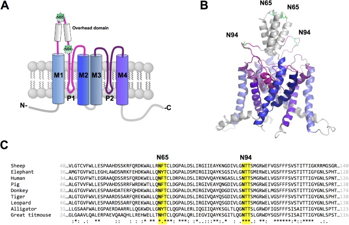
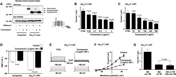
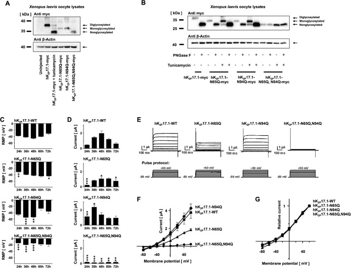
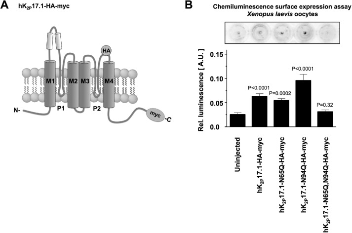
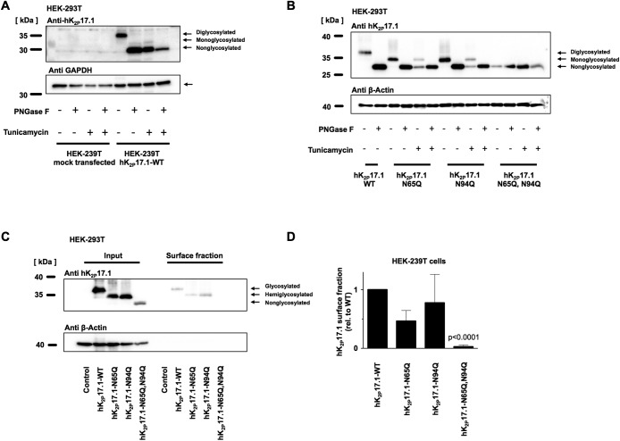
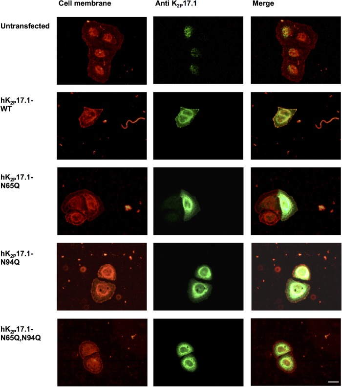
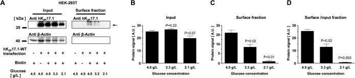
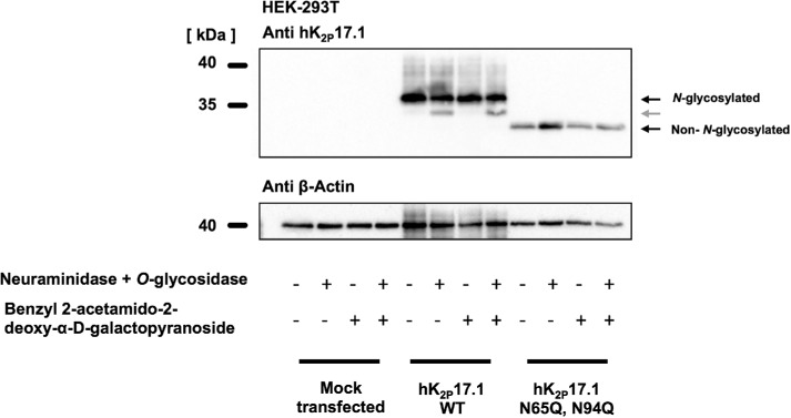
Two pore-domain potassium (K2P) channels mediate potassium background currents that stabilize the resting membrane potential and facilitate action potential repolarization. In the human heart, hK2P17.1 channels are predominantly expressed in the atria and Purkinje cells. Reduced atrial hK2P17.1 protein levels were described in patients with atrial fibrillation or heart failure. Genetic alterations in hK2P17.1 were associated with cardiac conduction disorders. Little is known about posttranslational modifications of hK2P17.1. Here, we characterized glycosylation of hK2P17.1 and investigated how glycosylation alters its surface expression and activity. Wild-type hK2P17.1 channels and channels lacking specific glycosylation sites were expressed in Xenopus laevis oocytes, HEK-293T cells, and HeLa cells. N-glycosylation was disrupted using N-glycosidase F and tunicamycin. hK2P17.1 expression and activity were assessed using immunoblot analysis and a two-electrode voltage clamp technique. Channel subunits of hK2P17.1 harbor two functional N-glycosylation sites at positions N65 and N94. In hemi-glycosylated hK2P17.1 channels, functionality and membrane trafficking remain preserved. Disruption of both N-glycosylation sites results in loss of hK2P17.1 currents, presumably caused by impaired surface expression. This study confirms diglycosylation of hK2P17.1 channel subunits and its pivotal role in cell-surface targeting. Our findings underline the functional relevance of N-glycosylation in biogenesis and membrane trafficking of ion channels.


Figures

FIGURE 1:
FIGURE 1:
Putative N-glycosylation sites of hK2P17.1. (A) Schematic two-dimensional membrane model of an hK2P17.1 subunit. Putative N-glycosylated asparagine residues 65 and 94 in the M1-P1 linker are highlighted. P, pore-forming domain; M, transmembrane domain; N, N-terminus; C, C-terminus; extracellular site top, intracellular site bottom. (B) Three-dimensional homology model of hK2P17.1, assembled as dimer illustrates that asparagine residues 65 and 94 are directed toward the extracellular site and therefore accessible to N-glycosylation. (C) Species conservation of N-glycosylation motives at asparagine residues 65 and 94. *, full conservation; :, conservative substitution; ., semi-conservative substitution.
FIGURE 2:
FIGURE 2:
N-glycosylation regulates current amplitude of hK2P17.1 channels expressed in Xenopus oocytes. (A) Immunoblot of Xenopus oocyte lysates heterologously expressing hK2P17.1-myc proteins under control conditions, in the presence of the N-glycosylation inhibitor tunicamycin or after cleavage of N-linked sugar moieties with PNGase F. Glyceraldehyde-3-phosphate dehydrogenase (GAPDH) served as loading control. Insert: schematic illustration of C-terminal myc-tagged hK2P17.1 subunits. (B) Dose–response curve of tunicamycin on outward potassium currents of Xenopus oocytes, heterologously expressing hK2P17.1 channels, 24 h after cRNA injection (n = 5–8). (C) Time course of tunicamycin-induced inhibition of hK2P17.1 currents, expressed in Xenopus oocytes. Measurements were performed 48 h after cRNA injection. Different time intervals of tunicamycin incubation (as provided) refer to time intervals directly before the measurement (i.e., 2 h of tunicamycin incubations means the start of the incubation period is 46 h after injection and TEVC measurements were carried out 48 h postinjection; n = 10–12). (D) Resting membrane potential (RMP) of uninjected Xenopus oocyte and cells expressing hK2P17.1 are depicted under control conditions (clear bars) and after 48 h of incubation with 2 µg/ml tunicamycin (black bars). (E) Families of hK2P17.1 current traces after 48 h of incubation with 2 µg/ml tunicamycin or after 48 h of incubation in the respective amount of DMSO (CTRL). (F) Corresponding mean step current amplitudes of the currents displayed in E are plotted as functions of test pulse potentials. (G) Upon 24 h of incubation with tunicamycin (TM), reversibility was probed by incubation in tunicamycin-free medium for another 24 h. Data are given as mean values ± SEM; pulse protocols and scale bars as well as p values of two-tailed Student’s t tests (vs. respective CTRL) are indicated above or below the bars.
FIGURE 3:
FIGURE 3:
Verification of hK2P17.1 glycosylation sites. (A) Lysates of Xenopus oocytes, expressing the indicated glutamine mutants of hK2P17.1-myc, were separated by SDS–PAGE followed by anti-myc immunoblotting. Elimination of N-glycosylation motives resulted in increased protein mobility. Tunicamycin was administered as indicated. β-Actin immunoreactivity served as loading control. (B) Glutamine mutants of hK2P17.1-myc, heterologously expressed in Xenopus oocytes, were treated with the N-glycosidase PNGase F or the N-glycosylation inhibitor tunicamycin as indicated, followed by SDS–PAGE and anti-myc immunoblotting. β-Actin signals served as loading control. (C, D) Xenopus oocytes were injected with cRNA of either WT hK2P17.1 or indicated glutamine mutants. Measurements were taken at different time points between 24 and 72 h. (C) Resting membrane potential (RMP) of the cells. (D) Outward potassium currents, measured at the end of a 500-ms +20-mV test pulse (n = 4–10). (E) Representative sets of macroscopic potassium current recordings in Xenopus oocytes expressing hK2P17.1-WT or glutamine mutants. Currents were elicited by application of the test pulse protocol as depicted at the bottom. Dotted lines indicate zero current levels. (F, G) Corresponding mean step current amplitudes are plotted as functions of test pulse potentials to compare mean current–voltage relationships of artificially di-, mono-, and nonglycosylated hK2P17.1 monomers. (F) Original current amplitudes. (G) Currents normalized to maximum currents at +60 mV (n = 6–9). Data are given as mean values ± SEM. *, p < 0.05; **, p < 0.01; ***, p < 0.001 for Bonferroni-corrected two-tailed Student’s t tests.
FIGURE 4:
FIGURE 4:
N-glycosylation regulates surface expression of hK2P17.1. (A) Schematic diagram of the hK2P17.1-myc-HA construct used in this experiment. An internal HA tag localized at the extracellular part of the P2-M4 interdomain was used for immunological detection of hK2P17.1 dimers at the surface of nonpermeabilized Xenopus oocytes. (B) Surface expression of WT hK2P17.1 and mutants was measured by HRP-mediated chemilumine­scence in Xenopus oocytes. Data are given as mean values ± SEM of n = 11– 29 cells, p values are indicated above the bars.
FIGURE 5:
FIGURE 5:
N-glycosylation of hK2P17.1 expressed in mammalian cells. (A) hK2P17.1-WT channel subunits were heterologously expressed in HEK-293T cells in the presence or absence of the N-glycosylation inhibitor tunicamycin. Cell lysates were digested with PNGase F to remove N-linked sugar moieties as indicated. Immunoblots for glyceraldehyde-3-phosphate dehydrogenase (GAPDH) served as loading control. (B) WT hK2P17.1 channel subunits and glutamine mutants were expressed in HEK-293T cells and treated as described in A. (C) Surface fractions of HEK-293T cells expressing indicated hK2P17.1 variants were isolated by surface biotinylation followed by streptavidin precipitation. (D) Mean optical densities of the surface blots were normalized to the signal of WT hK2P17.1. Data are provided as mean values ± SEM of three independent experiments.
FIGURE 6:
FIGURE 6:
N-glycosylation–dependent hK2P17.1 surface expression in HeLa cells. WT hK2P17.1 or glutamine mutants lacking either one or both N-glycosylation motives were expressed in HeLa cells. Cell membranes stained with Alexa 594-labeled wheat germ agglutinin are depicted in red. Immunostaining of hK2P17.1-variants is shown in green. Overlays demonstrate co-localization of di- and monoglycosylated hK2P17.1 subunits with the cell membrane. Nonglycosylated double-mutant channels cannot be detected at the cell membrane. Scale bar: 5 µm.
FIGURE 7:
FIGURE 7:
Effects of external glucose concentration on surface expression of hK2P17.1 in HEK-293T cells. (A) Representative immunoblots of hK2P17.1-WT channels expressed in HEK-293T cells cultured under different glucose concentrations. Input fractions (left) are provided, as well as surface fractions (right) obtained via surface biotinylation and streptavidin precipitation. Transfection state, absence or presence of biotin, and external glucose concentration are displayed. (B) Mean hK2P17.1 protein signals in the input fractions of n = 3 independent experiments, quantified via densitometry. (C) Mean optical densities of hK2P17.1 protein signals in the respective surface fractions. (D) Ratio of hK2P17.1 protein signals of surface/input fractions. Data are presented as mean ± SEM. p values of two-tailed Student’s t tests vs. glucose 4.5 g/l are given as inserts.
FIGURE 8:
FIGURE 8:
In HEK-293 cells, hK2P17.1 is not modified by O-glycosylation. Wild-type K2P17.1 channels and glutamine mutants lacking N-glycosylation were heterologously expressed in HEK-293T cells. Immunoblots of hK2P17.1 after coincubation with the O-glycosylation inhibitor benzyl 2-acetamido-2-deoxy-α-d-galactopyranoside or after treatment of protein lysates with O-glycosidase and neuraminidase are shown. After treatment with O-glycosidase and neuraminidase, a mobility shift can only be observed in N-glycosylated subunits (gray arrow), arguing against O-glycosylation of hK2P17.1 channels.

References

    1. Arnold K, Bordoli L, Kopp J, Schwede T. (2006). The SWISS-MODEL Workspace: a web-based environment for protein structure homology modelling. Bioinformatics , 195–201. - PubMed
    1. Baycin-Hizal D, Gottschalk A, Jacobson E, Mai S, Wolozny D, Zhang H, Krag SS, Betenbaugh MJ. (2014). Physiologic and pathophysiologic consequences of altered sialylation and glycosylation on ion channel function. Biochem Biophys Res Commun , 243–253. - PMC - PubMed
    1. Biasini M, Bienert S, Waterhouse A, Arnold K, Studer G, Schmidt T, Kiefer F, Cassarino TG, Bertoni M, Bordoli L, Schwede T. (2014). SWISS-MODEL: modelling protein tertiary and quaternary structure using evolutionary information. Nucleic Acids Research , W252–W258. - PMC - PubMed
    1. Brohawn SG, del Mármol J, MacKinnon R. (2012). Crystal structure of the human K2P TRAAK, a lipid- and mechano-sensitive K+ ion channel. Science , 436–441. - PMC - PubMed
    1. Chai S, Wan X, Ramirez-Navarro A, Tesar PJ, Kaufman ES, Ficker E, George AL, Deschênes I. (2018). Physiological genomics identifies genetic modifiers of long QT syndrome type 2 severity. J Clin Invest , 1043–1056. - PMC - PubMed

Publication types

Substances

LinkOut - more resources