. 2019 Jun 1;30(12):1505-1522.
doi: 10.1091/mbc.E19-03-0167. Epub 2019 Apr 10.

Structure and function of Spc42 coiled-coils in yeast centrosome assembly and duplication

Affiliations

Structure and function of Spc42 coiled-coils in yeast centrosome assembly and duplication

Amanda C Drennan et al. Mol Biol Cell. .

Abstract

Centrosomes and spindle pole bodies (SPBs) are membraneless organelles whose duplication and assembly is necessary for bipolar mitotic spindle formation. The structural organization and functional roles of major proteins in these organelles can provide critical insights into cell division control. Spc42, a phosphoregulated protein with an N-terminal dimeric coiled-coil (DCC), assembles into a hexameric array at the budding yeast SPB core, where it functions as a scaffold for SPB assembly. Here, we present in vitro and in vivo data to elucidate the structural arrangement and biological roles of Spc42 elements. Crystal structures reveal details of two additional coiled-coils in Spc42: a central trimeric coiled-coil and a C-terminal antiparallel DCC. Contributions of the three Spc42 coiled-coils and adjacent undetermined regions to the formation of an ∼145 Å hexameric lattice in an in vitro lipid monolayer assay and to SPB duplication and assembly in vivo reveal structural and functional redundancy in Spc42 assembly. We propose an updated model that incorporates the inherent symmetry of these Spc42 elements into a lattice, and thereby establishes the observed sixfold symmetry. The implications of this model for the organization of the central SPB core layer are discussed.


Figures
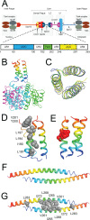
FIGURE 1:
FIGURE 1:
Conserved regions of Spc42 adopt distinct coiled-coil structures in vitro. (A) Schematic of protein organization within the SPB (top) and Spc42 coiled-coil regions (ACC, antiparallel coiled-coil; DCC, dimeric coiled-coil; TCC, trimeric coiled-coil) as well as undetermined regions (UR1–UR4) (bottom). (B–E) TCC. (B) Structure of Spc42181–211 TCC (rainbow), attached to the trimeric fusion protein 3H5I (pink, teal and green). (C) Alignment of four trimers of the Spc42181–211 asymmetric unit. (D) Stabilizing hydrophobic residues of the TCC are shown as gray spheres. (E) Phosphomimetic residues T193E and S195E of the TCC are shown as red spheres. (F, G) ACC. (F) Structure of Spc42246–305 ACC. (G) Stabilizing hydrophobic residues of the ACC are shown as gray spheres.
FIGURE 2:
FIGURE 2:
Assembly of Spc42 variant constructs on lipid monolayers in vitro. (A) Schematic of Spc42 variant constructs composed of three coiled-coil regions (DCC, TCC, and ACC) along with four undetermined regions (UR1–UR4). H6 (black pentagon) and a stabilizing globular domain (3H5I, Gp7, Sumo, 3K2N, or GCN4) were fused to the N-termini of Spc42 variants, and each was independently assayed for 2D array formation on a lipid monolayer. Nomenclature for these constructs is defined on the right. (B, C) EM images of Spc42 arrays for various constructs. (B) Spc42 hexameric 2D crystals were selected from broad-view images (Supplemental Figure S2A) for FFT computation (Supplemental Figure S2B). The 2D class averages of single particles from respective Spc42 constructs were obtained from broad-view images using EMAN2.2 (Supplemental Figure S2C). (C) All particles of a given Spc42 variant construct were aligned to a single class average and subsequently averaged. (D) The final averaged images of aligned particles have sixfold symmetry. Line profiles for each of the three directions (see black trace) were globally fit (pink traces) to obtain the unit cell dimensions of the hexameric array (see text and Table 2).
FIGURE 3:
FIGURE 3:
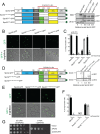
Multiple regions within the C-terminus are required for Spc42 oligomerization in vivo. (A) Left, schematic of Spc42-GFP (green star, GFP) variant constructs tested for superplaque formation following induction of overnight, mid–log phase YEP/2% raffinose cultures with 2% galactose for 3 h. Right, levels of overexpressed protein in whole-cell lysates determined by Western blotting with an anti-GFP antibody; histone H4 (HH4) serves as a loading control. Relative levels were normalized to the amount of GFP in an untagged control and in wild-type Spc42-GFP. (B) GFP fluorescence was analyzed by microscopy. Images are contrasted so that the small puncta present in mutants are visible. An overlay with the bright-field image is shown below. Scale bar: 2 µm. (C) The ability of Spc42-GFP or mutants to incorporate into a superplaque, judged by one or two easily visible spots of fluorescence in large budded yeast cells, was quantitated in each sample. n = 100, in 3 experiments. Error bars, SD. The p values were determined using Fisher’s test. All are statistically significant (p < 0.0001), unless indicated. (D) Left, schematic of two Spc42 C-terminal fragments expressed as GFP fusions. Right, expression was analyzed in whole-cell lysates as in A. (E, F) Superplaque formation was assayed as in B and C. (G) An empty vector, wild-type SPC42, and spc42∆304–363 were transformed into a strain containing a deletion of SPC42 covered by a URA3-based plasmid containing wild-type SPC42. The ability of each version of spc42 to rescue the SPC42 deletion was tested by plating fivefold serial dilutions of cells on 5-FOA plates. As a control, cells were stamped onto SC-URA plates. Shown are plates grown for 2 d at 30°C, although the mutant was inviable at all temperatures (unpublished data).
FIGURE 4:
FIGURE 4:
The TCC and UR3 are involved in SPB duplication and separation. (A) Schematic of Spc42 variant constructs. (B) Plasmid constructs were tested for their ability to rescue the SPC42 deletion as in Figure 3G. Plates were incubated at 23°C for 3 d or 37°C for 2 d. (C) The spc42∆185–245 and SPC42 cells were grown to mid–log phase in YPD at 23°C; the culture was then divided and shifted to 37°C for 4 h or kept at 23°C. Spindle morphology was analyzed by confocal imaging using GFP-Tub1 (microtubules, green) and Tub4-mCherry (SPBs, red). Cell outlines are based on bright-field images (white). Scale bar: 2 µm. (D) The percentage of large-budded cells from C that contained one, two, or more than two Tub4 foci were determined. An example from one of three independent experiments is shown, with at least 100 cells counted per sample. The p values were calculated using Fisher’s exact test. (E–H) Thin-section EM images of wild-type (E) and spc42∆185–245 (F–H) cells grown at 37°C as in C are shown. (E) Wild-type cell with two SPBs connected by a spindle. (F) Example of two serial sections of a SPB from spc42∆185–245 with an elongated bridge. A magnified image is shown on the right. (G, H) Two examples of duplicated side-by-side SPBs in spc42∆185–245 mutants. Note that in the magnified image in H, amorphous material is also present between the two poles, even though cytoplasmic microtubules emerge from each SPB. Arrowheads show cytoplasmic microtubules. Asterisks mark the position of nuclear pore complexes. Scale bars in E–H: 200 µm.
FIGURE 5:
FIGURE 5:
Conserved hydrophobic residues in the ACC interface are important for SPB function. (A) Schematic of Spc42 variant constructs. (B) Plasmid constructs were tested for their ability to rescue the SPC42 deletion as in Figure 4B. (C, D) Spindle morphology was analyzed by confocal imaging (C) using GFP-Tub1 (microtubules, green) and Tub4-mCherry (SPBs, red) in wild-type and spc42∆-V261D L268D I272D L283D cells grown at 30°C or shifted to 16°C for 15 h. (D) The percentage of large-budded cells from C that contained one, two, or more than two Tub4 foci were determined. An example from one of three independent experiments is shown, with at least 100 cells counted per sample. The p values were calculated using Fisher’s exact test. Scale bar: 2 µm. (E–G) The ability of Spc42V261D,L268D,I272D,L283D to form a superplaque was assayed as Figure 3, A–C, but with addition of 2% galactose for only 2 h. (E) Levels of overexpressed protein were analyzed as for Figure 3A. (F) GFP fluorescence was analyzed as for Figure 3B. Scale bar: 2 µm. (G) Incorporation of constructs into a superplaque were quantitated as for Figure 3C.
FIGURE 6:
FIGURE 6:
The DCC is essential for Spc42 stability. (A) Left, schematic of Spc42 variant constructs. Right, an empty vector, wild-type SPC42, and spc42∆54–141 were transformed into a strain containing a deletion of SPC42 covered by a URA3-based plasmid containing wild-type SPC42. The ability to rescue the SPC42 deletion was tested as for Figure 3G; the same phenotype was seen at all temperatures (unpublished data). (B) Schematic showing smaller deletions within the first coiled-coil as well as replacement constructs that have heptad repeats from the coiled-coil of myosin. An empty vector, wild-type SPC42, and the indicated deletions/replacements were transformed into a strain containing a deletion of SPC42 covered by a URA3-based plasmid containing wild-type SPC42 and tested for function as in Figure 3G. (C–E) Spc42∆60–94 or Spc42∆95–130 deletions and myosin replacements expressed as GFP fusions under the control of the GAL1 promoter were grown and induced as for Figure 5, E–G. (C) GFP fluorescence was analyzed as for Figure 3B. Scale bar: 2 µm. (D) Incorporation of constructs into a superplaque were quantitated as for Figure 3C. Overexpression of Spc42∆95–130-GFP resulted in a diffuse cytoplasmic signal with no visible puncta/foci, so it was not quantitated (ND) in D. (E) Levels of overexpressed protein from were analyzed as for Figure 3A. (F) Heptad repeats from the coiled-coil of myosin were added within the DCC to examine how increased length would affect Spc42 function, using a dilution assay as in A.
FIGURE 7:
FIGURE 7:
Evolution of interpretation of Spc42 architecture. (A) The trimer-of-dimers model, postulated before the structural data presented in this article, placed 12 Spc42 monomers in the hexagonal unit cell. In this model, the DCC, shown as blue spheres, form trimers of dimers, while the C-terminal regions of the protein, shown as green half-ellipsoids, dimerize with adjacent C-terminal regions (Muller et al., 2005; Viswanath et al., 2017). (B) Schematic representation of the P6 2D space group. (C) The TCC model incorporates the symmetry elements discerned from the crystallographic data presented here into a hexagonal lattice. In this model, the DCCs, shown as blue spheres, form dimers, while the TCCs, shown as green spheres, trimerize, and the ACCs, shown as yellow bars, dimerize in an antiparallel conformation. This orientation requires only 6 Spc42 monomers in the hexagonal unit cell. Undetermined regions (URs) of Spc42 are not included in this model.
FIGURE 8:
FIGURE 8:
TCC model of Spc42 lattice organization. (A) Top, edge-on view of the proposed organization of the DCC, TCC, and ACC. UR2–4 are depicted in gray. Here, the ACC is shown to provide an intramolecular interaction, in which two Spc42 polypeptide chains dimerize through the DCC and ACC, while the TCC networks with polypeptide chains outside of said dimers to establish the hexameric array (Supplemental Figure S2D). Alternatively, the ACC could provide an intermolecular interaction by instead making contacts between adjacent Spc42 dimers (Supplemental Figure S2E). Both interpretations incorporate the symmetry elements in similar ways, but with differing connectivity; current data do not distinguish between these models. (B) Overhead view of the model incorporating an intramolecular role for the ACC, overlaid on the small globular fusion protein Gp7. (Recall that these fusion proteins are necessary for improved solubility and stability of Spc42 constructs and do not affect the arrangement of Spc42.) Unit cell dimensions were determined from single-particle analysis of EM images (see Materials and Methods; Table 2). (C) Spc42 array reconstruction by Bullitt et al. (1997) is consistent with the trimeric coiled-coil model. (D, E) Structures of 3K2N (D) or GFP (E) are overlaid on the lattice to show that the fusion proteins can be readily accommodated in the lattice. Because these blocking domains lie beneath the segments of Spc42, it is not expected that they will interfere with the assembly of Spc42. Consistent with this conjecture, the lattice dimensions are independent of the size of the fusion domain.

Similar articles

Cited by

References

    1. Adams IR, Kilmartin JV. (1999). Localization of core spindle pole body (SPB) components during SPB duplication in Saccharomyces cerevisiae. J Cell Biol , 809–823. - PMC - PubMed
    1. Adams PD, Afonine PV, Bunkoczi G, Chen VB, Davis IW, Echols N, Headd JJ, Hung LW, Kapral GJ, Grosse-Kunstleve RW, et al (2010). PHENIX: a comprehensive Python-based system for macromolecular structure solution. Acta Crystallogr D Biol Crystallogr , 213–221. - PMC - PubMed
    1. Andreas MP, Ajay G, Gellings JA, Rayment I. (2017). Design considerations in coiled-coil fusion constructs for the structural determination of a problematic region of the human cardiac myosin rod. J Struct Biol , 219–228. - PMC - PubMed
    1. Bell JM, Chen M, Baldwin PR, Ludtke SJ. (2016). High resolution single particle refinement in EMAN2.1. Methods , 25–34. - PMC - PubMed
    1. Blommel PG, Fox BG. (2007). A combined approach to improving large-scale production of tobacco etch virus protease. Protein Expr Purif , 53–68. - PMC - PubMed

Publication types

MeSH terms

LinkOut - more resources