. 2019 Apr 10;15(4):e1008063.
doi: 10.1371/journal.pgen.1008063. eCollection 2019 Apr.

Precise staging of beetle horn formation in Trypoxylus dichotomus reveals the pleiotropic roles of doublesex depending on the spatiotemporal developmental contexts

Affiliations

Precise staging of beetle horn formation in Trypoxylus dichotomus reveals the pleiotropic roles of doublesex depending on the spatiotemporal developmental contexts

Shinichi Morita et al. PLoS Genet. .

Abstract

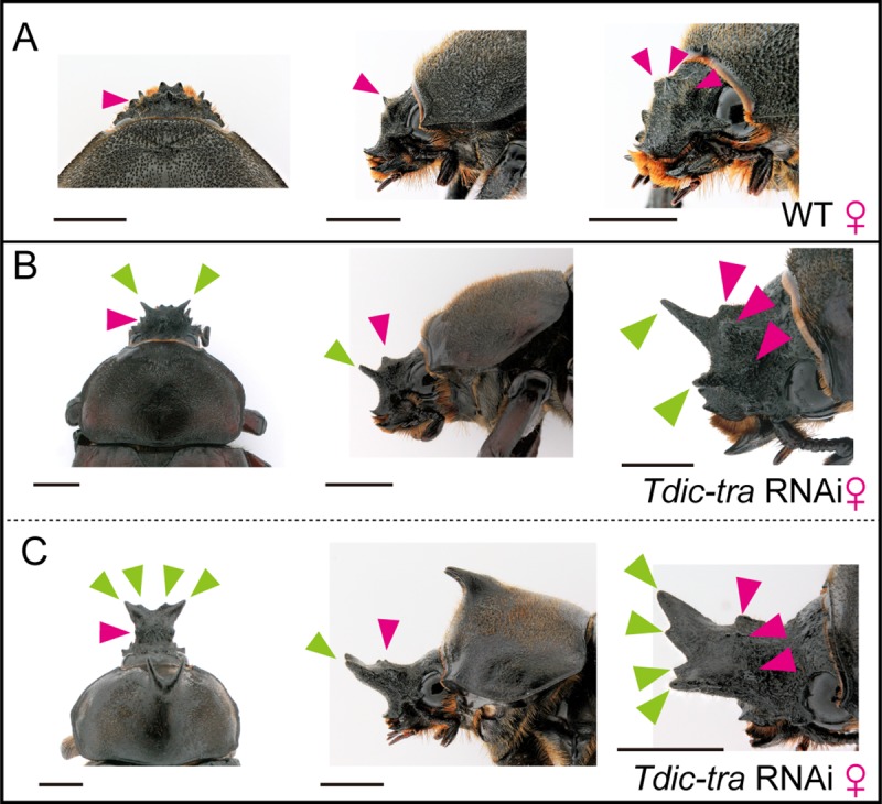
Many scarab beetles have sexually dimorphic exaggerated horns that are an evolutionary novelty. Since the shape, number, size, and location of horns are highly diverged within Scarabaeidae, beetle horns are an attractive model for studying the evolution of sexually dimorphic and novel traits. In beetles including the Japanese rhinoceros beetle Trypoxylus dichotomus, the sex differentiation gene doublesex (dsx) plays a crucial role in sexually dimorphic horn formation during larval-pupal development. However, knowledge of when and how dsx drives the gene regulatory network (GRN) for horn formation to form sexually dimorphic horns during development remains elusive. To address this issue, we identified a Trypoxylus-ortholog of the sex determination gene, transformer (tra), that regulates sex-specific splicing of the dsx pre-mRNA, and whose loss of function results in sex transformation. By knocking down tra function at multiple developmental timepoints during larval-pupal development, we estimated the onset when the sex-specific GRN for horn formation is driven. In addition, we also revealed that dsx regulates different aspects of morphogenetic activities during the prepupal and pupal developmental stages to form appropriate morphologies of pupal head and thoracic horn primordia as well as those of adult horns. Based on these findings, we discuss the evolutionary developmental background of sexually dimorphic trait growth in horned beetles.


Conflict of interest statement

The authors have declared that no competing interests exist.

Figures

Fig 1
Fig 1. Morphological change of horn primordia and the prepupal period in Trypoxylus dichotomus.
(A) (a) Schematic of the frontal view of larval head from T. dichotomus. Green, ocular (OC); Magenta, clypeolabrum; CL, clypeus; LB, labrum. (b), (c) Micro-CT images of prepupal head at 24 h APF from T. dichotomus. (b) The frontal view. Green arrowheads indicate the position of a sagittal section in (c). (c) The sagittal section of head. Head horn primordia were formed above the clypeus in the clypeolabrum (S1 Movie). (B) Morphological changes of head and thoracic horn primordia during larval-pupal development revealed by the time-lapse photography system. Both in males (the upper panels in the upper half) and in females (the upper panels in the lower half), horn primordia were formed in the clypeolabrum. In males, the frontal views are presented concerning the head horn primordia from 12 h APF to 36 h APF, and the anterodorsal views are presented concerning the head horn primordia from 48 h APF to 120 h APF. In females, the frontal views are presented demonstrated the head horn primordia from 12 h APF to 48 h APF, and the anterodorsal views are presented demonstrating the head horn primordia from 60 h APF to 120 h APF. The dorsal views are presented demonstrating thoracic horn primordia. APF, after pupal-chamber formation; HH, head horn primordia; TH, thoracic horn primordia; Orange arrowheads, the firstly bifurcated distal tips of a head horn; Green arrowheads, the secondly bifurcated distal tips of a head horn; Blue arrowheads, the long column shaped stalk of a head horn; Purple arrowheads, the bifurcated distal tips of a thoracic horn. Scale bars are 5 mm. (C) Snapshot images from time-lapse photography (S2 Movie). The panel on the left is a single raw image of time-lapse photography observing 64 larvae bottled in 100 ml plastic test tubes at the same time. The three panels on the right side are the magnified images of T. dichotomus at different developmental stages. (D) The durations of the prepupal periods in males and females estimated using the time-lapse photography system. Median, the upper/lower quantile, and minimum/maximum values were presented by a box- and whisker- plot. The median duration of prepupal periods in males and females were 131 hours and 129 hours, respectively. The mean duration of prepupal period in Tdic-tra RNAi females was 127 hours, which was slightly shorter (4 hours) than that of normally developed females.
Fig 2
Fig 2. Schematic representation of putative exon–intron structures and sex-specific splicing patterns of the sex determination genes.
(A) Schematics of putative exon-intron structures of the sex determination genes in T. dichotomus. Orange box, RRM (RNA binding motif); Green box, CAM (C, Ceratitis; A, Apis; M, Musca) domain [34, 47], which is conserved in all tra orthologs identified in other insects except for those in Drosophila species and the sandfly (Phlebotomus papatasi) [48]; Light blue box, Med29 (mediator complex subunit 29) domain; Brown box, DM (Doublesex/Mab-3 DNA-binding) domain; Yellow box, OD2 (Oligomerization dmain 2); Black bars, template sequences for dsRNA synthesis; Green bars, the amplified regions in qRT-PCR analysis; Light green bars, the amplified region to test sex-specific splicing variants; Magenta lines, translation start sites; Purple lines, stop codons. (B) Evaluation of expression and sex-specific splicing patterns in the sex determination genes in male and female prepupal head horn primordia. Template cDNAs were derived from prepupal horn primordia at 72 h APF. Tdic-RpL32 was used as an internal control for RT-PCR. Blue arrows, PCR products with male specific splicing variants; Magenta arrows, PCR products with female specific splicing variants; Black arrows, PCR products amplified in both sexes.
Fig 3
Fig 3. RNAi-mediated loss-of-function phenotypes of the sex determination genes.
(A) Representative individuals in each RNAi treatment in males (the left half) and females (the right half). Each dsRNA was injected into last-instar larvae. The negative control RNAi treatment (EGFP dsRNA) showed no morphological defects. The upper row, the dorsal views of adults; the second row, the lateral views of a head and a prothorax in adults; the third row, the ventral views of adults; the forth row, magnified views of orange squares (IPP, intercoxal process of prosternum) in the third row. Scale bars are 1 cm in the upper three rows and 1 mm in the fourth row. (B) Quantification of the relative head and thoracic horn length in RNAi-treated individuals. Relative head and thoracic horn length in RNAi-treated males and females are plotted in the blue dots and the magenta dots, respectively. The relative horn lengths were standardized by dividing the horn length by the body size in each RNAi treated individual and by the mean horn length of the EGFP RNAi-treated males. Differences in horn length among individual RNAi treatments were compared using Brunner-Munzel test. These p-values were adjusted by the Bonferroni correction. Asterisks denote significance: n.s., p > 0.05; *, p < 0.05. (C) Sex-specific splicing of Tdic-dsx in RNAi treatments targeting Tdic-Sxl, Tdic-tra, Tdic-tra2 and Tdic-ix. Blue arrowheads, male specific splicing patterns. Magenta arrowheads, female specific splicing patterns. Tdic-RpL32 was used as an internal control for RT-PCR.
Fig 4
Fig 4. Estimation of the onset of the Tdic-dsx dependent horn developmental program by Tdic-tra RNAi.
(A) Relationship between the timepoints of Tdic-tra dsRNA injection and the extent of sex transformation. The extent of the sex transformation (masculinization) phenotypes of Tdic-tra RNAi females were categorized into three classes: (i) prominent HH and TH were formed (< -7 h APF); (ii) only small HH was formed (5 h APF ~ 73 h APF); (iii) neither the HH nor TH was formed as in normal females (-3 h APF ~ 127h APF). HH, head horn primordia; TH, thoracic horn primordia. The dot plot panel was timepoints of Tdic-tra dsRNA injection before pupation in each class. The green dot, the magenta dot, and the blue dot indicate -3 h APF, -7 h APF and 73 h APF, respectively. Scale bars are 5 mm. (B) Time course of relative Tdic-tra and Tdic-dsxF mRNA expression level after Tdic-tra dsRNA injection at 24 h APF. mRNA expression levels at each timepoint were quantified by qRT-PCR. The expression levels of Tdic-tra were significantly decreased at 36 hours after injection (p<0.015). The expression level of Tdic-dsxF tended to be decreasing as time went on, but there were no statistically significant difference (p<0.056). (C) Summary of sex transformation phenotypes and the estimated onset of the Tdic-dsx dependent horn developmental program. The magenta line is the boundary timepoints of phenotype (i). Red arrowheads indicate the timepoints corrected by the mRNA degradation delay (36 hours), which estimated by qRT-PCR. APF, after puapal-chamber formation; Red arrowhead, the estimated onset of the developmental program for sexually dimorphic horn formation driven by Tdic-dsx.
Fig 5
Fig 5. Spatial and temporal expression of Tdic-Dsx protein in the head horn primordium during larval-pupal development.
(A-F) The head epidermis including the head horn primordium was stained with DAPI (magenta) to label nuclei and with anti-Tdic-Dsx antibody (green) to label Tdic-Dsx protein. Tdic-Dsx protein showed higher expression in the head primordial epidermis than in the surrounding head epidermis. Between Orange-Orange arrowheads is the head horn primordium. Between Orange-Blue arrowheads is the surrounding head epidermis. (A-C) Tdic-Dsx expression pattern in 12 h APF, Scale bars are 500 μm. (A’-C’) A higher magnification of the head horn primordium, respectively. Tdic-Dsx protein localization was cytoplasmic. Scale bars are 50 μm. (D-F) Tdic-Dsx expression pattern in 36 h APF, Scale bars are 500 μm. (D’-F’) A higher magnification of the head horn primordium, respectively. Tdic-Dsx protein localization were nuclei. Scale bars are 50 μm. (G) The epidermal regions used for qRT-PCR. The 3D volume image was reconstructed from sequential micro CT images. Blue, head horn primordium (HH); Purple, the head epidermis (non-HH). (H) The relative Tdic-dsx expression level in head horn primordia and head epidermis at 36 h APF in both sexes. Asterisks denote significance: n.s., p > 0.05; *, p < 0.05. HH, head horn primordium epidermis; non-HH, the head epidermis.
Fig 6
Fig 6. Horn formation phenotypes induced by Tdic-tra RNAi at late prepupal stages.
(A-C) Comparison of wild type female head and ectopic intermediate sexual transformation head horn in female heads induced by Tdic-tra RNAi treatments. (A) A wild type female head. (B) Small ectopic head horn formation in a female head. (C) Middle-sized ectopic head horn formation in a female head. Three small protrusions are formed in clypeolabrum on the wild type female head (A-C, magenta arrowheads), whereas ectopic small- or middle-sized ectopic head horns were formed in the region anterior to the three small protrusions (B, C, green arrowheads) in Tdic-tra RNAi treatments. Scale bars are 5 mm.
Fig 7
Fig 7. Horn formation phenotypes induced by Tdic-dsx RNAi at late prepupal stages.
(A-C) Thoracic horn remodeling during pupal-adult development in males. (A) A wild type male thoracic horn. (B) Almost complete reduction of a thoracic horn with an early Tdic-dsx RNAi treatment (-85 h APF). (B’) dorsolateral view. (C) Unremodelled thoracic horn formed by a late Tdic-dsx RNAi treatment (-13 h APF). APF, after puapal-chamber formation. Scale bars are 5 mm.
Fig 8
Fig 8. A regulatory model for the formation of sexually dimorphic horns in T. dichotomus.
In males, default splicing generates Tdic-dsxM, whereas in females, Tdic-tra (a female isoform, traF) and Tdic-tra2, generates Tdic-dsxF by female-specific splicing of Tdic-dsx pre-mRNA. Prepupal Tdic-dsxM expression in males positively regulates the growth of head and thoracic horn primordia, whereas prepupal Tdic-dsxF in females suppresses the horn growth activity. In pupa, Tdic-dsxM regulates thoracic horn remodeling, but is dispensable for remodeling of head horn. GRN, gene regulatory network.

Similar articles

Cited by

References

    1. Eberhard W.G., 1980. Horned beetles. Sci. Am. 242, 166–183. http://www.jstor.org/stable/24966285
    1. Emlen D.J., Marangelo J., Ball B., Cunningham C.W., 2007. Diversity in the weapons of sexual selection: horn evolution in the beetle genus Onthophagus (coleoptera: scarabaeidae). Evolution 59, 1060–1084. 10.1111/j.0014-3820.2005.tb01044.x - DOI - PubMed
    1. Busey H.A., Zattara E.E., Moczek A.P., 2016. Conservation, innovation, and bias: Embryonic segment boundaries position posterior, but not anterior, head horns in adult beetles. J. Exp. Zool. B. Mol. Dev. Evol. 326, 271–279. 10.1002/jez.b.22682 - DOI - PubMed
    1. McCullough E.L., Tobalske B.W., Emlen D.J., 2014. Structural adaptations to diverse fighting styles in sexually selected weapons. Proc. Natl. Acad. Sci. U.S.A. 111, 14484–14488. 10.1073/pnas.1409585111 - DOI - PMC - PubMed
    1. Siva-Jothy M.T., 1987. Mate securing tactics and the cost of fighting in the Japanese horned beetle, Allomyrina dichotoma L. (Scarabaeidae). J. Ethol. 5, 165–172. 10.1007/BF02349949 - DOI

Publication types

Substances