. 2019 Apr 10;14(4):e0215017.
doi: 10.1371/journal.pone.0215017. eCollection 2019.

Structure-based molecular characterization and regulatory mechanism of the LftR transcription factor from Listeria monocytogenes: Conformational flexibilities and a ligand-induced regulatory mechanism

Affiliations

Structure-based molecular characterization and regulatory mechanism of the LftR transcription factor from Listeria monocytogenes: Conformational flexibilities and a ligand-induced regulatory mechanism

Choongdeok Lee et al. PLoS One. .

Abstract

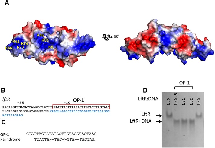
Listeria monocytogenes is a foodborne pathogen that causes listeriosis and can lead to serious clinical problems, such as sepsis and meningitis, in immunocompromised patients and neonates. Due to a growing number of antibiotic-resistant L. monocytogenes strains, listeriosis can steadily become refractory to antibiotic treatment. To develop novel therapeutics against listeriosis, the drug resistance mechanism of L. monocytogenes needs to be determined. The transcription factor LftR from L. monocytogenes regulates the expression of a putative multidrug resistance transporter, LieAB, and belongs to the PadR-2 subfamily of the PadR family. Despite the functional significance of LftR, our molecular understanding of the transcriptional regulatory mechanism for LftR and even for the PadR-2 subfamily is highly limited. Here, we report the crystal structure of LftR, which forms a dimer and protrudes two winged helix-turn-helix motifs for DNA recognition. Structure-based mutational and comparative analyses showed that LftR interacts with operator DNA through a LftR-specific mode as well as a common mechanism used by the PadR family. Moreover, the LftR dimer harbors one intersubunit cavity in the center of the dimeric structure as a putative ligand-binding site. Finally, conformational flexibilities in the LftR dimer and in the cavity suggest that a ligand-induced regulatory mechanism would be used by the LftR transcription factor.


Conflict of interest statement

The authors have declared that no competing interests exist.

Figures

Fig 1
Fig 1. LftR crystal structure and its sequence alignment with PadR-1 and PadR-2 subfamily members.
(A) Monomeric structure of LftR in rainbow-colored ribbons (N-terminus, blue; C-terminus, red). The N-terminal and C-terminal ends of LftR are labeled as “N” and “C,” respectively. Secondary structure elements are labeled (H, helix; β, β-strand). The wHTH motif contains the H2 and H3 helices, the β1 and β2 strands, and their connecting loops. The non-LftR residues that were introduced to design the recombinant protein expression construct are in grey. (B) Sequence alignment of LftR homologs. The secondary structures of LftR are shown above the LftR sequence. The LftR residues involved in dimerization are highlighted with green stars. The LftR residues that participate in DNA binding are highlighted by dots and labeled with their residue numbers above the sequences (LftR-specific DNA-binding residue, K9, red; PadR family-conserved residues, orange; positively charged patch residues of LftR, blue). The LftR residues that sterically clash with ethidium bromide in the complex model of LftR and ethidium bromide are underlined. (C) The dimeric structure of LftR is shown as ribbons (rainbow and light cyan). The intersubunit cavity of the LftR dimer is delineated by a dotted circle in the left figure.
Fig 2
Fig 2. Dimerization contacts of LftR.
One LftR subunit is shown as gray ribbons with surface representation, and its dimerization interface residues are highlighted by cyan surfaces and sticks. The other LftR subunit is shown as yellow ribbons, and its dimerization interface residues are depicted as yellow sticks. Hydrogen bonds are represented by dotted lines. (A) Dimerization interface at the N-terminal region of LftR. (B) Dimerization interface generated by the LftR H4 helix.
Fig 3
Fig 3. Operator DNA recognition by LftR.
(A) Electrostatic surface potentials of the LftR dimer (negative charge, red; positive charge, blue). A positive patch is observed on the bottom of the LftR dimer structure, and its positively charged residues are labeled in one LftR subunit (left). The orientation of the left figure is identical to the left figure in Fig 1C. (B) Putative operator DNA sequence (OP-1) of LftR located upstream of the LftR-encoding region (light blue). (C) Sequence analysis of the OP-1 operator site. Palindrome site sequences are indicated under the OP-1 sequence. (D) Analysis by electrophoretic mobility shift assay (EMSA) of the interaction between LftR and the operator DNA (OP-1). For the EMSA, a constant amount of LftR protein (3 μg, ~240 pmol) was incubated with operator DNA at different molar ratios indicated above the gel. The gel was stained with Coomassie brilliant blue to visualize protein bands. An uncropped gel image is shown in S3 Fig.
Fig 4
Fig 4. DNA recognition mode of LftR in comparison with BsPadR.
(A) Structure of a complex between BsPadR (green and light green) and its operator DNA (magenta). The Y20, Y42, and L65 residues of BsPadR were shown to be critical in DNA binding and are represented by sticks with labels in the figure. (B) Homology-based structural model of the LftR-DNA complex (LftR, cyan and light cyan; DNA, magenta). The LftR Y28, Y48, and L69 residues that correspond to BsPadR Y20, Y42, and L65, respectively, are shown as sticks with labels. The LftR-specific DNA-binding residue K9 is also depicted with sticks. (C, D) Fluorescent polarization (FP) analysis for the interactions of wildtype and mutant LftR proteins with 28-mer OP-1 dsDNA.
Fig 5
Fig 5. Structural diversity and intersubunit cavity of LftR.
(A) The LftRΔNtH structure (cyan and light cyan) was superimposed on the LftRCtH structure (magenta and light magenta) using the wHTH motifs in one subunit. (B) Intersubunit cavity located in the center of the LftRCtH dimer structure. The structure of the LftRCtH dimer is shown as magenta and light magenta ribbons. The intersubunit cavity of LftR is represented by yellow mesh and highlighted by a dashed circle. The structures of the LmrR-ligand complexes (LmrR-riboflavin, PDB accession code 4ZZD; LmrR-daunomycin, PDB accession code 3F8F) were overlaid on the LftR structure, and the ligand molecules of LmrR are shown as sticks (riboflavin, blue; daunomycin, green) with the LftRCtH dimer.
Fig 6
Fig 6. Intersubunit cavity of LftR as a putative ligand-binding site.
(A) LftR dimers (the LftRΔNtH structure, cyan and gray ribbons; the LftRCtH structure, magenta and light magenta ribbons) shown with an ethidium bromide molecule (red sticks), which was identified as an artificial ligand of LftR. (B) Steric clashes of LftR residues with ethidium bromide. Ethidium bromide is shown as red transparent spheres, and the LftRΔNtH structure is displayed in cyan and gray ribbons. LftR residues involved in steric clashes with the ethidium bromide molecule are shown as sticks with labels.

References

    1. Freitag NE, Port GC, Miner MD. Listeria monocytogenes—from saprophyte to intracellular pathogen. Nat Rev Microbiol. 2009;7(9):623–8. 10.1038/nrmicro2171 - DOI - PMC - PubMed
    1. Tilney LG, Portnoy DA. Actin filaments and the growth, movement, and spread of the intracellular bacterial parasite, Listeria monocytogenes. J Cell Biol. 1989;109(4 Pt 1):1597–608. - PMC - PubMed
    1. Ireton K, Rigano LA, Polle L, Schubert WD. Molecular mechanism of protrusion formation during cell-to-cell spread of Listeria. Front Cell Infect Microbiol. 2014;4:21 10.3389/fcimb.2014.00021 - DOI - PMC - PubMed
    1. Cossart P, Toledo-Arana A. Listeria monocytogenes, a unique model in infection biology: an overview. Microbes Infect. 2008;10(9):1041–50. 10.1016/j.micinf.2008.07.043 . - DOI - PubMed
    1. Swaminathan B, Gerner-Smidt P. The epidemiology of human listeriosis. Microbes Infect. 2007;9(10):1236–43. 10.1016/j.micinf.2007.05.011 . - DOI - PubMed

Publication types

MeSH terms