. 2019 Jun 28;70(12):3139-3151.
doi: 10.1093/jxb/erz162.

Cytokinin inhibits cotton fiber initiation by disrupting PIN3a-mediated asymmetric accumulation of auxin in the ovule epidermis

Affiliations

Cytokinin inhibits cotton fiber initiation by disrupting PIN3a-mediated asymmetric accumulation of auxin in the ovule epidermis

Jianyan Zeng et al. J Exp Bot. .

Abstract

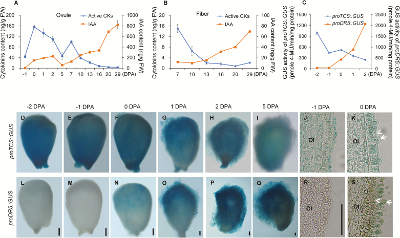
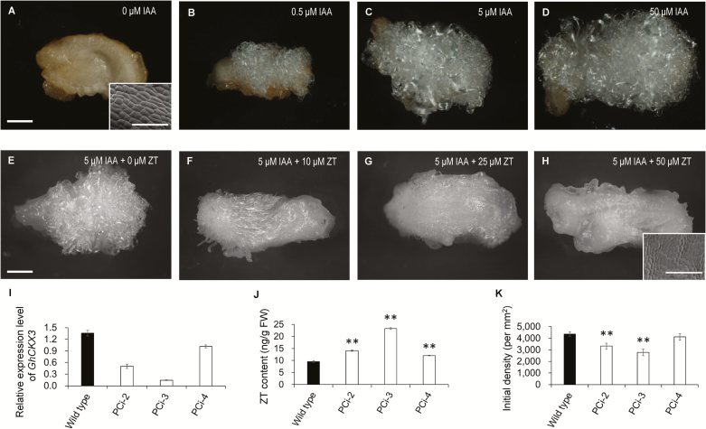
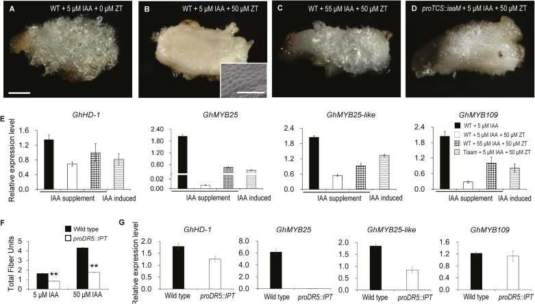
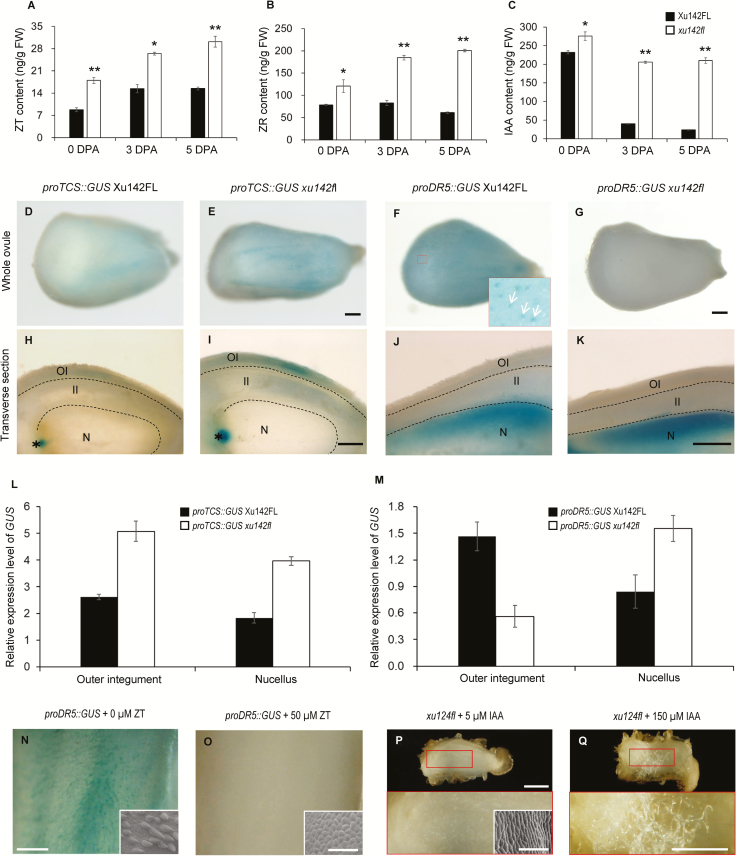
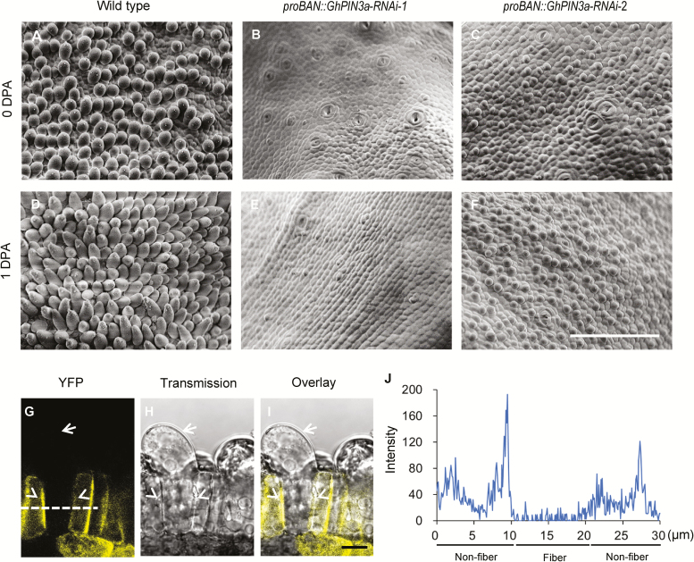
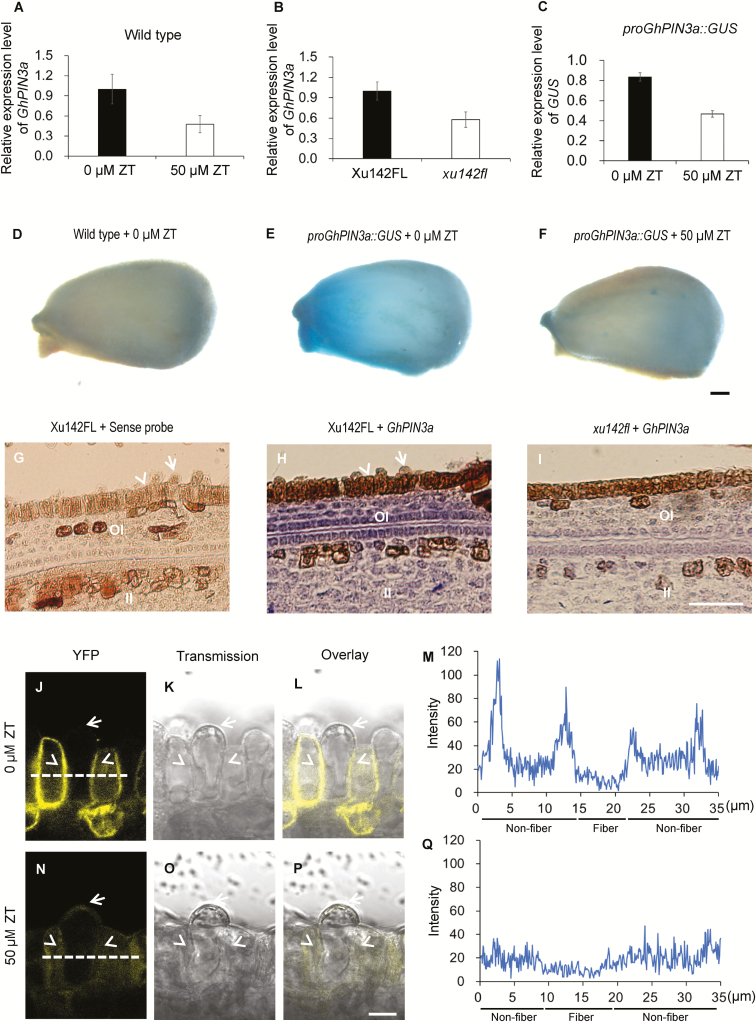
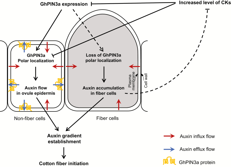
Auxin-dependent cell expansion is crucial for initiation of fiber cells in cotton (Gossypium hirsutum), which ultimately determines fiber yield and quality. However, the regulation of this process is far from being well understood. In this study, we demonstrate an antagonistic effect between cytokinin (CK) and auxin on cotton fiber initiation. In vitro and in planta experiments indicate that enhanced CK levels can reduce auxin accumulation in the ovule integument, which may account for the defects in the fiberless mutant xu142fl. In turn, supplementation with auxin can recover fiber growth of CK-treated ovules and mutant ovules. We further found that GhPIN3a is a key auxin transporter for fiber-cell initiation and is polarly localized to the plasma membranes of non-fiber cells, but not to those of fiber cells. This polar localization allows auxin to be transported within the ovule integument while specifically accumulating in fiber cells. We show that CKs antagonize the promotive effect of auxin on fiber cell initiation by undermining asymmetric accumulation of auxin in the ovule epidermis through down-regulation of GhPIN3a and disturbance of the polar localization of the protein.

Keywords: Auxin; GhPIN3a; cotton; cytokinin; fiber initiation; polar auxin transport.


Figures

Fig. 1.
Fig. 1.
Levels of cytokinins (CKs) and IAA in developing cotton ovules and fibers. (A, B) LC-MS/MS determination of contents of CKs and IAA in developing (A) ovules and (B) fibers. Active CKs represent the sum of contents of ZT, ZR, iP, iPR, and DZ. Ovules sampled from bolls at 0– 5 d post anthesis (DPA) contained fibers (because of technical limitations it is difficult to separate fibers from young ovules). Ovules sampled from bolls at –1, and 7–29 DPA were fiber-free. (C) GUS activity assay of proTCS::GUS and proDR5::GUS transgenic ovules. Data are means (±SD) of three biological replicates. (D–S) GUS staining of ovules of transgenic proTCS::GUS (D–I) and proDR5::GUS (L–Q) during cotton fiber initiation, together with paraffin-embedded sections of stained ovules harvested at –1 DPA and 0 DPA (J, K, R, S). Arrows indicate fiber cells and arrowheads indicate non-fiber cells in the ovule epidermis. OI, outer integument. Scale bars are 250 µm in ovule images or 50 µm in section images.
Fig. 2.
Fig. 2.
Auxin promotes fiber initiation in cotton, whilst cytokinin (CK) inhibits initiation. (A–D) Fiber growth on ovules cultured with different concentration of IAA. Ovules at –1 d post anthesis (DPA) were cultured in medium containing 0, 0.5, 5, or 50 μM IAA together with 0.5 μM GA3 for 15 d. (E–H) Fiber growth on ovules cultured with different concentration of ZT. Ovules at -1 DPA were cultured in the medium containing 0, 10, 25 or 50 μM ZT together with 0.5 μM GA3 and 5 μM IAA for 15 d. Fiber growth was observed using a stereo-microscope (scale bars are 1 mm) and a SEM (scale bars are 100 μm). (I) Transcription levels of GhCKX3 in proPV::antisenseGhCKX3 (PCi) transformants and the wild-type. Ovules at 0 DPA were used for RT-qPCR assays. Transcription levels (in arbitrary units) are normalized to that of GhHIS3. Data are means (±SD) of three repeats. (J) Content of ZT in ovules at 0 DPA. Data are means (±SD) of three biological replicates. (K) Initial fiber densities on the ovule surface at 0 DPA. Data are means (±SD) of nine ovules. Significant differences compared with the wild-type were determined using Student’s t-test (**P<0.01).
Fig. 3.
Fig. 3.
Antagonistic effect of cytokinin (CK) and auxin on fiber initiation in cotton. (A–D) IAA antagonizes the inhibitory effect of ZT on fiber initiation. Ovules at –1 d post anthesis (DPA) were cultured in medium containing different concentrations of IAA and ZT together with 0.5 μM GA3 for 15 d. Fiber growth was observed using a stereo-microscope (scale bars are 1 mm) and a SEM (scale bars are 100 μm). (E) Transcription levels of genes associated with fiber initiation in the ovules shown in (A–D). (F) Cytokinins antagonize the promotive effect of IAA on fiber production. Total fiber units were measured based on 20 ovules of proDR5::IPT transgenic and wild-type ovules cultured with 5 μM or 50 μM IAA together with 0.5 μM GA3 for 15 d. Data are means (±SD) of three biological replicates. Significant differences compared with the wild-type were determined using Student’s t-test (**P<0.01). (G) Transcription levels of genes associated with fiber initiation in proDR5::IPT and wild-type ovules cultured with 50 μM IAA and 0.5 μM GA3 for 3 d and assess using RT-qPCR assays. Transcription levels (in arbitrary units) are normalized to that of GhHIS3. Data are means (±SD) of three repeats. TiaaM, ovules from proTCS:iaaM transgenic cotton.
Fig. 4.
Fig. 4.
Cytokinins inhibit cotton fiber initiation through interfering with auxin accumulation in the ovule epidermis. (A–C) Contents of ZT (A), ZR (B), and IAA (C) in ovules of the fiberless mutant xu142fl and wild-type Xu142FL. Ovules at 0, 3, and 5 d post anthesis (DPA) were sampled. Ovules of Xu142FL contained fibers. Data are means (±SD) of three biological replicates. Significant differences between means were determined using Student’s t-test (*P<0.05; **P<0.01). (D, E, H, I) Distribution patterns of proTCS::GUS in ovules of xu142fl and Xu142FL. OI, outer integument; II, inner integument; N, nucellus. Asterisks indicate the chalazal region. Scale bars are 250 μm. (F, G, J, K) Distribution patterns of proDR5::GUS in ovules of xu142fl and Xu142FL. The inset image in (F) is an enlargement of the region indicated by the red square. Arrows indicate fiber cells. Scale are represent 250 μm. (L) Transcription levels of GUS in the outer integument and nucellus of ovules of proTCS::GUS xu142fl and proTCS::GUS Xu142FL. (M) Transcription levels of GUS in the outer integument and nucellus of ovules of proDR5::GUS xu142fl and proDR5::GUS Xu142FL. The outer integument and nucellus were separated from ovules at 0 DPA. Transcription levels (in arbitrary units) are normalized to that of GhHIS3. Data are means (±SD) of three repeats. (N, O) GUS staining of a proDR5::GUS Xu142FL ovule treated with ZT (N) and the control (O). proDR5:GUS Xu142FL ovules at –1 DPA were cultured with 50 μM ZT for 3 d and then used for GUS staining or SEM observation. Scale bars are 500 μm and 100 μm for the insets. (P, Q) Fiber growth of xu142fl ovules was recovered by IAA. Ovules of xu142fl at –1 DPA were cultured with 5 μM IAA (P) or 150 μM IAA (Q) for 15 d. The lower images are enlargements of the regions indicated with red squares in the upper images. The inset in (P) is a SEM image of the ovule surface. Scale bars are 1 mm (upper images), 250 μm (lower images), and 100 μm (SEM image).
Fig. 5.
Fig. 5.
GhPIN3a regulates cotton fiber initiation. (A–F) Fiber initiation on the ovule surface of proBAN::GhPIN3a-RNAi transgenic plants (B, C, E, F) and the wild-type (A, D). Ovules in the middle of each locule were observed using SEM. The scale bar is 100 μm. DPA, d post anthesis. (G–I) GhPIN3a::YFP localization in ovule epidermal cells at 0 DPA. GhPIN3a::YFP is localized to the plasma membrane in non-fiber cells but not in fiber cells. Arrows indicate fiber cells, arrowheads indicate non-fiber cells. The scale bar is 10 μm. (J) Fluorescence intensity along the dashed line in (G).
Fig. 6.
Fig. 6.
Cytokinins inhibit auxin accumulation in the ovule epidermis of cotton through disturbing GhPIN3a-mediated polar auxin transport. (A) GhPIN3a transcription levels in ZT-treated ovules and the control. Wild-type ovules at 0 d post anthesis (DPA) were treated with 50 μM ZT for 12 h, and then used for RT-qPCR assays. (B) GhPIN3a transcription levels in the outer integument of ovules of the fiberless xu142fl mutant and the Xu142FL wild-type at 0 DPA. (C) GUS transcription levels in ZT-treated proGhPIN3a::GUS ovules and the control. proGhPIN3a::GUS ovules at 0 DPA were treated with 50 μM ZT for 12 h and then used for RT-qPCR assays. Transcription levels (in arbitrary units) are normalized to that of GhHIS3. Data are means (±SD) of three repeats. (D–F) GUS staining of ovules of the wild-type control (D), proGhPIN3a::GUS (E), and ZT-treated proGhPIN3a::GUS (F). Ovules at 0 DPA were treated with or without 50 μM ZT for 12 h. The scale bar is 250 μm. (G–I) GhPIN3a RNA in situ hybridization in the integument of ovules of xu142fl (I) and Xu142FL (H). Transcription of GhPIN3a was repressed in xu142fl as compared with that in Xu142FL. Sections (10 μm) from ovules at 0 DPA were used for in situ hybridization with the GhPIN3a antisense probe. The sense probe was used as the negative control (G). OI, outer integument; II, inner integument. The scale bar is 20 μm. (J–Q) GhPIN3a::YFP localization in the ovule epidermal layer. The polar localization of GhPIN3a::YFP in non-fiber cells was impaired in ovules treated with ZT (N, P, Q) as compared with the control (J, L, M). Ovules at 0 DPA were sectioned by hand and then immersed in 50 μM ZT. After 30 min incubation in the dark, the samples were washed with distilled water and then mounted for observation. Fluorescence intensity along the dashed lines in (J) and (N) was shown in (M) and (Q), respectively. The scale bars is 10 μm. Arrows indicate fiber cells, and arrowheads indicate non-fiber cells in the ovule epidermis.
Fig. 7.
Fig. 7.
A model showing the antagonistic effect between cytokinins (CKs) and auxin on cotton fiber initiation. An auxin gradient is established in the cotton ovule epidermis by the asymmetric localization of the auxin transporter. GhPIN3a localizes to the plasma membrane of non-fiber cells and facilitates auxin efflux in the ovule integument. However, for reasons yet unknown, this polar localization is absent in neighboring fiber cells, which leads to auxin accumulation in the cell. Increased level of CKs may undermine the establishment of the auxin gradient in the epidermal cells through down-regulating the expression of GhPIN3a and disturbing the polar localization of the protein. The result is that fiber initiation is inhibited. On the other hand, auxin promotes fiber initiation. Augmenting IAA can antagonize the negative effect of CKs and give rise to fiber initiation on the fiberless mutant or on CK-treated ovules.

Comment in

References

    1. Adamowski M, Friml J. 2015. PIN-dependent auxin transport: action, regulation, and evolution. The Plant Cell 27, 20–32. - PMC - PubMed
    1. Armengot L, Marquès-Bueno MM, Jaillais Y. 2016. Regulation of polar auxin transport by protein and lipid kinases. Journal of Experimental Botany 67, 4015–4037. - PMC - PubMed
    1. Beasley CA. 1973. Hormonal regulation of growth in unfertilized cotton ovules. Science 179, 1003–1005. - PubMed
    1. Beasley CA, Ting IP. 1973. The effects of plant growth substances on in vitro fiber development from fertilized cotton ovules. American Journal of Botany 60, 130–139.
    1. Beasley CA, Ting IP. 1974. Effects of plant growth substances on in vitro fiber development from unfertilized cotton ovules. American Journal of Botany 61, 188–194.

Publication types