. 2019 Apr 9;20(7):1749.
doi: 10.3390/ijms20071749.

Anticancer Activities of Thymus vulgaris L. in Experimental Breast Carcinoma in Vivo and in Vitro

Affiliations

Anticancer Activities of Thymus vulgaris L. in Experimental Breast Carcinoma in Vivo and in Vitro

Peter Kubatka et al. Int J Mol Sci. .

Abstract

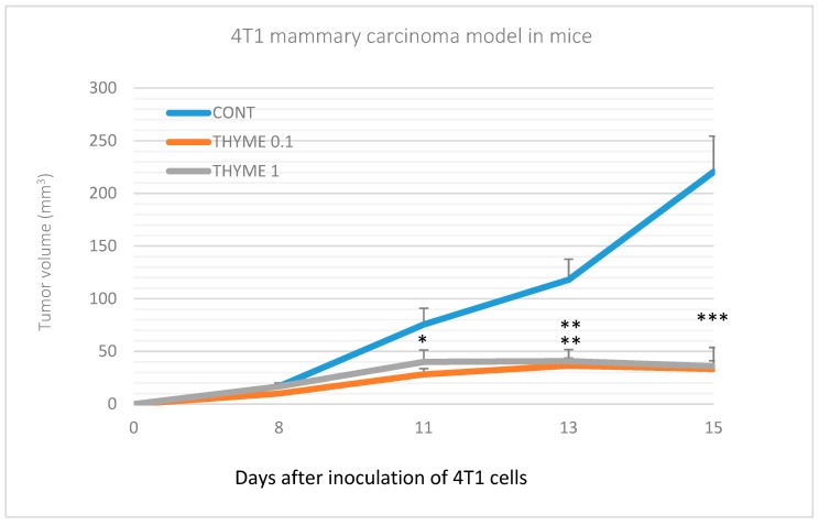
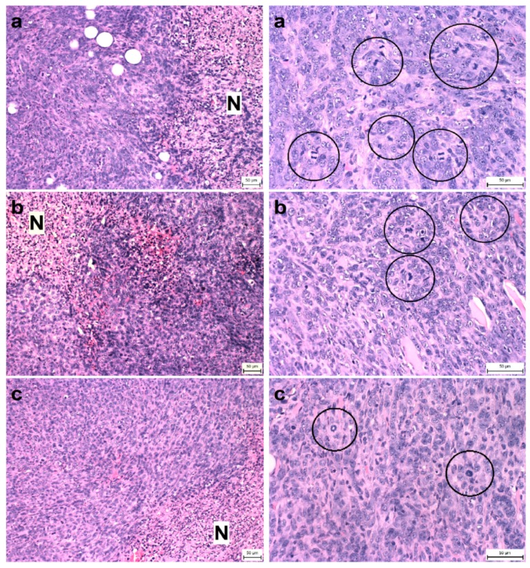
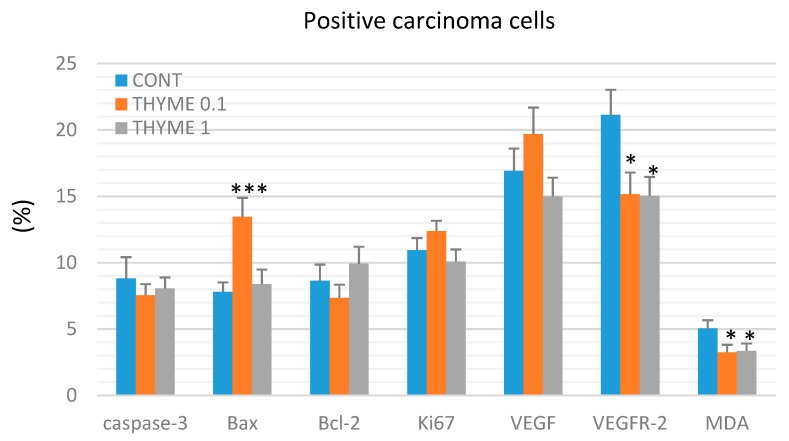
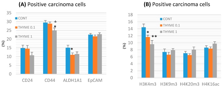
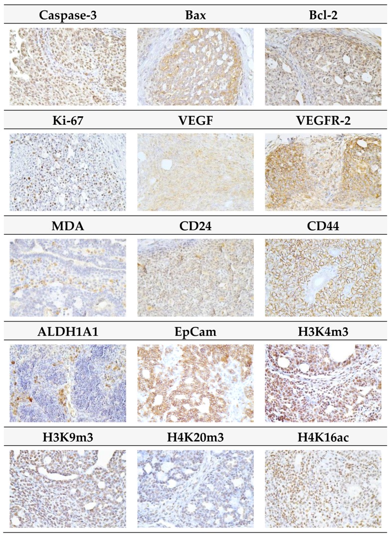
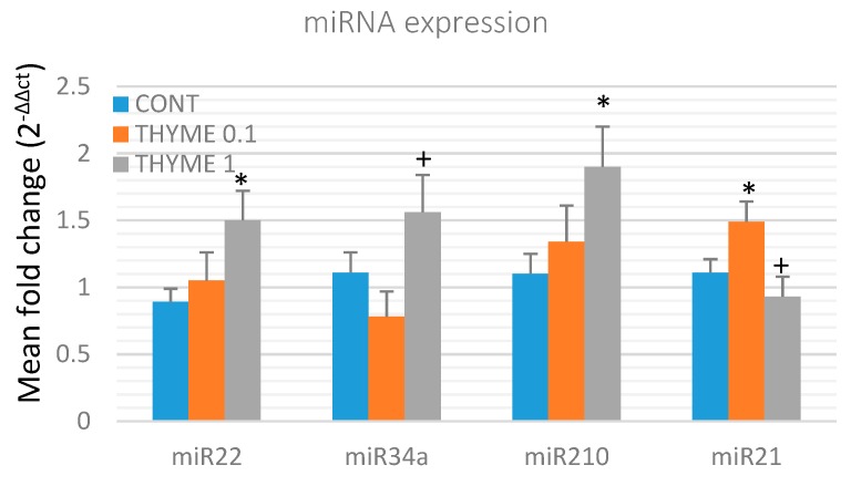
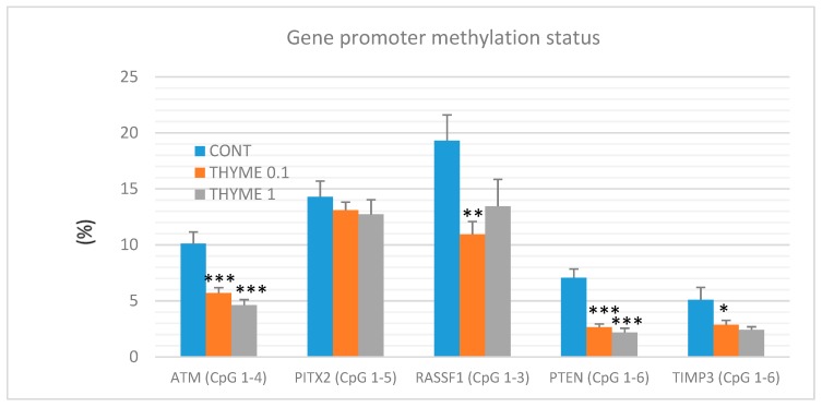
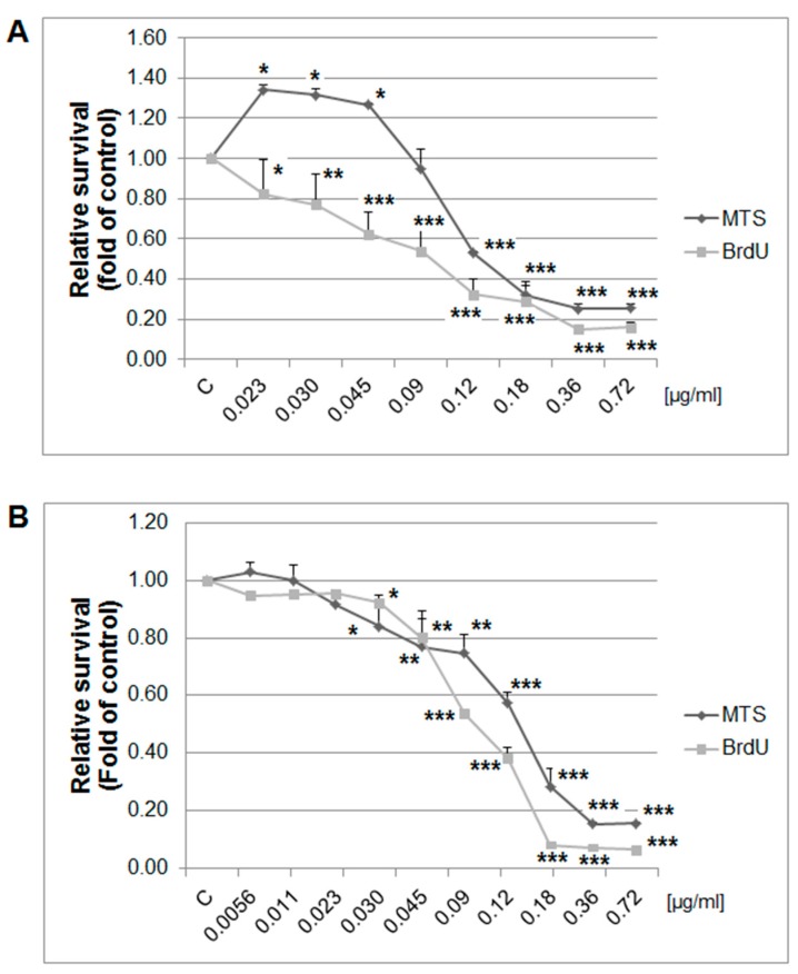
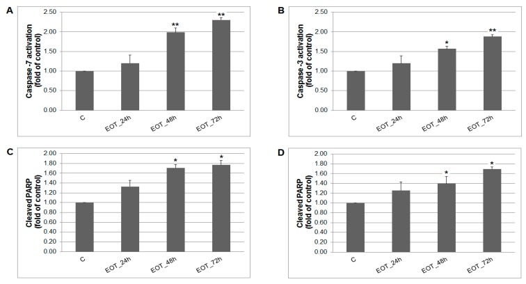
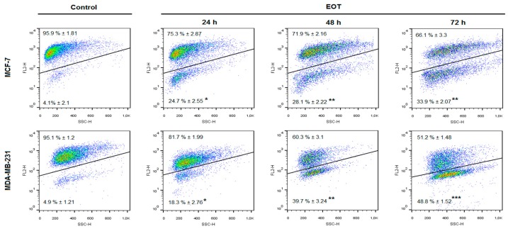
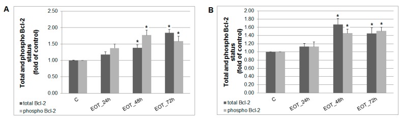
Naturally-occurring mixtures of phytochemicals present in plant foods are proposed to possess tumor-suppressive activities. In this work, we aimed to evaluate the antitumor effects of Thymus vulgaris L. in in vivo and in vitro mammary carcinoma models. Dried T. vulgaris (as haulm) was continuously administered at two concentrations of 0.1% and 1% in the diet in a chemically-induced rat mammary carcinomas model and a syngeneic 4T1 mouse model. After autopsy, histopathological and molecular analyses of rodent mammary carcinomas were performed. In addition, in vitro evaluations using MCF-7 and MDA-MB-231 cells were carried out. In mice, T. vulgaris at both doses reduced the volume of 4T1 tumors by 85% (0.1%) and 84% (1%) compared to the control, respectively. Moreover, treated tumors showed a substantial decrease in necrosis/tumor area ratio and mitotic activity index. In the rat model, T. vulgaris (1%) decreased the tumor frequency by 53% compared to the control. Analysis of the mechanisms of anticancer action included well-described and validated diagnostic and prognostic markers that are used in both clinical approach and preclinical research. In this regard, the analyses of treated rat carcinoma cells showed a CD44 and ALDH1A1 expression decrease and Bax expression increase. Malondialdehyde (MDA) levels and VEGFR-2 expression were decreased in rat carcinomas in both the T. vulgaris treated groups. Regarding the evaluations of epigenetic changes in rat tumors, we found a decrease in the lysine methylation status of H3K4me3 in both treated groups (H3K9m3, H4K20m3, and H4K16ac were not changed); up-regulations of miR22, miR34a, and miR210 expressions (only at higher doses); and significant reductions in the methylation status of four gene promoters-ATM serin/threonine kinase, also known as the NPAT gene (ATM); Ras-association domain family 1, isoform A (RASSF1); phosphatase and tensin homolog (PTEN); and tissue inhibitor of metalloproteinase-3 (TIMP3) (the paired-like homeodomain transcription factor (PITX2) promoter was not changed). In vitro study revealed the antiproliferative and proapoptotic effects of essential oils of T. vulgaris in MCF-7 and MDA-MB-231 cells (analyses of 3-(4,5-dimethylthiazol-2-yl)-5-(3-carboxymethoxyphenyl)-2-(4-sulfophenyl)-2H-tetrazolium) (MTS); 5-bromo-20-deoxyuridine (BrdU); cell cycle; annexin V/PI; caspase-3/7; Bcl-2; PARP; and mitochondrial membrane potential). T. vulgaris L. demonstrated significant chemopreventive and therapeutic activities against experimental breast carcinoma.

Keywords: MCF-7 cells; MDA-MB-231 cells; Thymus vulgaris; angiogenesis; apoptosis; cancer stem cells; cell proliferation; epigenetics; mammary carcinogenesis; predictive and preventive medicine; rat.


Conflict of interest statement

The authors declare no conflict of interest.

Figures

Figure 1
Figure 1
Tumor volume of 4T1 mammary gland cancer in mouse model during the experiment. Data are expressed as mean ± SEM. Significant differences: * p < 0.05, ** p < 0.01, *** p < 0.001 versus CONT.
Figure 2
Figure 2
Proportion of necrosis in tumor tissue (N/TT) (left column) and mitotic activity index (MAI) (right column) after treatment with T. vulgaris in 4T1 tumors in Balb/c mice. Tumor necrosis (N) mitotic figures (in circles) are showed in experimental groups: (a), control group; (b), THYME 0.1; (c), THYME 1.0; H&E staining magnifications: ×200 (N/TT), ×400 (MAI).
Figure 3
Figure 3
Immunohistochemical evaluation of caspase-3 (cytoplasmic), Bax, Bcl-2, Ki67, VEGFA, VEGFR-2, and MDA expression in rat mammary carcinoma cells after the administration of T. vulgaris in two doses. Data are expressed as mean ± SEM. Significant difference, * p < 0.05, *** p < 0.001 versus CONT. The figure represents the expression of proteins quantified as the average percentage of antigen positive area in standard fields (0.5655 mm2) of tumor hotspot areas. The values of protein expression were compared between treated (THYME 0.1, THYME 1) and non-treated (control) carcinoma cells of female rats; at least 60 images for one marker were analyzed.
Figure 4
Figure 4
Immunoexpression of cancer stem-cell (A) and epigenome (B) markers in rat mammary carcinoma cells after treatment with T. vulgaris. Data are expressed as mean ± SEM. Significant difference: * p < 0.05, ** p < 0.01 versus CONT, + p < 0.05 versus THYME 0.1. The values of protein expression were compared between treated (THYME 0.1, THYME 1) and non-treated (control) carcinoma cells of female rats; at least 60 images for one marker were analyzed.
Figure 5
Figure 5
Representative images of expression of caspase-3, Bax, Bcl-2, Ki67, VEGFA, VEGFR-2, MDA, CD24, CD44, ALDH1A1, EpCam, H3K4m3, H3K9m3, H4K20m3, and H4K16ac in rat mammary carcinoma cells. For detection, polyclonal caspase-3 antibody (Bioss, Woburn, MA, USA), polyclonal Bax and Bcl-2 antibodies (Santa Cruz Biotechnology, Paso Robles, CA, USA), monoclonal Ki67 antibody (Dako, Glostrup, Denmark), monoclonal VEGFA and VEGFR-2 antibodies (Santa Cruz Biotechnology, Paso Robles, CA, USA), polyclonal CD24 antibody (GeneTex, Irvine, CA, USA), polyclonal CD44 antibody (Boster, Pleasanton, CA, USA), polyclonal ALDH1A1 antibody (ThermoFisher, Rockford, IL, USA), polyclonal MDA, EpCAM, H3K4m, H3K9m3, and H4K20m3 antibodies (Abcam, Cambridge, MA, USA) and monoclonal H4K16ac antibody (Abcam, Cambridge, MA, USA) were used; final magnifications: ×400.
Figure 6
Figure 6
Relative miRNA expression of miR22, miR34a, miR210, and miR21 in rat mammary carcinomas. MiR-191-5p was selected as the internal control miRNA to normalize the cDNA levels of the samples. Data are expressed as mean ± SEM. Significant difference, * p < 0.05 versus CONT, + p < 0.05 versus THYME 0.1.
Figure 7
Figure 7
Total DNA promoter methylation status of ATM, PITX2, RASSF1A, PTEN, and TIMP3 genes in rat mammary carcinomas. Total promoter methylation status was taken from all evaluated CpG areas of ATM, PITX2, RASSF1A, PTEN, and TIMP3 in mammary carcinomas in the control and treated groups. Significant difference, * p < 0.05, ** p < 0.01, *** p < 0.001 versus control.
Figure 8
Figure 8
Relative survival of MCF-7 (A) and MDA-MB-231 (B) cells treated with EOT (0.72–0.023/0.0056 µg/mL) and analyzed by MTS and 5-bromo-20-deoxyuridine (BrdU) incorporation assays. Data were obtained from three independent experiments and significant differences were marked as * p < 0.05, ** p < 0.01, and *** p < 0.001 versus control cells (untreated).
Figure 9
Figure 9
The effect of EOT treatment (0.13 or 0.12 µg/mL) on: (A) caspase-7 (MCF-7), (B) caspase-3 activation (MDA-MB-231), and PARP cleavage in both cell lines (C,D) analyzed by flow cytometry. Data were obtained from three independent experiments and significant differences are shown: * p < 0.05, ** p < 0.01 versus control cells (untreated).
Figure 10
Figure 10
The effects of EOT treatment (0.13 or 0.12 µg/mL) on changes of mitochondrial membrane potential (MMP) in MCF-7 and MDA-MB-231 cells. Data were obtained from three independent experiments and significant differences are shown: * p < 0.05, ** p < 0.01, *** p < 0.001 versus control cells (untreated).
Figure 11
Figure 11
Distribution and activity of anti-apoptotic mitochondria-associated protein Bcl-2 after EOT treatment (0.13 or 0.12 µg/mL) in MCF-7 (A) and MDA-MB-231 (B) cells analyzed by flow cytometry. Results are expressed as the mean ± SD of three independent experiments and significant differences are given as * p < 0.05 versus untreated control.
Figure 12
Figure 12
GC/MS chromatogram of EOT with the most abundant peaks. Numbering of peaks is explained in Table 7.

References

    1. Li Y., Li S., Meng X., Gan R.Y., Zhang J.J., Li H.B. Dietary Natural Products for Prevention and Treatment of Breast Cancer. Nutrients. 2017;9:728. doi: 10.3390/nu9070728. - DOI - PMC - PubMed
    1. Takagi A., Kano M., Kaga C. Possibility of breast cancer prevention: Use of soy isoflavones and fermented soy beverage produced using probiotics. Int. J. Mol. Sci. 2015;16:10907–10920. doi: 10.3390/ijms160510907. - DOI - PMC - PubMed
    1. Shapira N. The potential contribution of dietary factors to breast cancer prevention. Eur. J. Cancer Prev. 2017;2:385–395. doi: 10.1097/CEJ.0000000000000406. - DOI - PMC - PubMed
    1. Giacosa A., Barale R., Bavaresco L., Gatenby P., Gerbi V., Janssens J., Johnston B., Kas K., La Vecchia C., Mainguet P., et al. Cancer prevention in Europe: The Mediterranean diet as a protective choice. Eur. J. Cancer Prev. 2013;22:90–95. - PubMed
    1. Ranaware A.M., Banik K., Deshpande V., Padmavathi G., Roy N.K., Sethi G., Fan L., Kumar A.P., Kunnumakkara A.B. Magnolol: A Neolignan from the Magnolia Family for the Prevention and Treatment of Cancer. Int. J. Mol. Sci. 2018;19:2362. doi: 10.3390/ijms19082362. - DOI - PMC - PubMed

MeSH terms