Review
. 2017 Jul 25;9(8):303.
doi: 10.3390/polym9080303.

Smart Sensor Systems for Wearable Electronic Devices

Affiliations
Review

Smart Sensor Systems for Wearable Electronic Devices

Byeong Wan An et al. Polymers (Basel). .

Abstract

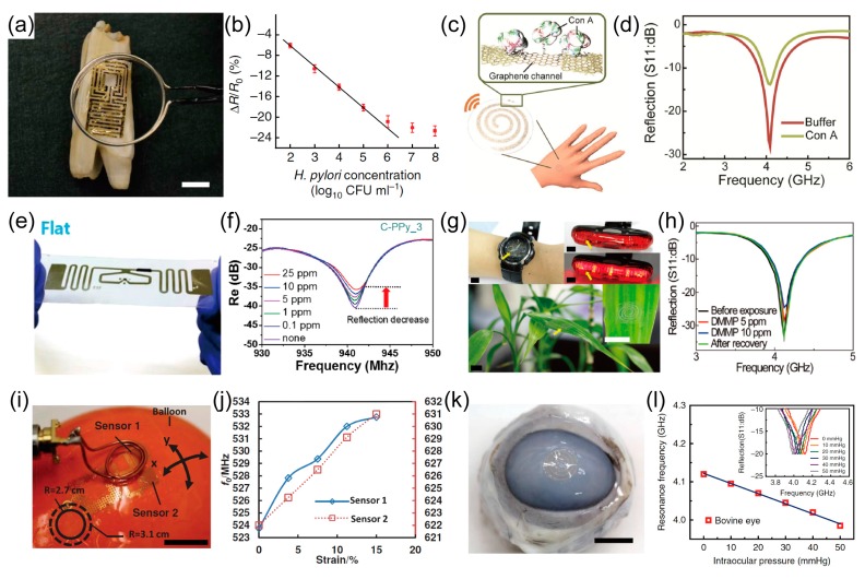
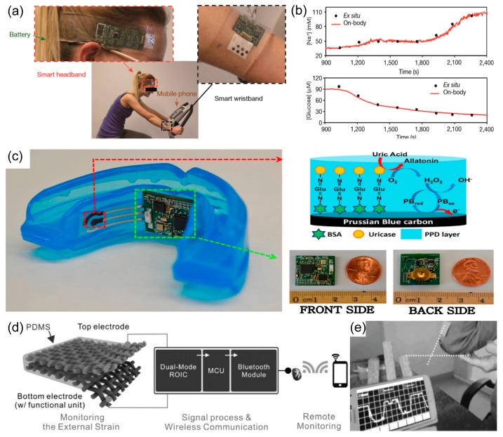
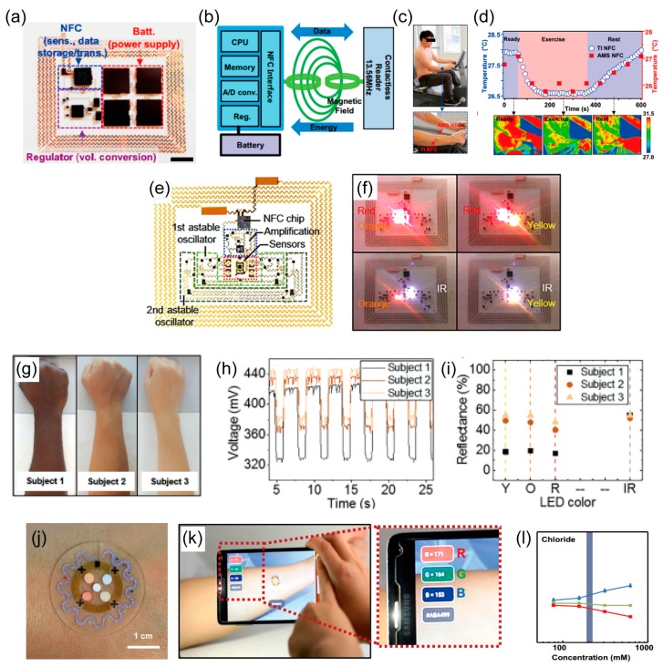
Wearable human interaction devices are technologies with various applications for improving human comfort, convenience and security and for monitoring health conditions. Healthcare monitoring includes caring for the welfare of every person, which includes early diagnosis of diseases, real-time monitoring of the effects of treatment, therapy, and the general monitoring of the conditions of people's health. As a result, wearable electronic devices are receiving greater attention because of their facile interaction with the human body, such as monitoring heart rate, wrist pulse, motion, blood pressure, intraocular pressure, and other health-related conditions. In this paper, various smart sensors and wireless systems are reviewed, the current state of research related to such systems is reported, and their detection mechanisms are compared. Our focus was limited to wearable and attachable sensors. Section 1 presents the various smart sensors. In Section 2, we describe multiplexed sensors that can monitor several physiological signals simultaneously. Section 3 provides a discussion about short-range wireless systems including bluetooth, near field communication (NFC), and resonance antenna systems for wearable electronic devices.

Keywords: healthcare; smart sensor; stretchable electronics; wearable electronics; wireless sensor.


Conflict of interest statement

The authors declare no conflict of interest.

Figures

Figure 1
Figure 1
Wearable temperature sensors. (a) Image of a 4 × 4 temperature coefficient of resistance (TCR) sensor array after application to the skin deformed by pinching the skin in a twisting motion (scale bar 8 mm). (b) Temperature of the palm measured with an infrared camera (blue) and a sensor array (red, offset for clarity) during mental and (c) physical stimulus tests. Reprinted with permission from Ref. [42]. Copyright 2016, Nature Publishing Group. (d) Schematic diagram and representative image of the stretchable graphene thermistors at twisted states (scale bar 1 cm). (e) Images of the stretchable graphene thermistor at 0% and 50% strains (scale bar 1 cm). (f) Resistance variation with temperature (30 to 100 °C) within 0% to 50% strains (step 10%). Reprinted with permission from Ref. [31]. Copyright 2015, American Chemical Society.
Figure 2
Figure 2
Wearable pressure sensors. (a) Cross-sectional schematic illustration of the pressure sensor and its connections to an associated transistor. (b) Photograph of the pressure sensor placed on a wrist and neck for measuring fast transients in the blood pressure (scale bars 1 cm and 2 cm). Reprinted with permission from Ref. [3]. Copyright 2014, Nature Publishing Group. (c) Images of pressure sensor printed on the commercial elastomeric patch. The sensor array is composed of four channels of pressure sensors (scale bars 1 cm). Reprinted with permission from Ref. [39]. Copyright 2014, John Wiley and Sons. (d) Photograph showing the skin-attachable sensor directly above the artery of the wrist (scale bar 3 cm). (e) Measurement of the physical force of a heartbeat under normal and exercise conditions. Reprinted with permission from Ref. [36]. Copyright 2014, Nature Publishing Group. (f) Schematic image of pressure-sensitive graphene FETs with air-dielectric layers. (g) Plot of normalized drain current changes versus applied pressure. (inset indicates relative change in the field effect mobility under applied pressure). Reprinted with permission from Ref. [33]. Copyright 2017, Nature Publishing Group.
Figure 3
Figure 3
Wearable strain sensors. (a) Schematic illustration of the cross-section of the strain sensor consisting of the three-layer stacked nano hybrid structure of polyurethane-poly(3,4-ethylenedioxythiophene) polystyrenesulfonate (PU-PEDOT:PSS)/single-wall carbon nanotube (SWCNT)/PU-PEDOT:PSS on a polydimethylsiloxane (PDMS) substrate. (b) Time-dependent ΔR/R0 responses of the sensor attached to the forehead when the subject was crying. Reprinted with permission from Ref. [34]. Copyright 2015, American Chemical Society. (c) Optical micrograph of a graphene woven fabrics (GWFs)-PDMS-tape composite film (scale bar 0.1 mm). (d) Relative change of resistance between 0% and 0.2% strain. Reprinted with permission from Ref. [86]. Copyright 2014, John Wiley and Sons. (e) Schematic illustration of stretchable capacitor with transparent electrode (top) and photograph of the same device reversibly adhered to a backlit liquid-crystal display (bottom) (scale bar 1 cm). (f) Change in capacitance ΔC/C0 versus strain ε (top) and ΔC/C0 versus time t over four cycles of stretching (bottom). Reprinted with permission from Ref. [69]. Copyright 2011, Nature Publishing Group. (g) Schematic image of multicore-shell printing process for fiber-type capacitive strain sensor. (h) Normalized decay time output of the sensor for different walking speeds up to 4 mph. Reprinted with permission from Ref. [92]. Copyright 2015, John Wiley and Sons.
Figure 4
Figure 4
Wearable gas sensors and its integrated systems. (a) Photograph of transparent and flexible single-layer graphene (SLG) sensor channel-bilayer graphene (BLG) heater on a polyethersulfone (PES) substrate (scale bar 7 mm). (b)Temperature distribution along transverse (x-axis) and longitudinal (y-axis) direction of sensor-heater device structured as laterally intercalated SLG sensor channel (6 mm width) between BLG heaters (7 mm width) with applied 1.7 W of electric power. Here the red dot and blue dot are temperature profiles of thermal image in inset along x-axis and y-axis with origin at center on channel, respectively. Inset: Spatial temperature distribution of graphene heaters (7 mm width) which intercalate 6 mm width graphene sensor with applied 1.7 W. Here three broken squares indicate center channel and side heaters area, respectively (scale bar 7 mm). (c) Recovering time constant τr as a function of heater temperature. Inset: the recovering curves of the ΔR/R0 as a function of time under different temperature range from room temperature to 250 °C. (d) The relative resistance variation ΔR/R0 of SLG channels as a function of time including recovery step with 100 to 165 °C heating under different NO2 gas concentration from 40 to 0.5 ppm. Reprinted with permission from Ref. [37] Copyright 2014, John Wiley and Sons. (e) Response curves of the sensor to NO2 of different concentrations. Inset: The sensor response depends linearly on NO2 concentration. (f) Response curves of the sensor to 50 ppm of NO2 when tested under different bending angles. (g) Response curves of the sensor tested before and after bending 1000 and 5000 times (bending angle = 50°). Reprinted with permission from Ref. [114] Copyright 2014, John Wiley and Sons. (h) Photograph of microfabricated flexible room temperature ionic liquid (RTIL) based gas sensor (scale bars 1 cm and 2 mm, respectively). (i) Current versus time curve at various oxygen concentrations when the potential is held at −1.4 V vs. Au. Nitrogen is the background gas. Oxygen concentration steps up from 0% to 21% and steps down from 21% to 0%. Reprinted with permission from Ref. [117] Copyright 2013, IEEE. (j) Schematic illustration of the preparation of the PDA/MoS2 film and the sensor upon exposure to DMF vapor. (k) UV-vis spectra of polydiacetylene (PDA)/MoS2 composites with an increased ratio of MoS2 to PDA in the absence and presence of 0.1% DMF vapor. (l) UV-vis spectra of PDA/MoS2 films exposed to N,N-dimethylformamide (DMF) vapor with different concentrations. (m) UV-vis spectra of the PDA/MoS2 film upon exposure to different (5%) vapors, in comparison with 2% DMF vapor. (n) Flexible transparent wrist strap with DMF sensing ability. Reprinted with permission from Ref. [110] Copyright 2017, Royal Society of Chemistry.
Figure 4
Figure 4
Wearable gas sensors and its integrated systems. (a) Photograph of transparent and flexible single-layer graphene (SLG) sensor channel-bilayer graphene (BLG) heater on a polyethersulfone (PES) substrate (scale bar 7 mm). (b)Temperature distribution along transverse (x-axis) and longitudinal (y-axis) direction of sensor-heater device structured as laterally intercalated SLG sensor channel (6 mm width) between BLG heaters (7 mm width) with applied 1.7 W of electric power. Here the red dot and blue dot are temperature profiles of thermal image in inset along x-axis and y-axis with origin at center on channel, respectively. Inset: Spatial temperature distribution of graphene heaters (7 mm width) which intercalate 6 mm width graphene sensor with applied 1.7 W. Here three broken squares indicate center channel and side heaters area, respectively (scale bar 7 mm). (c) Recovering time constant τr as a function of heater temperature. Inset: the recovering curves of the ΔR/R0 as a function of time under different temperature range from room temperature to 250 °C. (d) The relative resistance variation ΔR/R0 of SLG channels as a function of time including recovery step with 100 to 165 °C heating under different NO2 gas concentration from 40 to 0.5 ppm. Reprinted with permission from Ref. [37] Copyright 2014, John Wiley and Sons. (e) Response curves of the sensor to NO2 of different concentrations. Inset: The sensor response depends linearly on NO2 concentration. (f) Response curves of the sensor to 50 ppm of NO2 when tested under different bending angles. (g) Response curves of the sensor tested before and after bending 1000 and 5000 times (bending angle = 50°). Reprinted with permission from Ref. [114] Copyright 2014, John Wiley and Sons. (h) Photograph of microfabricated flexible room temperature ionic liquid (RTIL) based gas sensor (scale bars 1 cm and 2 mm, respectively). (i) Current versus time curve at various oxygen concentrations when the potential is held at −1.4 V vs. Au. Nitrogen is the background gas. Oxygen concentration steps up from 0% to 21% and steps down from 21% to 0%. Reprinted with permission from Ref. [117] Copyright 2013, IEEE. (j) Schematic illustration of the preparation of the PDA/MoS2 film and the sensor upon exposure to DMF vapor. (k) UV-vis spectra of polydiacetylene (PDA)/MoS2 composites with an increased ratio of MoS2 to PDA in the absence and presence of 0.1% DMF vapor. (l) UV-vis spectra of PDA/MoS2 films exposed to N,N-dimethylformamide (DMF) vapor with different concentrations. (m) UV-vis spectra of the PDA/MoS2 film upon exposure to different (5%) vapors, in comparison with 2% DMF vapor. (n) Flexible transparent wrist strap with DMF sensing ability. Reprinted with permission from Ref. [110] Copyright 2017, Royal Society of Chemistry.
Figure 5
Figure 5
Wearable electrochemical ion sensors and its integrated systems. (a) Schematic representation of the tailor-made stretchable materials and manufacturing process. (b) Photograph of the printed sensors on different common textiles and typical time trace plots for potassium and sodium. Reprinted with permission from Ref. [122]. Copyright 2016, John Wiley and Sons. (c) Photograph of a wearable flexible integrated sensing array and the open circuit potential responses of Na+ (d) and K+ (e) sensor, respectively. Reprinted with permission from Ref. [38]. Copyright 2016, Nature Publishing Group. (f) Water-gate characterization at different pH levels. (g) Photograph of monolithic device structures transferred onto the epidermis of an insect. (scale bar 5 mm). Reprinted with permission from Ref. [126]. Copyright 2012, Nature Publishing Group. (h) A fully integrated wearable sensing system on a subject’s arm and general performance of Ca2+ (i) and pH (j) sensors. Reprinted with permission from Ref. [127]. Copyright 2016, American Chemical Society.
Figure 6
Figure 6
Wearable biosensors (a) The images of the temporary transfer-tattoo biosensor attached on deltoid. (b) Amperometric response as a function of lactate concentration for the sensor at 37 °C. Inset: profiles of current at different lactate concentrations. Reprinted with permission from [136]. Copyright 2013, American Chemical Society. (c) Conceptual images of conformally contacted devices on an artificial eye for glucose sensing in tears are shown. Thin-film sensors remained in contact with skin even during tension and relaxation (scale bars 10 mm). (d) Scanning electron microscope image of a representative device (thickness of 1.7 μm) on an artificial PDMS skin replica indicating conformal contact between the device and the substrate (scale bar 500 μm). (e) Representative responses of In2O3 sensors to physiologically relevant d-glucose concentrations found in human diabetic tears (lower range) and blood (upper range). Inset: data from five devices. Error bars represent standard deviations of the means. Reprinted with permission from Ref. [40]. Copyright 2013, American Chemical Society. (f) The preparation route of the 4-boronobenzaldehyde (4-BBA)-modified poly(vinyl alcohol) (PVA) gelated colloidal crystal array (GCCA)-lens. (g) The diffraction response at low glucose concentration. Insert: the photograph of the GCCA-lens sample. Reprinted with permission from Ref. [144]. Copyright 2017, MDPI AG.
Figure 7
Figure 7
Wearable multiplexed sensors. (a) Photograph of a wearable flexible integrated sensing array (FISA) on a subject’s wrist, integrating the multiplexed sweat sensor array and the wireless flexible, printed circuit board (FPCB). (b) Schematic of the sensor array (including glucose, lactate, sodium, potassium and temperature sensors) for multiplexed perspiration analysis. GOx and LOx, glucose oxidase and lactate oxidase. (c) System-level interference studies of the sensor array. Reprinted with permission from Ref. [38]. Copyright 2014, Nature Publishing Group. (d) Optical image of the fabricated ion sensor on a cellular substrate. Ion sensor mounted on a rabbit heart model constructed from agarose gel (inset). (scale bar 2 mm) Reprinted with permission from Ref. [149]. Copyright 2014, John Wiley and Sons. (e) Photograph of the fabricated smart band. (f) Output AC signal with and without touching. (g) Normalized resistance change as a function of temperature. Reprinted with permission from Ref. [62]. Copyright 2014, John Wiley and Sons. (h) Photographs of the wearable bio-integrated system. Inset: Wearable 10 × 10 RRAM array on the hydrocolloid side of the patch (scale bars 5 mm). (i) Plot of percentage change in resistance versus strain for calculation of the gauge factor (j) Temperature distribution measurement of the heater on the skin patch using an infrared camera. Reprinted with permission from Ref. [99]. Copyright 2014, Nature Publishing Group.
Figure 8
Figure 8
Wearable sensors integrated with resonance antenna. (a) Optical image of the graphene-based wireless sensor transferred onto the surface of a tooth (scale bar: 1 cm). (b) graphene resistance change versus concentration of H. (a,b) Reproduced with permission from Ref. [159]. Copyright 2012, Nature Publishing Group; (c) Schematic of the biosensor attached to the skin on the back of a human hand; (d) Frequency response of the reflection coefficient of the antenna on the plastic substrates after buffer and Con A treatment. (c,d) Reproduced with permission from Ref. [12]. Copyright 2015, John Wiley and Sons; (e) Photographs of the RFID tag sensor; (f) Change in the reflectance properties. (e,f) Reproduced with permission from Ref. [163] Copyright 2016, American Chemical Society. (g) Optical photos of wearable gas sensors integrated with resonance antenna transferred onto various substrates (wristwatch, light of bicycle, and a leaf of live plant) (scale bars: 1 cm). (h) change in reflection coefficient (S11) of the wireless sensor on the leaf at varied DMMP vapor concentrations (before exposure, 5 ppm of DMMP, 10 ppm of DMMP, and after recovery). (g,h) Reproduced with permission from Ref. [18]. Copyright 2016, Royal Society of Chemistry. (i) Image of a wireless epidermal sensor attached onto the surface of a balloon to simulate measurement of lymphedema. Scale bar, 1 cm. (j) Change in resonance frequencies of strain sensors under the expansion of the balloon. (i,j) Reproduced with permission from Ref. [160]. Copyright 2014, John Wiley and Sons. (k) Photograph of the sensor transferred onto the contact lens worn by a bovine eyeball. Scale bar, 1 cm. (l) Frequency response of the intraocular pressure sensor on the bovine eye from 5 mmHg to 50 mmHg (Inset: the corresponding reflection coefficients of the sensor). (k,l) Reproduced with permission from Ref. [32]. Copyright 2017, Nature Publishing Group.
Figure 9
Figure 9
Bluetooth integrated wearable sensors. (a) Photographs of wearing a wearable sensor in band types during a stationary cycling; (b) Real-time reading data of the sodium and glucose measured with wearable sensors; (a,b) Reproduced with permission from Ref. [38]. Copyright 2016, Nature Publishing Group; (c) Photographs of the wireless SUA biosensor module with Bluetooth and the operation principles of mouthguard biosensor; (c) Reproduced with permission from Ref. [29]. Copyright 2015, Elsevier; (d) Schematic illustration of a flexible textile-based strain sensor with wireless monitoring system; (e) Photographs of the flexible textile-based strain sensor with Bluetooth module and real-time operation via wireless communication with mobile phone. (d,e) Reproduced with permission from Ref. [83]. Copyright 2016, John Wiley and Sons.
Figure 10
Figure 10
Near field communication (NFC)-enabled wearable sensor. (a) Image of the NFC-enabled wearable sensor system composed of the NFC devices, batteries, and a power regulator. Scale bar, 5 mm; (b) Schematic illustration of the entire operation system; (c) Photo images of the NFC-enabled wearable sensor attached onto the human skin during exercise; (d) Temperature data and IR images at the stage of the before, during, and after exercise (which correspond to the “ready”, “exercise”, “rest”) recorded by using NFC-enabled wearable sensor with a battery module (AMS NFC, red squares) and with the battery-integrated system (TI NFC, open circle). (ad) Reproduced with permission from Ref. [176]. Copyright 2016, National Academy of Sciences; (e) Image of a NFC-enabled wearable sensor system including four pulsed LEDs (red, IR, orange, and yellow), two oscillators, amplifier and sensors; (f) Photo images of the NFC-enabled wearable sensor system during operation; (g) Images of subjects with different skin colors; (h,i) Wirelessly measured data and calculated reflectance value of the different skin colors. (ei) Reproduced with permission from Ref. [157]. Copyright 2016, American Association for the Advancement of Science; (j) Optical image of a fabricated device mounted on the forearm; (k) Pictures demonstrating NFC between a sweat monitoring device and a smartphone to launch software for image capture and analysis; (l) Standard calibration curves between normalized %RGB value and concentration of markers for quantitative analysis (error bars show s.d. (N = 3)). (jl) Reproduced with permission from Ref. [177]. Copyright 2016, American Association for the Advancement of Science.

References

    1. Bonato P. Wearable sensors/systems and their impact on biomedical engineering. IEEE Eng. Med. Biol. Mag. 2003;22:18–20. doi: 10.1109/MEMB.2003.1213622. - DOI - PubMed
    1. Wang X., Gu Y., Xiong Z., Cui Z., Zhang T. Electronic Skin: Silk-Molded Flexible, Ultrasensitive, and Highly Stable Electronic Skin for Monitoring Human Physiological Signals (Adv. Mater. 9/2014) Adv. Mater. 2014;26:1309. doi: 10.1002/adma.201470054. - DOI - PubMed
    1. Dagdeviren C., Su Y., Joe P., Yona R., Liu Y., Kim Y.-S., Huang Y., Damadoran A.R., Xia J., Martin L.W., et al. Conformable amplified lead zirconate titanate sensors with enhanced piezoelectric response for cutaneous pressure monitoring. Nat. Commun. 2014;5:4496. doi: 10.1038/ncomms5496. - DOI - PubMed
    1. Pang C., Koo J.H., Nguyen A., Caves J.M., Kim M.-G., Chortos A., Kim K., Wang P.J., Tok J.B.-H., Bao Z. Highly skin-conformal microhairy sensor for pulse signal amplification. Adv. Mater. 2015;27:634–640. doi: 10.1002/adma.201403807. - DOI - PubMed
    1. Pang C., Lee C., Suh K.-Y. Recent advances in flexible sensors for wearable and implantable devices. J. Appl. Polym. Sci. 2013;130:1429–1441. doi: 10.1002/app.39461. - DOI