. 2019 Dec;10(1):121-132.
doi: 10.1080/21655979.2019.1605812.

Targeting MIAT reduces apoptosis of cardiomyocytes after ischemia/reperfusion injury

Affiliations

Targeting MIAT reduces apoptosis of cardiomyocytes after ischemia/reperfusion injury

Longying Chen et al. Bioengineered. 2019 Dec.

Abstract

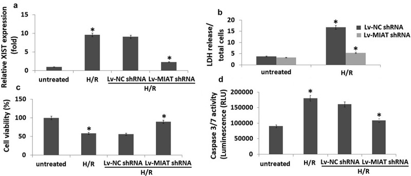
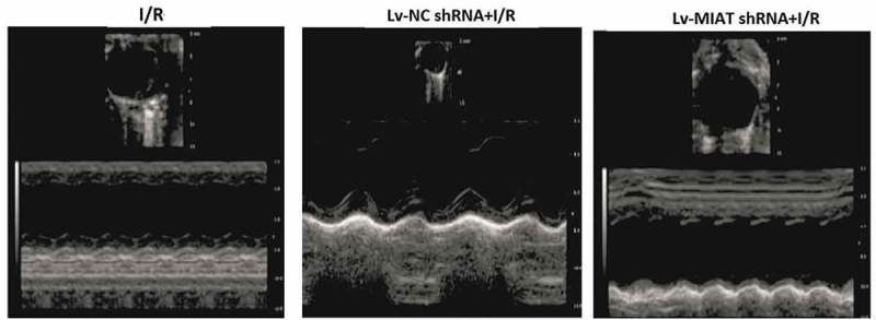
This study aims to investigate the role of targeting lncRNA myocardial infarction-associated transcript (MIAT) in protection against hypoxia/reoxygenation (H/R) injury in H9c2 cells in vitro and myocardial ischemia/reperfusion (I/R) injury in vivo by regulating expression of NF-kB and p53 upregulated modulator of apoptosis (PUMA). H9C2 cells were infected with lentivirus expressing the short-hairpin RNA direct against human MIAT gene (Lv-MIAT shRNA) or lentivirus expressing scrambled control (Lv-NC shRNA) or PUMA siRNA or p65 siRNA or their control siRNA respectively. Then the H9c2 cells were infected with Lv-shRNA to 2 hours of hypoxia (H) and 24 hour of reoxygenation (R). 100 ul of Lv-MIAT shRNA (1 × 10⁸ PFU) or Lv-NC shRNA was transfected into mouse hearts, then the hearts were subjected to I/R (1h/72 h). We discovered targeting MIAT remarkably enhanced H9c2 cell viability, decreased H/R-induced cell apoptosis and LDH leakage and significantly decreased I/R-induced myocardial infarct size, reduced myocardial apoptosis and enhanced the heart function. Targeting MIAT downregulated p65 nuclear translocation, NF-κB activity and anti-apoptotic protein cleaved-caspase-3, Bax, and upregulated anti-apoptotic protein Bcl-2 induced by H/R or I/R. Our study suggests that targeting MIAT may protect against H9c2 cardiomyoblasts H/R injury or myocardial I/R injury via inhibition of cell apoptosis, mediated by NF-κB and PUMA signal pathway.

Keywords: Hypoxia/reoxygenation; apoptosis; ischaemia-reperfusion; lncRNA myocardial infarction-associated transcript; nuclear factor kappa B; p53 upregulated modulator of apoptosis.


Figures

Figure 1.
Figure 1.
Targeting MIAT mediated cardioprotective effects in H/R-induced H9C2 cardiomyocytes apoptosis. H9C2 cells were infected with Lv- MIAT shRNA or Lv-NC shRNA 24 h, then subjected to H/R (2/24 h). a, MIAT expression was detected in H9C2 cells by qRT-PCR. b, The release of LDH (cell injury marker) in H9C2 cells. c, cell viability was detected by MTT. d, Cell apoptosis was detected by caspase-3/7 activity assay. *P < 0.05 compared with indicated groups.
Figure 2.
Figure 2.
H/R induced p65 nucleus translocation and PUMA signal expression in H9C2 cells. a, Expression of p65, PUMA, bax, bcl-2 and cleaved-caspase-3 were detected by Western blot assay; b, NF-KB activity was detected by EMSA assay; c, Cell viability was detected by MTT; D, Cell apoptosis was detected by caspase-3/7 activity assay. *P < 0.05 compared with indicated groups.
Figure 3.
Figure 3.
Targeting MIAT mediated cardioprotective effects in I/R-induced cardiac injury. a, Representative TTC staining (example slices from one animal in each group) shows infarcted areas in white and non-infarcted areas in red. b, Representative H&E-stained histological images (×200 magnification) of the myocardial sections from different treatment groups, *p < 0.01.
Figure 4.
Figure 4.
Effect of targeting MIAT on cardiac functions with echocardiographic evaluation. Representative echocardiograph recorded from a mouse after I/R in different groups of treatment. vs Lv-NC shRNA, **p < 0.01; vs I/R, *p < 0.05.
Figure 5.
Figure 5.
Targeting MIAT protects against I/R-induced cell apoptosis via inhibition of p65 nucleus translocation and PUMA signal expression. a, Representative pictures of sections stained with TUNEL (×400). TUNEL-positive nuclei are shown in green. Blue fluorescence indicated total cardiomyocyte nuclei. Quantitative analysis (percentage of apoptotic cells versus total) is shown in histogram. b, NF-KB activity was detected by EMSA assay; c, Western blots of p65 nucleus translocation, cleaved caspase-3,PUMA and Bax in I/R tissues in the presence or absence of Lv-MIAT shRNA. Vs Lv-NC shNA, **p < 0.01; Vs I/R, *p < 0.05

References

    1. D J H, Yellon DM.. Ischaemic conditioning and reperfusion injury. Nat Rev Cardiol. 2016;13:193–209. - PubMed
    1. Hausenloy DJ. Novel targets and future strategies for acute cardioprotection: position paper of the European society of cardiology working group on cellular biology of the heart. Cardiovasc Res. 2017;113:564–585. - PubMed
    1. Pu J, Yuan A, Shan P. Cardiomyocyte-expressed farnesoid-X-receptor is a novel apoptosis mediator and contributes to myocardial ischaemia/reperfusion injury. Eur Heart J. 2013;34:1834–1835. - PMC - PubMed
    1. Kopp F, Mendell JT. Functional classification and experimental dissection of long noncoding RNAs. Cell. 2018;172:393–407. - PMC - PubMed
    1. Duan Z, Yu AM. Bioengineered non-coding RNA agent (BERA) in action. Bioengineered. 2016;7:411–417. - PMC - PubMed

MeSH terms