. 2019 Apr 11;14(1):98.
doi: 10.1186/s13018-019-1115-z.

Progression of local kyphosis after conservative treatment for compressive cervical spine fracture with spinal cord injury

Affiliations

Progression of local kyphosis after conservative treatment for compressive cervical spine fracture with spinal cord injury

Kazuya Yokota et al. J Orthop Surg Res. .

Abstract

Introduction: Compressive-flexion type cervical spine fracture is typically accompanied by apparent dislocation of the facet joints, undesirable cervical alignment, and devastating neurological dysfunction, which provides strong rationale for rendering prompt operative treatment. However, the validity of conservative treatment for compressive-flexion cervical spine injury in cases with preserved congruity of the facet joints has yet to be elucidated. The purpose of this study is to evaluate the long-term outcome of cervical alignment following conservative treatment for compressive-flexion cervical spine injury with preserved congruity of the facet joints.

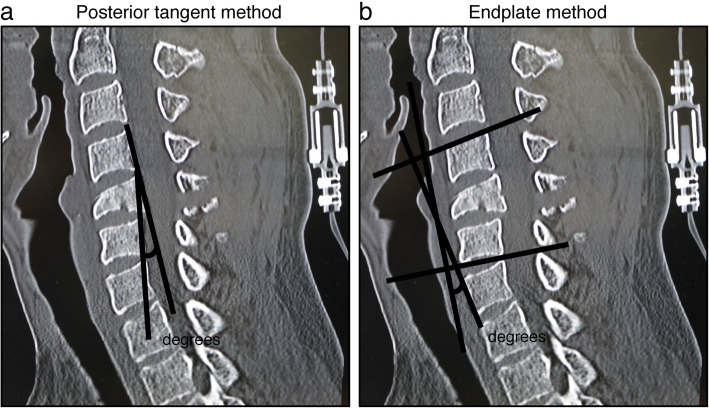
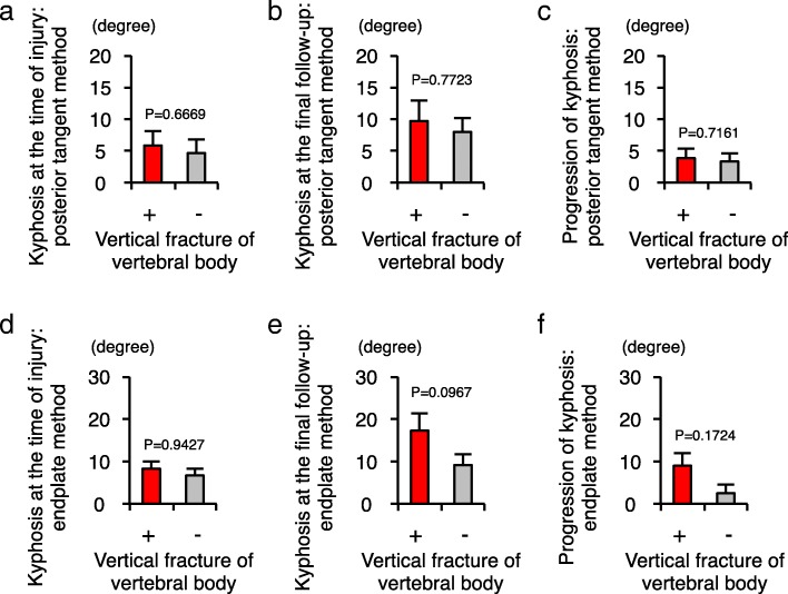
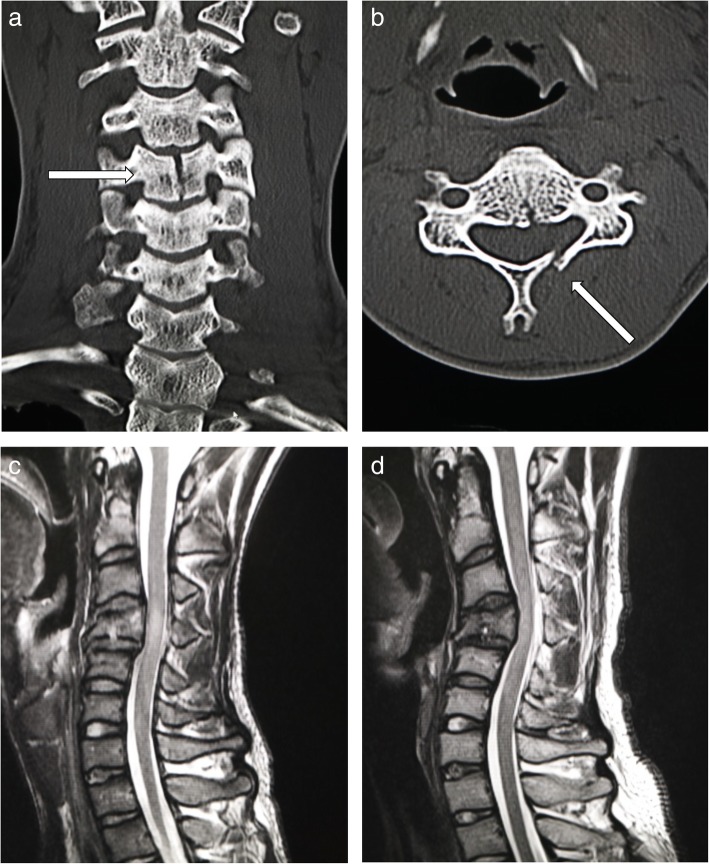
Methods: A total of 662 patients who experienced spinal cord injury from 2007 to 2017 were included and underwent retrospective review in a single institute. Thirteen patients were identified as receiving conservative therapy following compressive-flexion cervical spine fractures with spinal cord injury. Clinical and radiological results were collected, including vertical fractures of the vertebral column, laminar fractures, progression of local kyphosis, and neurological status. The degree of the local cervical kyphosis was evaluated with two methods: the posterior tangent method and the endplate method.

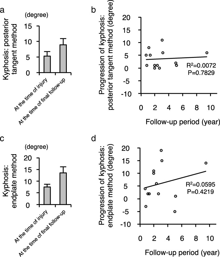
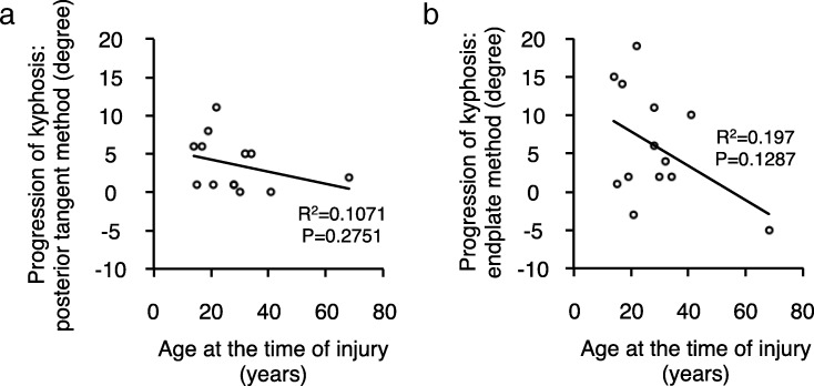
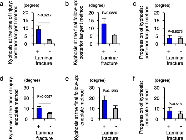
Results: All 13 patients were male, and the mean age at the time of injury was 28.4 years. The mean follow-up period was 3 years. Although none of the patients presented neurological deterioration after the injury, the degree of local kyphosis was increased at the time of final follow-up compared to what was observed at the time of injury. Patient age at the time of injury and concurrent vertical fracture of vertebral body could have been influencing factors for the progression of the kyphosis. While laminar fracture affected the kyphosis at the time of injury, it was not a strong influencing factor of the overall progression of local kyphosis.

Conclusions: The conservative option for the compressive-flexion cervical injury allowed us to treat without exacerbating neurological symptoms as long as the facet joints are preserved. However, in terms of cervical alignment, surgical stabilization may have been desirable for these patients. Notably, the younger patients and the patients with vertical fracture of the cervical vertebral column in this type of injury required closer observation to help prevent the progression of local kyphosis.

Keywords: Cervical spinal cord injury; Laminar fracture; Local kyphosis; Vertebral fracture.


Conflict of interest statement

Ethics approval and consent to participate

This study was approved by the Ethical Review Board of Japan Labor Health and Welfare Organization Spinal Injuries Center. We had all the necessary consent from the patients involved in the study, including consent to participate in the study where appropriate.

Consent for publication

Written informed consent for publication of their clinical details and/or clinical images was obtained from the patient.

Competing interests

The authors declare that they have no competing interests.

Publisher’s Note

Springer Nature remains neutral with regard to jurisdictional claims in published maps and institutional affiliations.

Figures

Fig. 1
Fig. 1
A radiographic example of a patient included in this study. a The patient suffered from compressive-flexion vertebral fracture with preserved cervical alignment. b The vertical fracture of the vertebra and the right laminar fracture occurred concurrently in this patient
Fig. 2
Fig. 2
Radiographic measurements for local cervical kyphosis. a Diagram depicting the measurement technique for local kyphosis using the posterior tangent method. The angle between the lines drawn along the posterior vertebral body was measured. b Diagram depicting the measurement technique for local kyphosis using the endplate method. The angle of intersection between a line drawn along the superior endplate of the suprajacent vertebra and a line drawn along the inferior endplate of the infrajacent vertebra was measured
Fig. 3
Fig. 3
The overall progression of local kyphosis after compressive cervical spine fractures. a The results of the local kyphosis at the time of injury (n = 13) and at the time of final follow-up (n = 13) measured by the posterior tangent method. b The relationship between the progression of local kyphosis measured by the posterior tangent method and the duration of the follow-up period. c The results of the local kyphosis at the time of injury (n = 13) and at the time of final follow-up (n = 13) measured by the endplate method. d The relationship between the progression of local kyphosis measured by the endplate method and the duration of the follow-up period. There is little correlation between the progression of local cervical kyphosis and the duration of the follow-up period
Fig. 4
Fig. 4
Moderate negative correlation between the age at the time of injury and the progression of local kyphosis. a The relationship between the progression of local kyphosis measured by the posterior tangent method and the age at the time of injury. b The relationship between the progression of local kyphosis measured by the endplate method and the age at the time of injury. Moderate negative correlation was found between the progression of kyphosis and the age at the time of injury
Fig. 5
Fig. 5
Laminar fractures affect the initial kyphotic alignment after injury and do not affect the progression of local kyphosis. a Comparison of local cervical kyphosis between the two groups of patients with (n = 6) and without (n = 7) laminar fracture, as measured by the posterior tangent method at the time of injury. b Comparison of local cervical kyphosis between the two groups of patients with and without laminar fracture, as measured by the posterior tangent method at the time of final follow-up. c Comparison of the progression of local cervical kyphosis between the two groups of patients with and without laminar fracture as measured by the posterior tangent method. d Comparison of the local cervical kyphosis between the two groups of patients with and without laminar fracture as measured by the endplate method at the time of injury. e Comparison of local cervical kyphosis between the two groups of patients with and without laminar fracture as measured by the endplate method at the time of final follow-up. f Comparison of the progression of local cervical kyphosis between the two groups of patients with and without laminar fracture as measured by the endplate method
Fig. 6
Fig. 6
Vertical fracture of the vertebral body could be an influencing factor for the progression of local kyphosis. a Comparison of the local cervical kyphosis between the two groups of patients with (n = 7) and without (n = 6) vertical fracture of the vertebral body, as measured by the posterior tangent method at the time of injury. b Comparison of the local cervical kyphosis between the two groups of patients with and without vertical fracture of the vertebral body as measured by the posterior tangent method at the time of final follow-up. c Comparison of the progression of local cervical kyphosis between the two groups of patients with and without vertical fracture of the vertebral body as measured by the posterior tangent method. d Comparison of the local cervical kyphosis between the two groups of patients with and without vertical fracture of the vertebral body as measured by the endplate method at the time of injury. e Comparison of the local cervical kyphosis between the two groups of patients with and without vertical fracture of the vertebral body as measured by the endplate method at the time of final follow-up. f Comparison of the progression of local cervical kyphosis between the two groups of patients with and without vertical fracture of the vertebral body as measured by the endplate method
Fig. 7
Fig. 7
No significant difference in the progression of local kyphosis between the two orthosis treatments. a Comparison of the local cervical kyphosis between the group of patients treated with neck brace (n = 7) and the group of patients treated with halo vest (n = 6) as measured by the posterior tangent method at the time of injury. b Comparison of the local cervical kyphosis between the group of patients treated with neck brace and the group of patients treated with halo vest as measured by the posterior tangent method at the time of final follow-up. c Comparison of the progression of local cervical kyphosis between the group of patients treated with neck brace and the group of patients treated with halo vest as measured by the posterior tangent method. d Comparison of the local cervical kyphosis between the group of patients treated with neck brace and the group of patients treated with halo vest as measured by the endplate method at the time of injury. e Comparison of the local cervical kyphosis between the group of patients treated with neck brace and the group of patients treated with halo vest as measured by endplate method at the time of final follow-up. f Comparison of the local cervical kyphosis between the group of patients treated with neck brace and the group of patients treated with halo vest as measured by the endplate method
Fig. 8
Fig. 8
A representative case with progression of local kyphosis. a, b Computed tomography (CT) images at the time of injury showed the vertebral vertical fracture and left laminar fracture at the C4 level. c Cervical spinal cord injury was confirmed by T2-weighted magnetic resonance imaging (MRI) at the time of injury. d MRI at the time of final follow-up (1110 days after injury) showed the progression of local cervical kyphosis from 17 to 36°, measured by the endplate method. His neurological finding remained at AIS grade D and improved to modified Frankel grade D3

Similar articles

Cited by

References

    1. Pickett GE, Campos-Benitez M, Keller JL, Duggal N. Epidemiology of traumatic spinal cord injury in Canada. Spine (Phila Pa 1976) 2006;31:799–805. doi: 10.1097/01.brs.0000207258.80129.03. - DOI - PubMed
    1. Allen BL, Jr, Ferguson RL, Lehmann TR, O’Brien RP. A mechanistic classification of closed, indirect fractures and dislocations of the lower cervical spine. Spine (Phila Pa 1976) 1982;7:1–27. doi: 10.1097/00007632-198201000-00001. - DOI - PubMed
    1. Raniga SB, Menon V, Al Muzahmi KS, Butt S. MDCT of acute subaxial cervical spine trauma: a mechanism-based approach. Insights Imaging. 2014;5:321–338. doi: 10.1007/s13244-014-0311-y. - DOI - PMC - PubMed
    1. Vaccaro AR, Koerner JD, Radcliff KE, Oner FC, Reinhold M, Schnake KJ, Kandziora F, Fehlings MG, Dvorak MF, Aarabi B, et al. AOSpine subaxial cervical spine injury classification system. Eur Spine J. 2016;25:2173–2184. doi: 10.1007/s00586-015-3831-3. - DOI - PubMed
    1. Dogan S, Safavi-Abbasi S, Theodore N, Horn E, Rekate HL, Sonntag VK. Pediatric subaxial cervical spine injuries: origins, management, and outcome in 51 patients. Neurosurg Focus. 2006;20:E1. doi: 10.3171/foc.2006.20.2.2. - DOI - PubMed

Publication types

MeSH terms