. 2019 Mar 26:13:254.
doi: 10.3389/fnins.2019.00254. eCollection 2019.

D-Serine Contributes to Seizure Development via ERK Signaling

Affiliations

D-Serine Contributes to Seizure Development via ERK Signaling

Tie Ma et al. Front Neurosci. .

Erratum in

Abstract

A seizure is one of the leading neurological disorders. NMDA receptor-mediated neuronal excitation has been thought to be essential for epileptogenesis. As an endogenous co-agonist of the NMDA receptor, D-serine has been suggested to play a role in epileptogenesis. However, the underlying mechanisms remain unclear. In the current study, we investigated the effects of antagonizing two key enzymes in D-serine metabolism on the development of seizures and the downstream signaling. Our results showed that serine racemase (SR), a key enzyme in regulating the L-to-D-serine conversion, was significantly up-regulated in hippocampal astrocytes in rats and patients who experienced seizure, in comparison with control rats and patients. L-aspartic acid β-hydroxamate (LaaβH), an inhibitor of SR, significantly prolonged the latencies of seizures, shortened the durations of seizures, and decreased the total EEG power in rats. In contrast, D-amino acid oxidase inhibitor 5-chlorobenzo[d]isoxazol-3-ol (CBIO), which can increase D-serine levels, showed the opposite effects. Furthermore, our data showed that LaaβH and CBIO significantly affected the phosphorylation of Extracellular Signal-regulated Kinase (ERK). Antagonizing or activating ERK could significantly block the effects of LaaβH/CBIO on the occurrence of seizures. In summary, our study revealed that D-serine is involved in the development of epileptic seizures, partially through ERK signaling, indicating that the metabolism of D-serine may be targeted for the treatment of epilepsy.

Keywords: D-serine; ERK; astrocyte; epilepsy; hippocampus; serine racemase.


Conflict of interest statement

The authors declare that the research was conducted in the absence of any commercial or financial relationships that could be construed as a potential conflict of interest.

Figures

FIGURE 1
FIGURE 1
Expression of SR in astrocytes after seizure induction in rats. (A–D) Double immunostaining of GFAP/SR in the hippocampi of control rats (A,A’,A”), pilocarpine treated rats (B,B’,B”), pilocarpine + MK801 treated rats (C,C’,C”), and pilocarpine + LaaβH treated rats (D, D’, D”). (E) Quantification of SR/GFAP-positive cells in the hippocampi of rats treated with pilocarpine, MK801, pilocarpine + MK801, and pilocarpine + LaaβH. N = 3–5 rats per group. (F) Real-time RT-PCR of SR in the hippocampi of rats treated with pilocarpine, MK801, pilocarpine + MK801, and pilocarpine + LaaβH. N = 3 rats per group. The expression level of SR was upregulated after seizure induction. MK801 had no significant effects on the induction of SR, and LaaβH could inhibit the up-regulation of SR by seizures. P < 0.05, ∗∗P < 0.01, ∗∗∗P < 0.001. Bars = 50 μm.
FIGURE 2
FIGURE 2
Expression of SR in TLE patients. (A–C) Double-immunostaining of GFAP/SR in the hippocampi of control and TLE patients. (D) Quantification of astrocytes expressing SR in the hippocampi of control and TLE patients. The expression of SR was up-regulated in the hippocampi of TLE patients. P < 0.05. Bars = 50 μm.
FIGURE 3
FIGURE 3
Effects of MK801, LaaβH and CBIO on the expression of D-serine in the hippocampi of rats. (A) Double-immunostaining for GFAP/ D-serine in the hippocampi of rats that experienced seizures and were treated with saline, LaaβH, MK801, or CBIO. (B) Quantification of the GFAP/D-serine double positive cells shown in (A). (C) HPLC analysis of D-serine levels in hippocampus of rats treated by saline, LaaβH, MK801, or CBIO. Notice the decrease of D-serine in the LaaβH-treated rats and increase of D-serine in the CBIO-treated rats. Inserts are magnified double positive cells. P < 0.05, ∗∗P < 0.01. Bars = 50 μm.
FIGURE 4
FIGURE 4
Effects of MK801, LaaβH and CBIO on the on the development of seizures. (A,B) Effects of LaaβH and MK801 on the latency to stage 3 seizures. (C) Effects of LaaβH and MK801 on the duration of stage 4 seizures. (D,E) Effects of CBIO on the latency to stage 3 seizures. (F) Effects of CBIO on the duration of stage 4 seizures. N = 5 rats per group. LaaβH could significantly inhibit the development of seizures, while CBIO could significantly stimulate the development of seizures. P < 0.05, ∗∗P < 0.01.
FIGURE 5
FIGURE 5
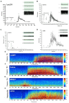
Effects of MK801, LaaβH, and CBIO on the EEG recordings. (A–D) Mean power of EEG recordings in rats treated with LaaβH (A), saline (B), MK801 (C), and CBIO (D). (E–H) Representative frequency images of EEG recordings in rats treated with LaaβH (E), saline (F), MK801 (G), and CBIO (H). N = 7–9 rats per group. Compared with the saline control, LaaβH could prolong the onset of seizure occurrence and reduce the mean power of the EEG, while CBIO could shorten the onset of seizure induction and increase the mean power of the EEG.
FIGURE 6
FIGURE 6
Effects of manipulating SR activity on the expression and phosphorylation of ERK, JNK, and AKT. (A) Western blots of p-ERK/ ERK in LaaβH, saline, and CBIO treated rats, and the quantification of p-ERK. (B) Western blots of p-JNK/JNK in LaaβH, saline, and CBIO treated rats, and the quantification of p-JNK. (C) Western blots of p-AKT/AKT in LaaβH, saline, and CBIO treated rats, and the quantification of p-AKT. N = 3 rats in each group. Notice that LaaβH decreased and CBIO increased the phosphorylation of ERK. ∗∗P < 0.01, ∗∗∗P < 0.001.
FIGURE 7
FIGURE 7
Effects of interfering p-ERK on seizure development in rats treated with D-serine modulators. (A) Effects of the ERK activator U46619 on seizure development and p-ERK/ERK expression in rats treated with LaaβH, compared with rats treated with LaaβH alone. N = 5 rats per group. (B) Effects of the ERK inhibitor PD98059 on seizure development and p-JNK/JNK expression in rats treated with CBIO, compared with rats treated with CBIO alone. N = 5 rats per group. Notice that the ERK activator could block the effects of LaaβH and the ERK inhibitor could block the effects of CBIO. P < 0.05.

Similar articles

Cited by

References

    1. Bado P., Madeira C., Vargas-Lopes C., Moulin T. C., Wasilewska-Sampaio A. P., Maretti L., et al. (2011). Effects of low-dose D-serine on recognition and working memory in mice. Psychopharmacology 218 461–470. 10.1007/s00213-011-2330-4 - DOI - PubMed
    1. Ballard T. M., Pauly-Evers M., Higgins G. A., Ouagazzal A. M., Mutel V., Borroni E., et al. (2002). Severe impairment of NMDA receptor function in mice carrying targeted point mutations in the glycine binding site results in drug-resistant nonhabituating hyperactivity. J. Neurosci. 22 6713–6723. 10.1523/JNEUROSCI.22-15-06713.2002 - DOI - PMC - PubMed
    1. Balu D. T., Li Y., Puhl M. D., Benneyworth M. A., Basu A. C., Takagi S., et al. (2013). Multiple risk pathways for schizophrenia converge in serine racemase knockout mice, a mouse model of NMDA receptor hypofunction. Proc. Natl. Acad. Sci. U.S.A. 110 E2400–E2409. 10.1073/pnas.1304308110 - DOI - PMC - PubMed
    1. Balu D. T., Takagi S., Puhl M. D., Benneyworth M. A., Coyle J. T. (2014). D-serine and serine racemase are localized to neurons in the adult mouse and human forebrain. Cell Mol. Neurobiol. 34 419–435. 10.1007/s10571-014-0027-z - DOI - PMC - PubMed
    1. Clasadonte J., Dong J., Hines D. J., Haydon P. G. (2013). Astrocyte control of synaptic NMDA receptors contributes to the progressive development of temporal lobe epilepsy. Proc. Natl. Acad. Sci. U.S.A. 110 17540–17545. 10.1073/pnas.1311967110 - DOI - PMC - PubMed