. 2019 Mar 26:13:278.
doi: 10.3389/fnins.2019.00278. eCollection 2019.

Levocarnitine Improves AlCl3-Induced Spatial Working Memory Impairment in Swiss albino Mice

Affiliations

Levocarnitine Improves AlCl3-Induced Spatial Working Memory Impairment in Swiss albino Mice

Md Mamun Al-Amin et al. Front Neurosci. .

Abstract

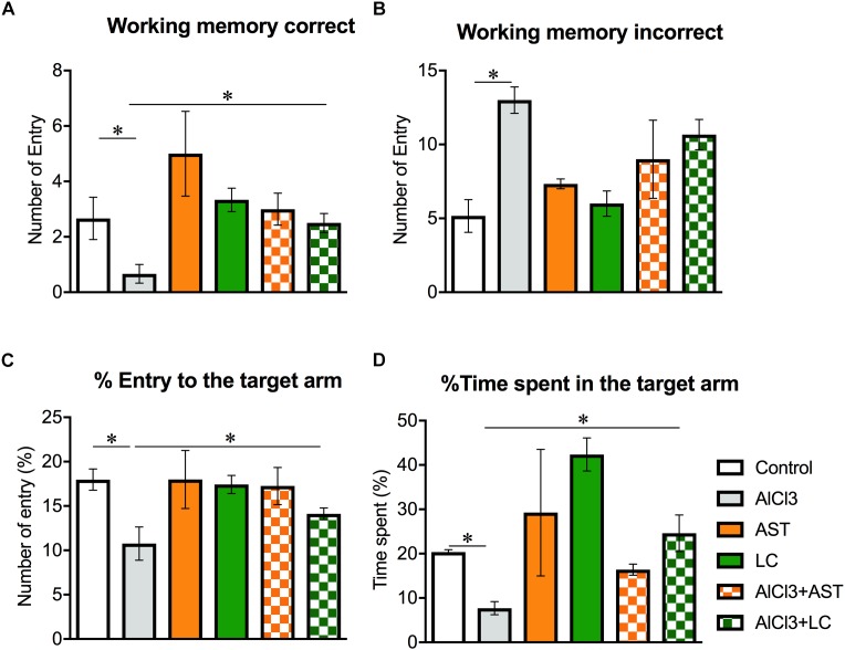
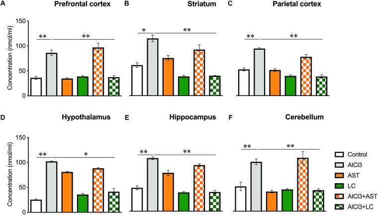
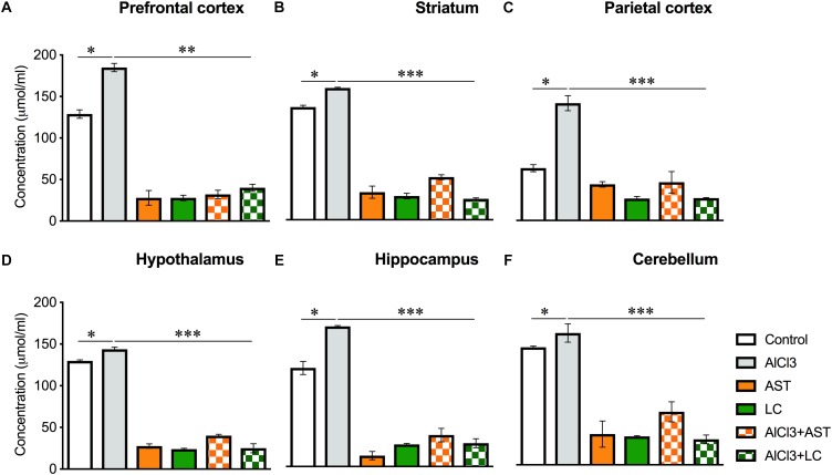
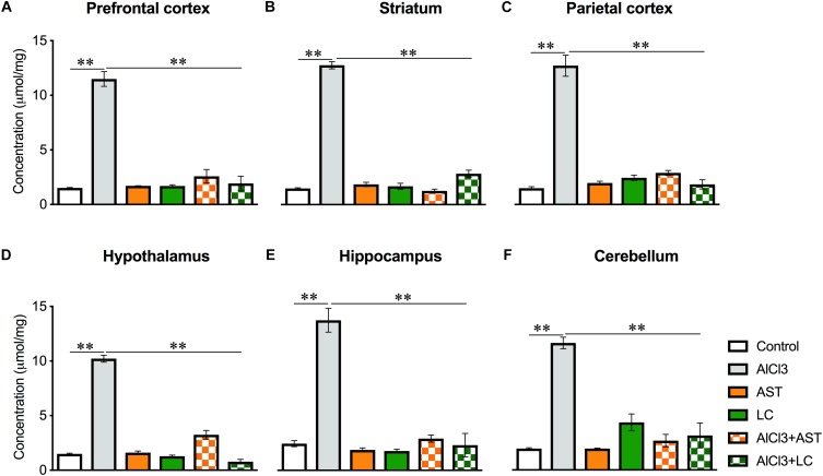
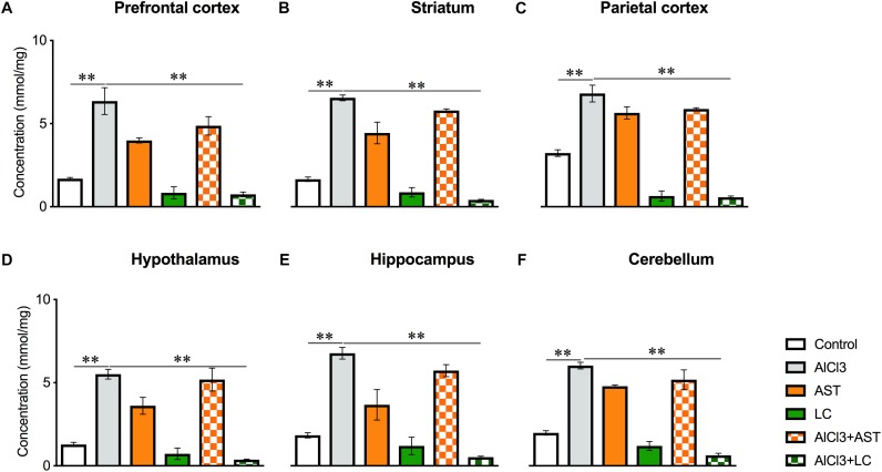
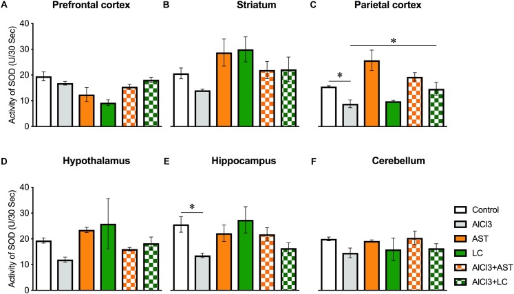
Background: Aluminum, a neurotoxic substance, causes oxidative stress induced-neurodegenerative diseases. Several lines of evidence suggest that levocarnitine has an antioxidant effect and also plays an important role in beta-oxidation of fatty acids. However, the role of levocarnitine in aluminum-induced neurotoxicity has not been well documented. Here we aimed to investigate the effect of levocarnitine on aluminum chloride (AlCl3)-induced oxidative stress and memory dysfunction. Methods: Male Swiss albino mice (n = 30) were treated with either control (saline) or AlCl3 or AlCl3 plus levocarnitine or levocarnitine or astaxanthin plus AlCl3 or astaxanthin alone. The spatial working memory was determined by radial arm maze (RAM). In addition, we measured the lipid peroxidation (MDA), glutathione (GSH), advanced oxidation of protein products (AOPP), nitric oxide (NO) and activity of superoxide dismutase (SOD) in the various brain regions including prefrontal cortex (PFC), striatum (ST), parietal cortex (PC), hippocampus (HIP) hypothalamus (HT) and cerebellum (CB). We used astaxanthin as a standard antioxidant to compare the antioxidant activity of levocarnitine. Results: The RAM data showed that AlCl3 treatment (50 mg/kg) for 2 weeks resulted in a significant deficit in spatial learning in mice. Moreover, aluminum exposure significantly (p < 0.05) increased the level of oxidative stress markers such as MDA, GSH, AOPP and NO in the various brain regions compared to the controls. In addition, combined administration of levocarnitine and AlCl3 significantly (p < 0.05) lowered the MDA, AOPP, GSH and NO levels in mice. Conclusion: Our results demonstrate that levocarnitine could serve as a potential therapeutic agent in the treatment of oxidative stress associated diseases as well as in memory impairment.

Keywords: Alzheimer’s disease; antioxidants; levocarnitine; neurotoxicity; oxidative stress markers; working memory.


Figures

Figure 1
Figure 1
Effect of levocarnitine and aluminum chloride on spatial working memory. The aluminum treated mice showed a lower working memory (A); a higher incorrect working memory (B); a lower percentage of entry to the target arm (C); and a higher number of arm entry (D). The combined (levocarnitine plus aluminum) treatment improved these parameters. The groups were control, AlCl3 (aluminum chloride) and AlCl3+LC (aluminum chloride and levocarnitine) and LC (levocarnitine). Data presented as mean ± SEM. n = 6 per group except AST (n = 3); and AlCL3+AST (n = 3); ∗∗p < 0.01, p < 0.05.
Figure 2
Figure 2
Treatment of levocarnitine and aluminum chloride on MDA (lipid peroxidation) (A–F); in the prefrontal cortex (PFC) (A), striatum (ST) (B), parietal cortex (PC) (C), hypothalamus (HT) (D), hippocampus (HP) (E) and cerebellum (CB) (F). The groups were control, AlCl3 (aluminum chloride), AST (astaxanthin), AST+AlCl3 (aluminum chloride and astaxanthin) and AlCl3+LC (aluminum chloride and levocarnitine) and LC (levocarnitine). Data presented as mean ± SEM. n = 6 per group except AST (n = 3); and AlCL3+AST (n = 3); ∗∗∗p < 0.001, ∗∗p < 0.01, p < 0.05.
Figure 3
Figure 3
Treatment of levocarnitine and aluminum chloride on advanced oxidation of protein produces (AOPP) (A–F); in the prefrontal cortex (PFC) (A), striatum (ST) (B), parietal cortex (PC) (C), hypothalamus (HT) (D), hippocampus (HP) (E) and cerebellum (CB) (F). The groups were control, AlCl3 (aluminum chloride), AST (astaxanthin), AST+AlCl3 (aluminum chloride and astaxanthin) and AlCl3+LC (aluminum chloride and levocarnitine) and LC (levocarnitine). Data presented as mean ± SEM. n = 6 per group except AST (n = 3); and AlCL3+AST (n = 3); ∗∗∗p < 0.001, ∗∗p < 0.01, p < 0.05.
Figure 4
Figure 4
Treatment of levocarnitine and aluminum chloride on glutathione (GSH) (A–F); in the prefrontal cortex (PFC) (A), striatum (ST) (B), parietal cortex (PC) (C), hypothalamus (HT) (D), hippocampus (HP) (E) and cerebellum (CB) (F). The groups were control, AlCl3 (aluminum chloride), AST (astaxanthin), AST+AlCl3 (aluminum chloride and astaxanthin) and AlCl3+LC (aluminum chloride and levocarnitine) and LC (levocarnitine). Data presented as mean ± SEM. n = 6 per group except AST (n = 3); and AlCL3+AST (n = 3); ∗∗∗p < 0.001, ∗∗p < 0.01, p < 0.05.
Figure 5
Figure 5
Treatment of levocarnitine and aluminum chloride on nitric oxide (NO) (A–F); in the prefrontal cortex (PFC) (A), striatum (ST) (B), parietal cortex (PC) (C), hypothalamus (HT) (D), hippocampus (HP) (E) and cerebellum (CB) (F). The groups were control, AlCl3 (aluminum chloride), AST (astaxanthin), AST+AlCl3 (aluminum chloride and astaxanthin) and AlCl3+LC (aluminum chloride and levocarnitine) and LC (levocarnitine). Data presented as mean ± SEM. n = 6 per group except AST (n = 3); and AlCL3+AST (n = 3); ∗∗∗p < 0.001, ∗∗p < 0.01, p < 0.05.
Figure 6
Figure 6
Treatment of levocarnitine and aluminum chloride on superoxide dismutase (SOD) (A–F); in the prefrontal cortex (PFC) (A), striatum (ST) (B), parietal cortex (PC) (C), hypothalamus (HT) (D), hippocampus (HP) (E) and cerebellum (CB) (F). The groups were control, AlCl3 (aluminum chloride), AST (astaxanthin), AST+AlCl3 (aluminum chloride and astaxanthin) and AlCl3+LC (aluminum chloride and levocarnitine) and LC (levocarnitine). Data presented as mean ± SEM. n = 6 per group except AST (n = 3); and AlCL3+AST (n = 3); ∗∗∗p < 0.001, ∗∗p < 0.01, p < 0.05.

References

    1. Akisu M., Ozmen D., Baka M., Habif S., Yalaz M., Arslanoglu S., et al. (2002). Protective effect of dietary supplementation with L-arginine and L-carnitine on hypoxia/reoxygenation-induced necrotizing enterocolitis in young mice. Biol. Neonate 81 260–265. 10.1159/000056757 - DOI - PubMed
    1. Al-Amin M. M., Akhter S., Hasan A. T., Alam T., Nageeb Hasan S. M., Saifullah A. R., et al. (2015a). The antioxidant effect of astaxanthin is higher in young mice than aged: a region specific study on brain. Metab. Brain Dis. 30 1237–1246. 10.1007/s11011-015-9699-4 - DOI - PubMed
    1. Al-Amin M. M., Rahman M. M., Khan F. R., Zaman F., Reza H. M. (2015b). Astaxanthin improves behavioral disorder and oxidative stress in prenatal valproic acid-induced mice model of autism. Behav. Brain Res. 286 112–121. 10.1016/j.bbr.2015.02.041 - DOI - PubMed
    1. Al-Amin M. M., Choudhury M. F. R., Chowdhury A. S., Chowdhury T. R., Jain P., Kazi M., et al. (2018a). Pretreatment with risperidone ameliorates systemic LPS-induced oxidative stress in the cortex and hippocampus. Front. Neurosci. 12:384. 10.3389/fnins.2018.00384 - DOI - PMC - PubMed
    1. Al-Amin M. M., Mahmud W., Pervin M. S., Islam S. M. R., Rahman M. A., Zinchenko A. (2018b). Astaxanthin ameliorates scopolamine-induced spatial memory via reduced cortical-striato-hippocampal oxidative stress. Brain Res. 10.1016/j.brainres.2018.12.014 [Epub ahead of print]. - DOI - PubMed