Review
. 2019 Mar 26:12:69.
doi: 10.3389/fnmol.2019.00069. eCollection 2019.

Schwann Cell Precursors; Multipotent Glial Cells in Embryonic Nerves

Affiliations
Review

Schwann Cell Precursors; Multipotent Glial Cells in Embryonic Nerves

Kristjan R Jessen et al. Front Mol Neurosci. .

Abstract

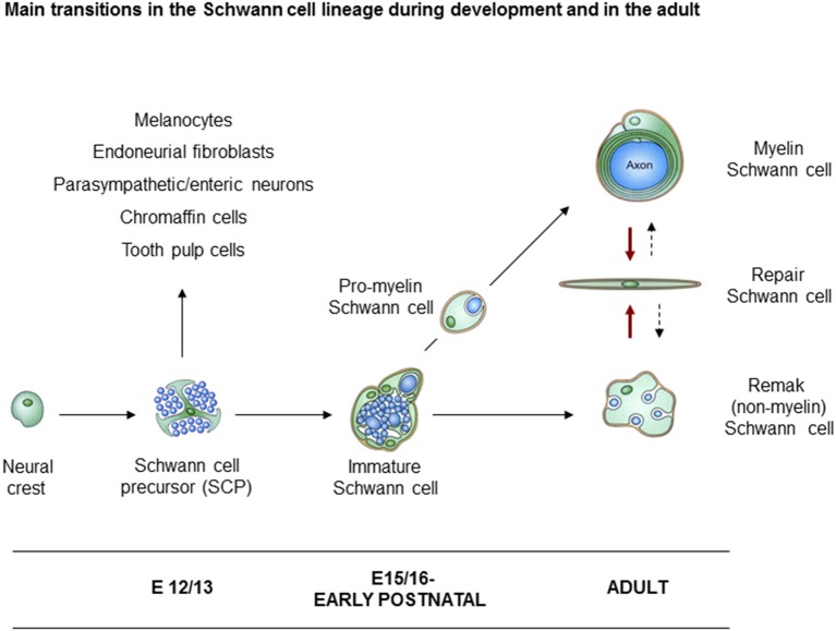
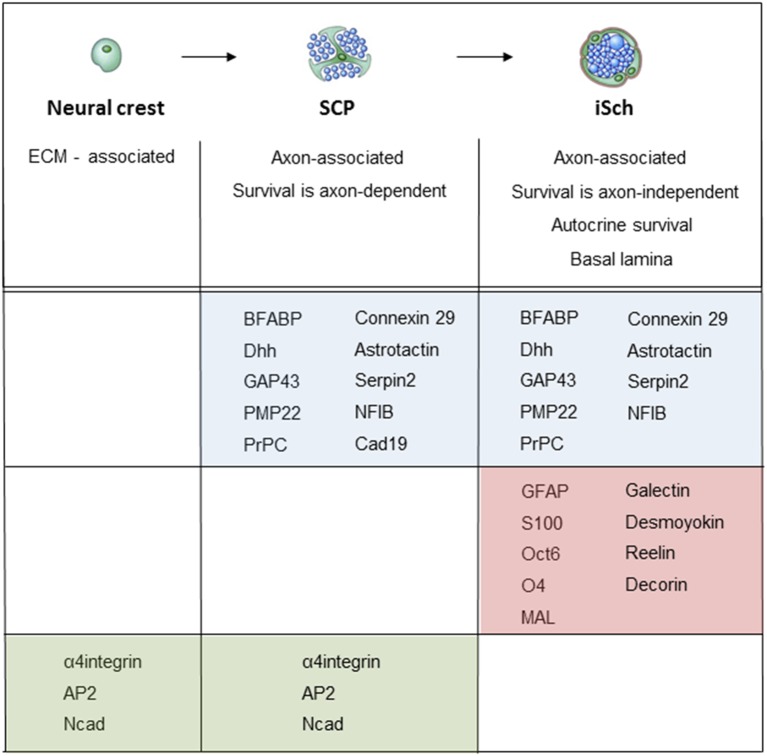
The cells of the neural crest, often referred to as neural crest stem cells, give rise to a number of sub-lineages, one of which is Schwann cells, the glial cells of peripheral nerves. Crest cells transform to adult Schwann cells through the generation of two well defined intermediate stages, the Schwann cell precursors (SCP) in early embryonic nerves, and immature Schwann cells (iSch) in late embryonic and perinatal nerves. SCP are formed when neural crest cells enter nascent nerves and form intimate relationships with axons, a diagnostic feature of glial cells. This involves large-scale changes in gene expression, including the activation of established glial cell markers. Like early glia in the CNS, radial glia, SCP retain developmental multipotency and contribute to other crest-derived lineages during embryonic development. SCP, as well as closely related cells termed boundary cap cells, and later stages of the Schwann cell lineage have all been implicated as the tumor initiating cell in NF1 associated neurofibromas. iSch are formed from SCP in a process that involves the appearance of additional differentiation markers, autocrine survival circuits, cellular elongation, a formation of endoneurial connective tissue and basal lamina. Finally, in peri- and post-natal nerves, iSch are reversibly induced by axon-associated signals to form the myelin and non-myelin Schwann cells of adult nerves. This review article discusses early Schwann cell development in detail and describes a large number of molecular signaling systems that control glial development in embryonic nerves.

Keywords: PNS; PNS glia; Schwann cell lineage; Schwann cell precursor; multipotent glia; nerve development; neural crest.


Figures

Figure 1
Figure 1
Main transitions in the Schwann cell lineage. A scheme illustrating key cell types involved in Schwann cell development, and in nerve repair. Also shown are the alternative cells that can develop from Schwann cell precursors (SCP) in addition to immature Schwann cells (iSch). Black uninterrupted arrows: normal developmental transitions. Red arrows: the Schwann cell injury response. Stippled arrows: post-repair formation of myelin and Remak cells (with permission from Jessen et al., 2015b).
Figure 2
Figure 2
Myelin protein zero (mpz) gene expression at the onset of Schwann cell development. (A) In situ hybridization showing mpz expression in iSch of embryonic (E16) rat nerves (nerve in developing limb arrowed). (B) In situ hybridization showing an initial appearance of mpz expression in scattered neural crest cells at the level with the ventral developing spinal cord (e.g., arrow). Strong mpz labeling is also seen in the notochord. Transverse section from an E9/10 rat embryo. (C) When axons emerge from the ventral spinal cord (shown by TuJ1 immunolabeling in the lower panel), mpz expressing cells cluster nearby (upper panel). Transverse section from an E10/11 rat embryo. (D) In E12/13 rat embryo, mpz expression is seen in nascent spinal nerves (asterisk), and in the ventral root and ventral and lateral aspects of the dorsal root ganglion (G) (with permission, modified from Lee et al., 1997).
Figure 3
Figure 3
The tight association between SCP and axons in developing peripheral nerves. This electron micrograph shows a transverse section through the sciatic nerve of an E14 rat embryo. Note the intimate association between large groups of axons and SCP, and the absence of significant extracellular spaces and connective tissue. Parts of three SCP are visible. Part of the nucleus (N) and the cell body of one of them is included in the field. Arrows point to the junctions between this cell and processes from two other SCP. Scale bar: 1.7 μm (with permission from Jessen and Mirsky, 2005b).
Figure 4
Figure 4
Architectural reorganization of developing nerves. (A) Electron micrograph of a transverse section through E14 rat sciatic nerve. SCP are embedded among the axons and at the surface of the nerve (big arrows). A dividing SCP is also seen (small tilted arrow). Note that connective tissue (yellow) surrounds the nerve, but is not found inside the nerve. (B) Electron micrograph of a transverse section through E18 rat sciatic nerve. iSch surround the collection of axons, forming compact groups (“families”; examples indicated by asterisks). Extensive connective tissue (yellow) containing blood vessels (arrow) is now found inside the nerve surrounding the families, as well as outside the nerve. Bracket indicates the developing perineurium (scale bar: 4 μm).
Figure 5
Figure 5
Molecular expression at the main stages of the embryonic Schwann cell lineage. Below the lineage drawing, main cell biological differences between each developmental each stage are indicated. The molecular markers of each stage divide into three groups: (i) markers that are upregulated at the crest/SCP transition, and maintained by iSCh (blue); (ii) markers that are upregulated at the SCP/iSch transition (pink); and (iii) markers that present on neural crest cells and SCP but are downregulated at the SCP/iSch transition (green). For references, see Jessen and Mirsky (2005a) where more detailed comments on gene expression patterns are provided.

References

    1. Adameyko I., Lallemend F., Aquino J. B., Pereira J. A., Topilko P., Müller T., et al. (2009). Schwann cell precursors from nerve innervation are a cellular origin of melanocytes in skin. Cell 139, 366–379. 10.1016/j.cell.2009.07.049 - DOI - PubMed
    1. Adameyko I., Lallemend F., Furlan A., Zinin N., Aranda S., Kitambi S. S., et al. (2012). Sox2 and Mitf cross-regulatory interactions consolidate progenitor and melanocyte lineages in the cranial neural crest. Development 39, 397–410. 10.1242/dev.065581 - DOI - PMC - PubMed
    1. Arthur-Farraj P. J., Latouche M., Wilton D. K., Quintes S., Chabrol E., Banerjee A., et al. (2012). c-Jun reprograms Schwann cells of injured nerves to generate a repair cell essential for regeneration. Neuron 75, 633–647. 10.1016/j.neuron.2012.06.021 - DOI - PMC - PubMed
    1. Arthur-Farraj P. J., Morgan C. C., Adamowicz M., Gomez-Sanchez J. A., Fazal S. V., Beucher A., et al. (2017). Changes in the coding and non-coding transcriptome and DNA methylome that define the Schwann cell repair phenotype after nerve injury. Cell Rep. 20, 2719–2734. 10.1016/j.celrep.2017.08.064 - DOI - PMC - PubMed
    1. Arthur-Farraj P. J., Wanek K., Hantke J., Davis C. M., Jayakar A., Parkinson D. B., et al. (2011). Mouse Schwann cells need both NRG1 and cyclic AMP to myelinate. Glia 59, 720–733. 10.1002/glia.21144 - DOI - PMC - PubMed