Case Reports
. 2019 Apr;78(4):128-131.

Management of Symptomatic Hepatic "Mega" Hemangioma

Affiliations
Case Reports

Management of Symptomatic Hepatic "Mega" Hemangioma

William A Ketchum et al. Hawaii J Med Public Health. 2019 Apr.

Abstract

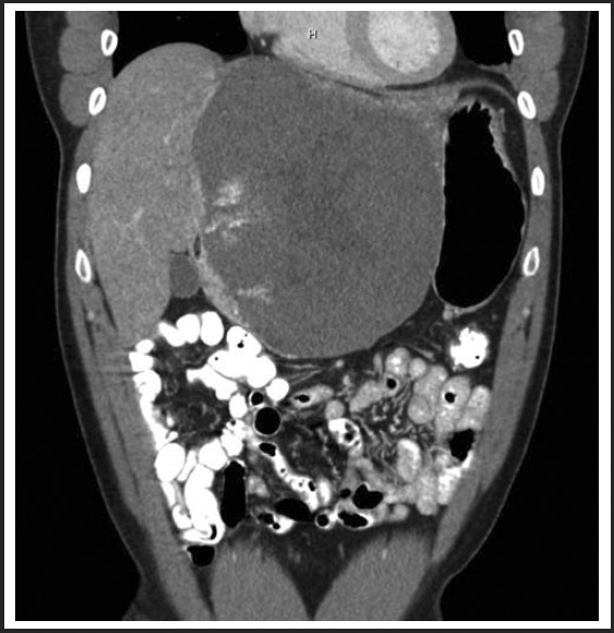
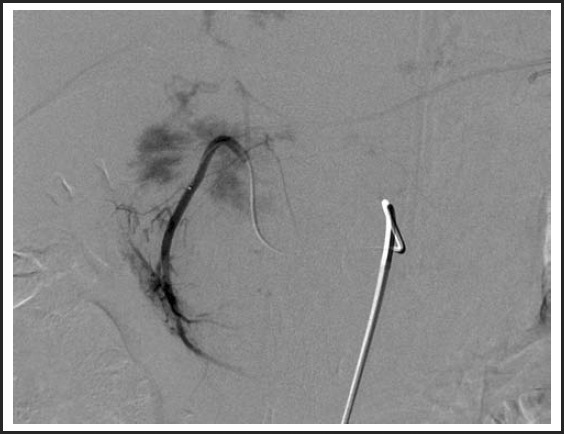
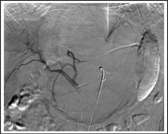
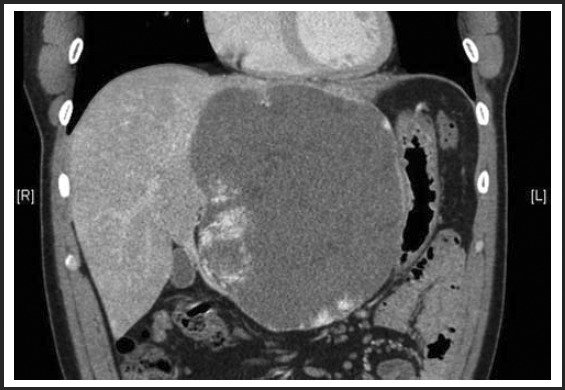
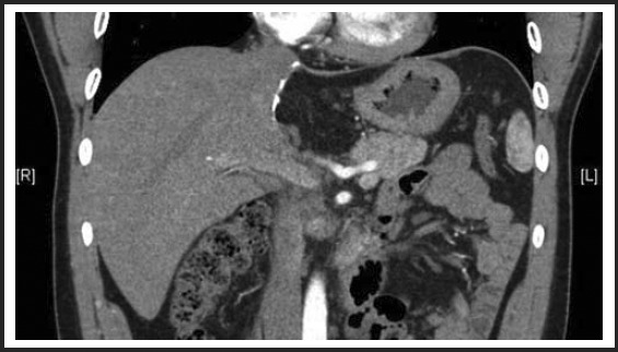
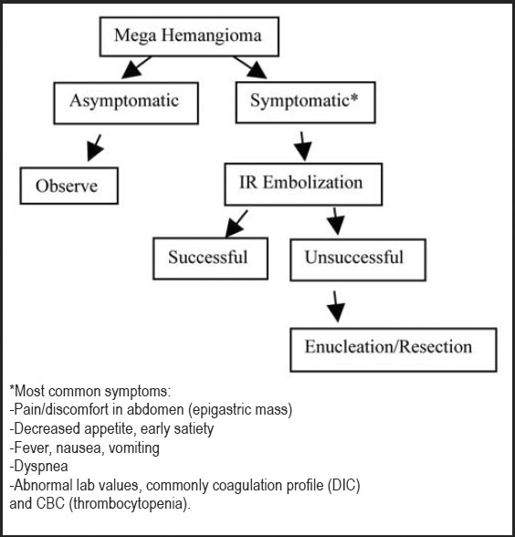
The majority of giant hepatic cavernous hemangiomas are asymptomatic and can safely be observed. However, when a lesion becomes symptomatic, affecting quality of life or cannot be distinguished from a malignancy, then operative therapy should be considered. We herein present a case of a symptomatic 12cm × 14cm × 17cm "mega" hemangioma (>10cm) of the left hepatic lobe. This lesion was initially refractory to transarterial embolization of the left hepatic artery, but was subsequently treated successfully with a left lateral extended hepatic segmentectomy (resection). We thus advocate a rational treatment algorithm for management of hepatic "mega" hemangiomas.

Keywords: hepatic cavernous hemangioma; liver resection; transarterial embolization.


Conflict of interest statement

None of the authors identify a conflict of interest.

Figures

Figure 1
Figure 1
12cm × 14cm low density heterogeneous mass within the left hepatic lobe on CT performed in the Fall of 2010. The mass demonstrated peripheral pooling of contrast initially with centripetal filling on delayed images using triphase IV contrast abdominal CT, consistent with a cavernous hemangioma.
Figure 2
Figure 2
Pre-embolization arteriogram.
Figure 3
Figure 3
Post-embolization of the left hepatic arterial trunk with 6ml of Bead Block 500–700 microspheres.
Figure 4
Figure 4
No significant change in size or appearance of mega hemangioma three months status post-embolization, measuring 12.3cm × 13.7cm × 17.2cm.
Figure 5
Figure 5
Six month status-post extended left lateral segmentectomy.
Figure 6
Figure 6
Proposed treatment algorithm for management of mega hepatic hemangioma (>10 cm).

References

    1. Bailey J, Di Carlo S, Blackwell J, Gomez D. Same day arterial embolization followed by hepatic resection for treatment of giant haemangioma. BMJ Case Rep. 2016 Feb 25; doi: 10.1136/bcr-2015-213259. - DOI - PMC - PubMed
    1. Hoekstra LT, Bieze M, Erdogan D, Roelofs JJTH, Beuers UHW, van Gulik TM. Management of giant liver hemangiomas: an update. Expert Review of Gastroenterology & Hepatology. 2013;7(3):263+. - PubMed
    1. Plackett TP, Lin-Hurtubise KM. Hepatic hemangiomas and parachuting. Aviat Space Environ Med. 2008 Oct;79(10):986–988. - PubMed
    1. Bajenaru N, Balaban V, Savulescu F, Campeanu I, Patrascu T. Hepatic hemangioma-review. Journal of Medicine and Life. 2015;8(Spec Issue):4–11. - PMC - PubMed
    1. Sharma A, Kaspar M, Siddiqui M, Kim J. Enucleation after Embolization of Liver Failure-Causing Giant Liver Hemangioma. Am J Case Rep. 2015 Aug 24;16:563–567. - PMC - PubMed

Publication types