

Inter-observer Variability in the Diagnosis of Proliferative Verrucous Leukoplakia: Clinical Implications for Oral and Maxillofacial Surgeon Understanding: A Collaborative Pilot Study
- PMID: 30972634
- PMCID: PMC7021885
- DOI: 10.1007/s12105-019-01035-z
Inter-observer Variability in the Diagnosis of Proliferative Verrucous Leukoplakia: Clinical Implications for Oral and Maxillofacial Surgeon Understanding: A Collaborative Pilot Study
Abstract
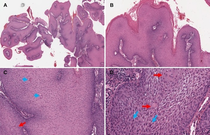
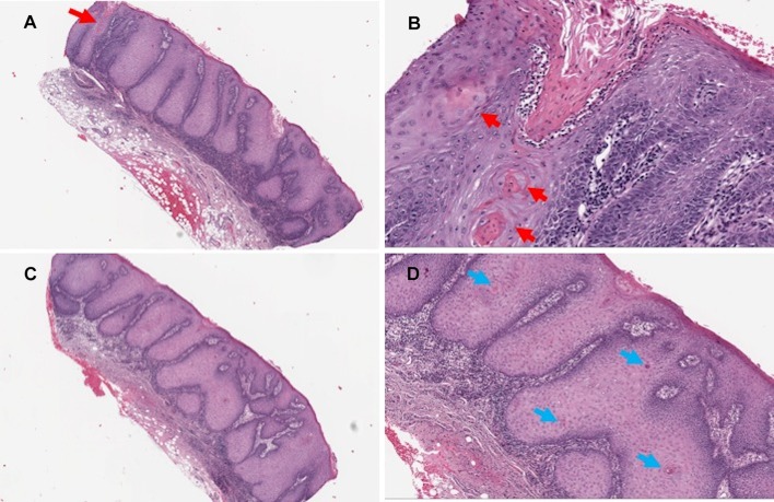
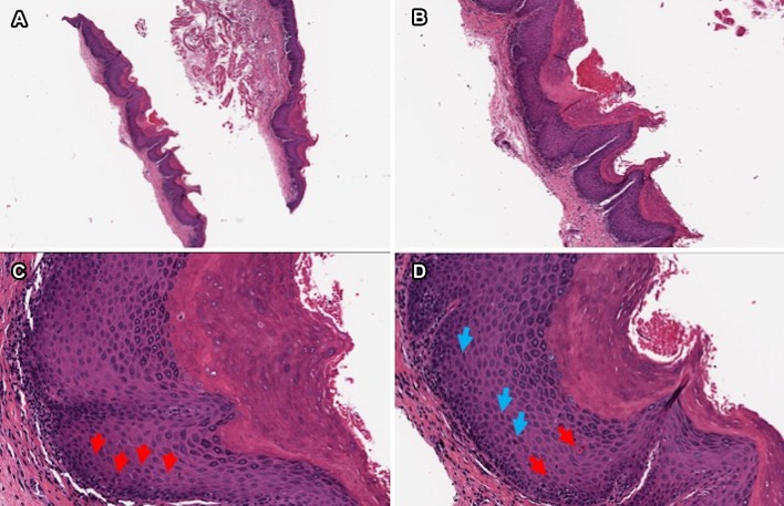
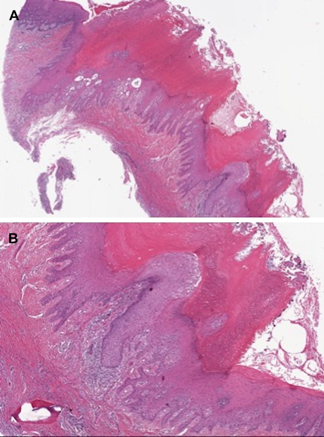
The use of diverse terminology may lead to inconsistent diagnosis and subsequent mistreatment of lesions within the proliferative verrucous leukoplakia (PVL) spectrum. The objectives of this study were: (a) to measure inter-observer variability between a variety of pathologists diagnosing PVL lesions; and (b) to evaluate the impact of diverse terminologies on understanding, interpretation, and subsequent treatment planning by oral and maxillofacial surgeons (OMFS). Six oral pathologists (OP) and six head and neck pathologists (HNP) reviewed 40 digitally scanned slides of PVL-type lesions. Inter-observer agreement on diagnoses was evaluated by Fleiss' kappa analysis. The most commonly used diagnostic terminologies were sent to ten OMFS to evaluate their resulting interpretations and potential follow-up treatment approaches. The overall means of the surgeons' responses were compared by Student t test. There was poor inter-observer agreement between pathologists on the diagnosis of PVL lesions (κ = 0.270), although there was good agreement (κ = 0.650) when diagnosing frankly malignant lesions. The lowest agreement was in diagnosing verrucous hyperplasia (VH) with/without dysplasia, atypical epithelial proliferation (AEP), and verrucous carcinoma (VC). The OMFS showed the lowest agreement on identical categories of non-malignant diagnoses, specifically VH and AEP. This study demonstrates a lack of standardized terminology and diagnostic criteria for the spectrum of PVL lesions. We recommend adopting standardized criteria and terminology, proposed and established by an expert panel white paper, to assist pathologists and clinicians in uniformly diagnosing and managing PVL spectrum lesions.
Keywords: Atypical epithelial proliferation; Inter-observer variability; Papillary squamous cell carcinoma; Proliferative verrucous leukoplakia; Verrucous hyperplasia.
Conflict of interest statement
All authors declare that they have no conflict of interest as it relates to this research project.
Figures


References
-
- Bagan JV, Murillo J, Poveda R, Gavalda C, Jimenez Y, Scully C. Proliferative verrucous leukoplakia: unusual locations of oral squamous cell carcinomas, and field cancerization as shown by the appearance of multiple OSCCs. Oral Oncol. 2004;40:440–443. doi: 10.1016/j.oraloncology.2003.10.008. - DOI - PubMed
-
- Feller L, Wood NH, Raubenheimer EJ. Proliferative verrucous leukoplakia and field cancerization: report of a case. J Int Acad Periodontol. 2006;8:67–70. - PubMed
-
- Fleiss JL. Statistical methods for rates and proportions. 1. London: John Wiley & Sons; 1981.
MeSH terms
LinkOut - more resources
Full Text Sources
Miscellaneous
