

Expression of programmed cell death protein 1 and T-cell immunoglobulin- and mucin-domain-containing molecule-3 on peripheral blood CD4+CD8+ double positive T cells in patients with chronic hepatitis C virus infection and in subjects who spontaneously cleared the virus
- PMID: 30972915
- PMCID: PMC6850126
- DOI: 10.1111/jvh.13108
Expression of programmed cell death protein 1 and T-cell immunoglobulin- and mucin-domain-containing molecule-3 on peripheral blood CD4+CD8+ double positive T cells in patients with chronic hepatitis C virus infection and in subjects who spontaneously cleared the virus
Abstract
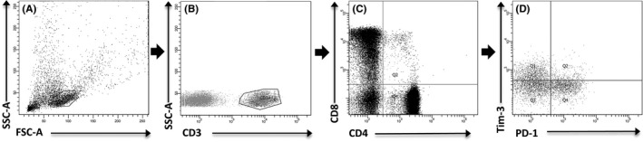
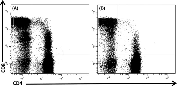
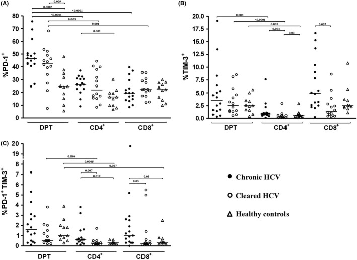
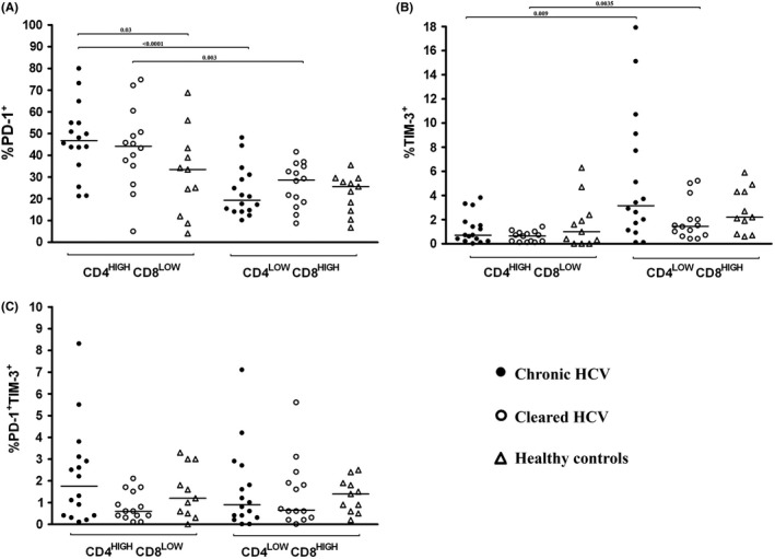
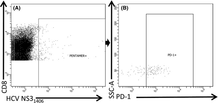
Chronic hepatitis C virus (HCV) infection is characterized by increased proportion of CD4+CD8+ double positive (DP) T cells, but their role in this infection is unclear. In chronic hepatitis C, immune responses to HCV become functionally exhausted, which manifests itself by increased expression of programmed cell death protein 1 (PD-1) and T-cell immunoglobulin- and mucin-domain-containing molecule-3 (Tim-3) on T cells. The aim of our study was to determine PD-1 and Tim-3 phenotype of DP T cells in subjects with naturally resolved and chronic HCV infection. Peripheral blood mononuclear cells from 16 patients with chronic infection and 14 subjects who cleared HCV in the past were stained with anti-CD3, anti-CD4, anti-CD8, anti-PD-1 and anti-Tim-3 antibodies and, in 12 HLA-A*02-positive subjects, MHC class I pentamer with HCV NS31406 epitope. In chronic and past HCV infection, proportions of total DP T cells and PD-1+ DP T cells were similar but significantly higher than in healthy controls. DP T cells were more likely to be PD-1+ than either CD4+ or CD8+ single positive (SP) T cells. HCV-specific cells were present in higher proportions among DP T cells than among CD8+ SP T cells in both patient groups. Furthermore, while the majority of HCV-specific DP T cells were PD-1+, the proportion of HCV-specific CD8+ T cells which were PD-1+ was 4.9 and 1.9 times lower (chronic and past infection, respectively). PD-1 and Tim-3 were predominantly expressed on CD4high CD8low and CD4low CD8high cells, respectively, and co-expression of both markers was uncommon.
Keywords: DP T cells; PD-1; Tim-3; hepatitis C virus.
© 2019 The Authors. Journal of Viral Hepatitis Published by John Wiley & Sons Ltd.
Conflict of interest statement
None declared.
Figures
References
-
- Takaki A, Wiese M, Maertens G, et al. Cellular immune responses persist and humoral responses decrease two decades after recovery from a single‐source outbreak of hepatitis C. Nat Med. 2000;6(5):578‐582. - PubMed
-
- Botarelli P, Brunetto MR, Minutello MA, et al. T‐lymphocyte response to hepatitis C virus in different clinical courses of infection. Gastroenterology. 1993;104(2):580‐587. - PubMed
-
- Seeff LB. Natural history of chronic hepatitis C. Hepatology. 2002;36(5 Suppl 1):S35‐46. - PubMed
Publication types
MeSH terms
Substances
LinkOut - more resources
Full Text Sources
Molecular Biology Databases
Research Materials
Miscellaneous
