. 2019 Aug;25(8):815-823.
doi: 10.1111/cns.13124. Epub 2019 Apr 11.

Prophylactic treatment of hyperbaric oxygen treatment mitigates inflammatory response via mitochondria transfer

Affiliations

Prophylactic treatment of hyperbaric oxygen treatment mitigates inflammatory response via mitochondria transfer

Trenton Lippert et al. CNS Neurosci Ther. 2019 Aug.

Abstract

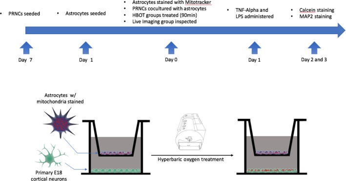
Aims: Hyperbaric oxygen therapy (HBOT) has been widely used as postinjury treatment; however, we investigate its ability to mitigate potential damage as a preconditioning option. Here, we tested the hypothesis that HBOT preconditioning mitigates cell death in primary rat neuronal cells (PRNCs) through the transfer of mitochondria from astrocytes.

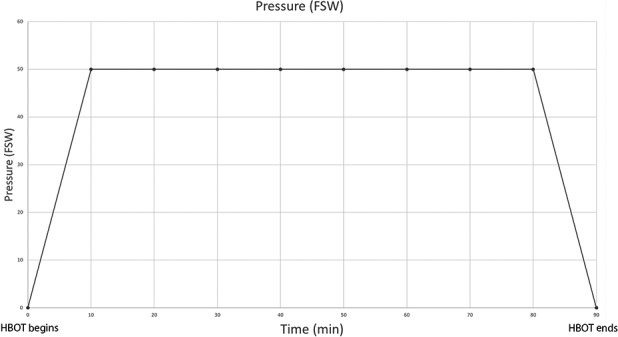
Methods: Primary rat neuronal cells were subjected to a 90-minute HBOT treatment at 2.5 absolute atmospheres prior to either tumor necrosis factor-alpha (TNF-alpha) or lipopolysaccharide (LPS) injury to simulate the inflammation-plagued secondary cell death associated with stroke and traumatic brain injury (TBI). After incubation with TNF-alpha or LPS, the cell viability of each group was examined.

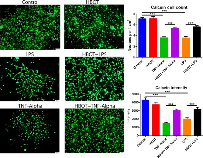
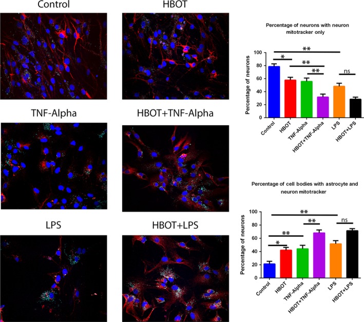
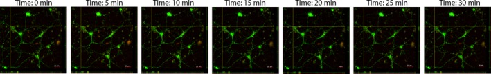
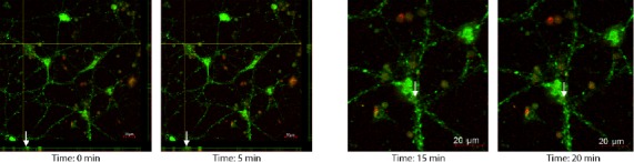
Results: There was a significant increase of cell viability accompanied by mitochondrial transfer in the injury groups that received HBOT preconditioning compared to the injury alone groups (44 ± 5.2 vs 68 ± 4.48, n = 20, P < 0.05). The transfer of mitochondria directly after HBOT treatment was visualized by capturing images in 5-minute intervals, which revealed that the robust transfer of mitochondria begins soon after HBOT and persisted throughout the treatment.

Conclusion: This study shows that HBOT preconditioning stands as a robust prophylactic treatment for sequestration of inflammation inherent in stroke and TBI, possibly facilitating the transfer of resilient mitochondria from astrocytes to inflammation-susceptible neuronal cells in mitigating cell death.

Keywords: hyperbaric; mitochondria transfer; preconditioning; stroke; traumatic brain injury.


Conflict of interest statement

The authors declare no conflicts of interest.

Figures

Figure 1
Figure 1
Experimental design. Timeline of experiment, including cell culture and immunocytochemistry. Hyperbaric oxygen preconditioning in vitro treatment. Astrocytes stained with Mitotracker are cocultured on top of E18 primary cortical neurons (PRNCs) in a growth medium. The cocultured plates were then placed in a hyperbaric oxygen chamber for treatment. Mitotracker of astrocytes was found in the primary E18 cortical neurons
Figure 2
Figure 2
Hyperbaric oxygen therapy course. Pressure graph of hyperbaric oxygen preconditioning. During the first 10 min, there is a constant increase of pressure until the chamber reaches 49.5 feet of seawater (FSW). This pressure is sustained for 70‐min, followed by a constant depressurization of the chamber over a 10‐min period. The total length of the preconditioning treatment is 90 min
Figure 3
Figure 3
Primary cortical neuron cell viability. Administration of Hyperbaric oxygen therapy (HBOT) precondition occurred 24 h prior to injury onset. Primary rat neuronal cells (PRNCs) cocultured with astrocytes were subjected to HBOT for 90 min in the preconditioning group. After the insult was introduced to the PRNCs cocultured with astrocytes for 24 h, the cell viability was analyzed by Calcein‐AM staining. Cell counting was performed and standardized per 1 cm2. Intensity of signal was also calculated as described in the materials and methods. *P < 0.05, **P < 0.01, ***P < 0.001. The scale bar corresponds to 20 μm
Figure 4
Figure 4
Mitochondrial transfer to primary cortical neurons. Migration of astrocyte mitochondria was tracked using Mitotracker staining of astrocytes prior to coculture with primary rat neuronal cells (PRNCs). Cocultures were subjected to the same preconditioning setting as previously described. Transfer of astrocyte mitochondria (cyan) was quantified by counting the number PRNCs containing cyan and dividing by the total number of PRNCs marked with MAP2 (red). DAPI was utilized to mark PRNC nuclei. *P < 0.05. ** P < 0.01. The scale bar corresponds to 20 μm
Figure 5
Figure 5
Live imaging of primary cortical neurons undergoing mitochondrial transfer. Rat E18 neuronal cells were harvested and seeded in poly‐D‐lysine coated (100 μg/mL) 6‐well plates at 1.5 × 106 cells/well in Dulbecco's Modified Eagle Media high glucose with 1% antibiotic/antimycotic for 24 h. The media was changed every 3 d, and the cells were subcultured at 90% confluency as needed. Twenty‐four hours prior to the preconditioning, U87 astrocytes were stained with MitoTracker Deep Red FM (500 nmol/L) according to manufacturer's protocol and seeded into coculture inserts at 0.5 × 106 cells/well. On the day of the experiment, rat E18 neuronal cells were stained with MitoTracker Green FM (200 nmol/L) according to manufacturer's protocol. The neuronal cells were then cocultured with U87 astrocytes for 3 h prior to Hyperbaric oxygen therapy (HBOT) administration. The cells were subjected to 70 min of HBOT at 2.5 ATA with 10 min pressurization and depressurization at a rate of 0.07 atm/min for a total of 90 min. Directly following HBOT treatment, the cocultured astrocytes were removed and the confocal z‐stacks live images were captured at 180× every 5 min for 30 min. Primary rat neuronal cell (PRNC) mitochondria: Green; Astrocyte mitochondria: Red. The scale bar corresponds to 20 μm
Figure 6
Figure 6
Live imaging of primary cortical neurons undergoing mitochondrial transfer. Rat E18 neuronal cells were harvested and seeded in poly‐D‐lysine coated (100 μg/mL) 6‐well plates at 1.5 × 106 cells/well in Dulbecco's Modified Eagle Media high glucose with 1% antibiotic/antimycotic for 24 h. The media was changed every 3 d, and the cells were subcultured at 90% confluency as needed. Twenty‐four hours prior to the preconditioning, U87 astrocytes were stained with MitoTracker Deep Red FM (500 nmol/L) according to manufacturer's protocol and seeded into coculture inserts at 0.5 × 106 cells/well. On the day of the experiment, rat E18 neuronal cells were stained with MitoTracker Green FM (200 nmol/L) according to manufacturer's protocol. The neuronal cells were then cocultured with U87 astrocytes for 3 h prior to Hyperbaric oxygen therapy (HBOT) administration. The cells were subjected to 70 min of HBOT at 2.5 ATA with 10 min pressurization and depressurization at a rate of 0.07 atm/min for a total of 90 min. Directly following HBOT treatment, the cocultured astrocytes were removed and the confocal z‐stacks live images were captured at 180×. Primary rat neuronal cell (PRNC) mitochondria: Green; Astrocyte mitochondria: Red. White arrows indicate the movement of astrocyte mitochondria into the PRNC during 5‐min intervals. The scale bar corresponds to 20 μm

References

    1. Borlongan CV, Burns J, Tajiri N, et al. Epidemiological survey‐based formulae to approximate incidence and prevalence of neurological disorders in the United States: a meta‐analysis. PLoS One. 2013;8(10):e78490. - PMC - PubMed
    1. Benjamin EJ, Blaha MJ, Chiuve SE, et al. Heart disease and stroke statistics‐2017 update: a report from the American Heart Association. Circulation. 2017;135(10):e146‐e603. - PMC - PubMed
    1. Taylor CA, Bell JM, Breiding MJ, Xu L. Traumatic brain injury‐related emergency department visits, hospitalizations, and deaths ‐ United States, 2007 and 2013. MMWR Surveill Summ. 2017;66(9):815‐16. - PMC - PubMed
    1. Ma VY, Chan L, Carruthers KJ. Incidence, prevalence, costs, and impact on disability of common conditions requiring rehabilitation in the United States: stroke, spinal cord injury, traumatic brain injury, multiple sclerosis, osteoarthritis, rheumatoid arthritis, limb loss, and back pain. Arch Phys Med Rehabil. 2014;95(5):986‐995.e1. - PMC - PubMed
    1. Borlongan CV, Chopp M, Steinberg GK, et al. Potential of stem/progenitor cells in treating stroke: the missing steps in translating cell therapy from laboratory to clinic. Regen Med. 2008;3(3):249‐250. - PMC - PubMed

Publication types