. 2019 Jun;23(6):4290-4300.
doi: 10.1111/jcmm.14320. Epub 2019 Apr 11.

The phytochemical polydatin ameliorates non-alcoholic steatohepatitis by restoring lysosomal function and autophagic flux

Affiliations

The phytochemical polydatin ameliorates non-alcoholic steatohepatitis by restoring lysosomal function and autophagic flux

Xiaoting Chen et al. J Cell Mol Med. 2019 Jun.

Abstract

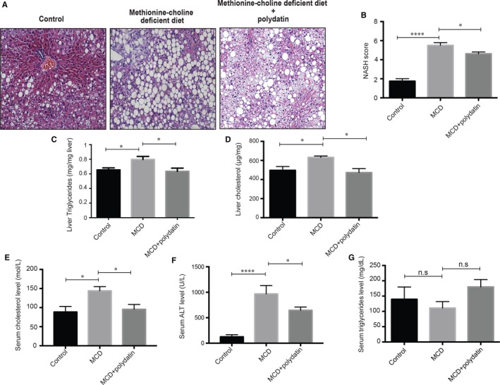
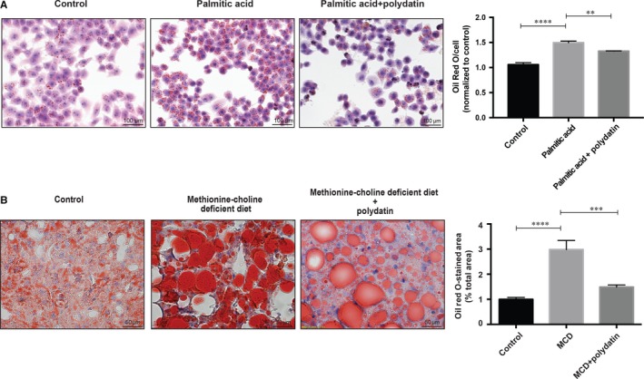
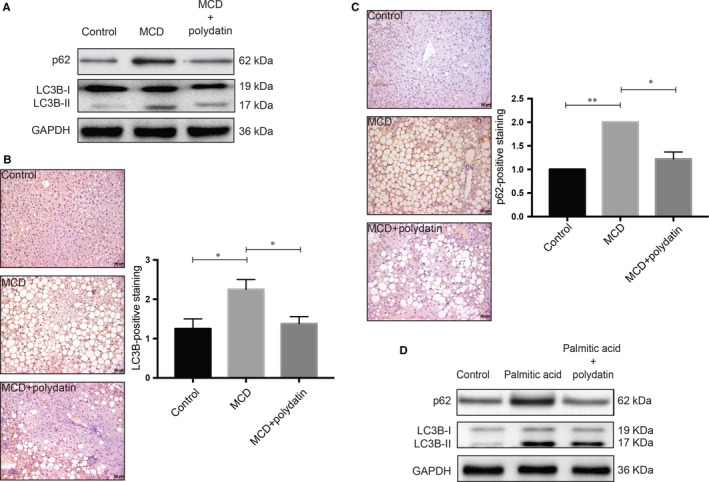
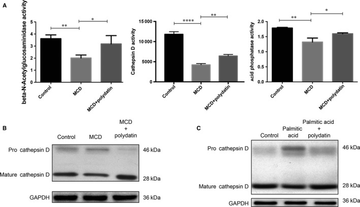
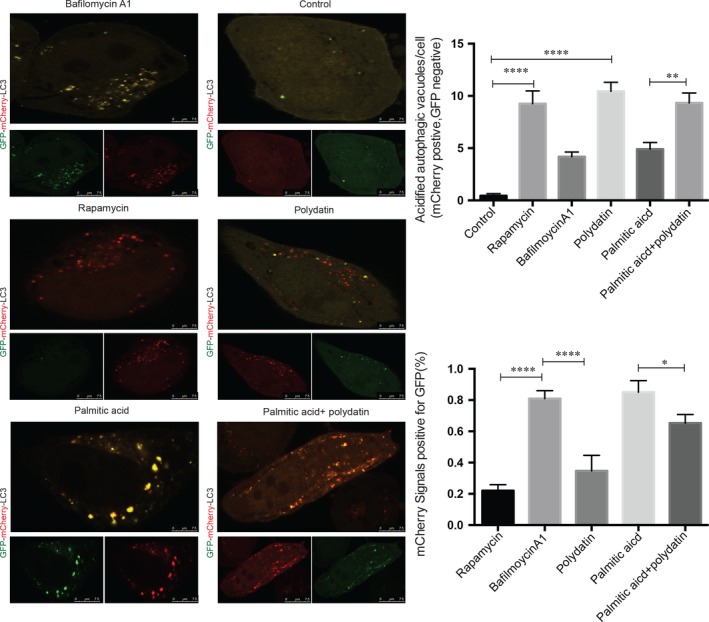
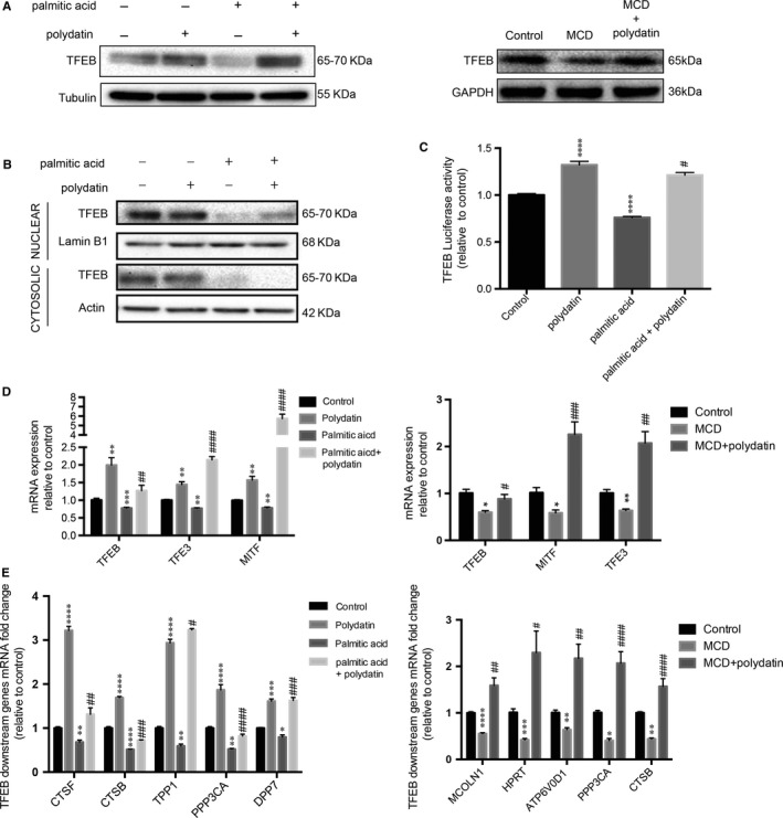
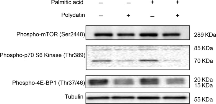
Impaired autophagic degradation of intracellular lipids is causally linked to the development of non-alcoholic steatohepatitis (NASH). Pharmacological agents that can restore hepatic autophagic flux could therefore have therapeutic potentials for this increasingly prevalent disease. Herein, we investigated the effects of polydatin, a natural precursor of resveratrol, in a murine nutritional model of NASH and a cell line model of steatosis. Results showed that oral administration of polydatin protected against hepatic lipid accumulation and alleviated inflammation and hepatocyte damage in db/db mice fed methionine-choline deficient diet. Polydatin also alleviated palmitic acid-induced lipid accumulation in cultured hepatocytes. In both models, polydatin restored lysosomal function and autophagic flux that were impaired by NASH or steatosis. Mechanistically, polydatin inhibited mTOR signalling and up-regulated the expression and activity of TFEB, a known master regulator of lysosomal function. In conclusion, polydatin ameliorated NASH through restoring autophagic flux. The polydatin-regulated autophagy was associated with inhibition of mTOR pathway and restoration of lysosomal function by TFEB. Our study provided affirmative preclinical evidence to inform future clinical trials for examining the potential anti-NASH effect of polydatin in humans.

Keywords: LC3; NAFLD; cathepsin D; lipophagy; p62.


Conflict of interest statement

The authors confirm that there is no conflict of interest.

Figures

Figure 1
Figure 1
Effects of polydatin on liver of methionine‐choline deficient (MCD) diet‐fed db/db mice. Polydatin ameliorated MCD diet‐induced hepatic damages. (A) Representative H&E staining from db/db mice fed control diet, MCD diet, or MCD diet with polydatin. Polydatin was administered by oral gavage at the dosage of 100 mg/kg every other day for 4 wk. Bars = 50 µm; (B) Non‐alcoholic steatohepatitis (NASH) activity score calculated for steatosis, lobular inflammation and ballooning; (C‐D) The content of liver triglyceride and hepatic total cholesterol from liver tissues were measured; (E‐G) Serum total cholesterol, aminotransferases (ALT) levels and triglycerides levels of mice. Mean ± SEM (n = 8 per group). *P < 0.05; ****P < 0.0001
Figure 2
Figure 2
Effects of polydatin on lipid accumulation in LO2 hepatocytes and methionine‐choline deficient (MCD) diet‐fed db/db mice. (A) Treatment with polydatin at the concentration of 24 μmol/L for 24 h alleviated palmitic acid (PA; 60 µg/mL)‐induced lipid accumulation. Representative Oil‐Red‐O staining of hepatocytes from different groups were shown. Oil Red O contents from different groups were determined by colorimetry. Mean ± SEM (n = 3). **P < 0.01; ****P < 0.0001. (B) Representative Oil red O staining from db/db mice fed control diet, MCD diet, or MCD diet with polydatin. Bars = 50 µm. Mean ± SEM (n = 8 per group). ***P < 0.001; ****P < 0.0001
Figure 3
Figure 3
Alleviation of steatosis/steatohepatitis‐induced impairment of autophagic flux by polydatin. Polydatin rectified the autophagic impairment as shown by reduced accumulation of LC3B‐II and p62/SQSTM1. (A) Protein levels of autophagy markers (LC3B and p62) in non‐alcoholic steatohepatitis (NASH) mouse liver samples were determined. Polydatin administered at the dosage of 100 mg/kg every other day for 4 weeks reduced the hepatic accumulation of LC3B‐II and p62 in NASH mice. Representative blots from three independent experiments were shown. (B and C) Immunohistochemical assessment of autophagy markers p62 and LC3B in liver tissues was performed. Liver tissue sections were stained using LC3B or p62 antibodies. For semi‐quantitative analysis of p62 and LC3 accumulation, the scores were rated as grades 0 (none), 1 (minor), 2 (moderate) and 3 (severe). More than 10 sections in each mouse were evaluated. (D) Protein levels of autophagy markers in palmitic acid (PA; 60 µg/mL)‐exposed human LO2 cells were measured. Polydatin (24 μmol/L) diminished the concomitant accumulation of LC3B‐II and p62 caused by PA exposure (24 h). Representative blots were selected from three independent experiments. Bars = 50 µm. Mean ± SEM. *P < 0.05; **P < 0.01
Figure 4
Figure 4
Reactivation of lysosomal enzyme activities by polydatin. Polydatin alleviated methionine‐choline deficient diet‐triggered and PA‐induced pro‐cathepsin D accumulation and reduction of lysosomal enzyme activities. (A) Hepatic lysosomal enzyme activities (β‐N‐acetylglucoseaminidase, acid phosphatase, and cathepsin (D) were measured. (B) Hepatic levels of pro‐cathepsin D and mature cathepsin D from non‐alcoholic steatohepatitis mouse model were determined by Western blots. (C) LO2 cells were exposed to palmitic acid (60 µg/mL) for 24 h in the absence or presence of polydatin (24 µmol/L). Pro‐cathepsin D and mature cathepsin levels were determined by Western blots. Representative blots were selected from three independent experiments. Mean ± SEM. *P < 0.05; **P < 0.01; ****P < 0.0001
Figure 5
Figure 5
Rectification of palmitic acid‐induced impairment of autolysosomal acidification by polydatin in LO2 hepatocytes. LO2 cells were transfected with mCherry‐GFP‐LC3 plasmid for 24 h followed by treatment with rapamycin (1.1 µmol/L), polydatin (24 µmol/L), bafilomycin A1 (200 µmol/L) or palmitic acid (60 µg/mL) in the absence or presence of polydatin (24 µmol/L) for additional 24 h. Acidified and non‐acidified LC3‐positive autophagosomes were visualized and counted under a confocal microscope. Mean ± SEM in three independent experiments.*P < 0.05; **P < 0.01; ****P < 0.0001
Figure 6
Figure 6
Restoration of transcription factor EB (TFEB) transcription activity, mRNA and protein levels by polydatin. (A) LO2 cells were treated with polydatin (24 µmol/L) alone or exposed to palmitic acid (PA; 60 µg/mL) for 24 h in the absence or presence of polydatin (24 µmol/L). Hepatic tissues were extracted from non‐alcoholic steatohepatitis (NASH) mouse model. TFEB levels were determined by Western blots. Representative blots were selected from three independent experiments. (B) The cytosolic and nuclear fractions were isolated and the expression levels of TFEB were evaluated by Western blots. Representative blots were selected from three independent experiments. (C) LO2 cells were transfected with vector only or 4X CLEAR (Coordinated Lysosomal Expression and Regulation) promoter–luciferase vector and exposed to polydatin (24 µmol/L) alone or PA (60 µg/mL) in the absence or presence of polydatin (24 µmol/L) for 24 h. Afterwards, luciferase activity of TFEB was assayed. (D) Relative quantitative PCR analysis of mRNA levels of MiTF/TFE family members in LO2 cells treated with polydatin (24 µmol/L) alone or exposed to palmitic acid (60 µg/mL) in the absence or presence of polydatin (24 µmol/L) for 24 h or in hepatic tissues of NASH mouse model. (E) Relative quantitative PCR analysis of mRNA levels of TFEB target genes in LO2 cells treated with polydatin (24 µmol/L) alone or exposed to PA (60 µg/mL) in the absence or presence of polydatin (24 µmol/L) for 24 h and in hepatic tissues of NASH mouse model. Mean ± SEM in three independent experiments. *P < 0.05; **P < 0.01; ***P < 0.001;****P < 0.0001 when comparing control and polydatin or PA; comparing control diet and methionine‐choline deficient (MCD) diet. # P < 0.05; ## P < 0.01; ### P < 0.001; #### P < 0.0001 when comparing PA and the combination of PA and polydatin; comparing MCD diet and MCD diet with the administration of polydatin
Figure 7
Figure 7
Inhibition of mTOR pathway by polydatin in LO2 hepatocytes. Polydatin‐enhanced autophagic flux was accompanied by inhibition of mTOR. LO2 cells were treated with polydatin (24 µmol/L) alone or exposed to palmitic acid (PA; 60 µg/mL) for 24 h in the absence or presence of polydatin (24 µmol/L). Phosphorylation levels of mTOR (Ser2448) or its related proteins p70‐S6K (Thr389) and 4E‐BP1 (Thr37/46) were determined by Western blots. Representative blots of three independent experiments are shown

References

    1. Yeh MM, Brunt EM. Pathology of nonalcoholic fatty liver disease. Am J Clin Pathol. 2007;128(5):837‐847. - PubMed
    1. Yu J, Shen J, Sun TT, Zhang X, Wong N. Obesity, insulin resistance, NASH and hepatocellular carcinoma. Semin Cancer Biol 2013;23(6 Pt B):483‐491. - PubMed
    1. Neuschwander‐Tetri BA. Nontriglyceride hepatic lipotoxicity: the new paradigm for the pathogenesis of NASH. Curr Gastroenterol Rep. 2010;12(1):49‐56. - PubMed
    1. Wu W, Coffelt SB, Cho Ch, et al. The autophagic paradox in cancer therapy. Oncogene. 2012;31(8):939‐953. - PubMed
    1. Trocoli A, Djavaheri‐Mergny M. The complex interplay between autophagy and NF‐kappaB signaling pathways in cancer cells. Am J Cancer Res. 2011;1(5):629‐649. - PMC - PubMed

Publication types

LinkOut - more resources