. 2019 Sep 1;44(17):1177-1185.
doi: 10.1097/BRS.0000000000003054.

The Role of Type I Diabetes in Intervertebral Disc Degeneration

Affiliations

The Role of Type I Diabetes in Intervertebral Disc Degeneration

Fabrizio Russo et al. Spine (Phila Pa 1976). .

Abstract

Study design: An experimental laboratory study.

Objective: To investigate the pathogenesis of intervertebral disc degeneration (IDD) in a murine model of type 1 diabetes mellitus (DM), namely nonobese diabetic (NOD) mouse.

Summary of background data: IDD is a leading contributor of low back pain, which represents one of the most disabling symptoms within the adult population. DM is a chronic metabolic disease currently affecting one in 10 adults in the United States. It is associated with an increased risk of developing IDD, but the underlying process remains poorly understood.

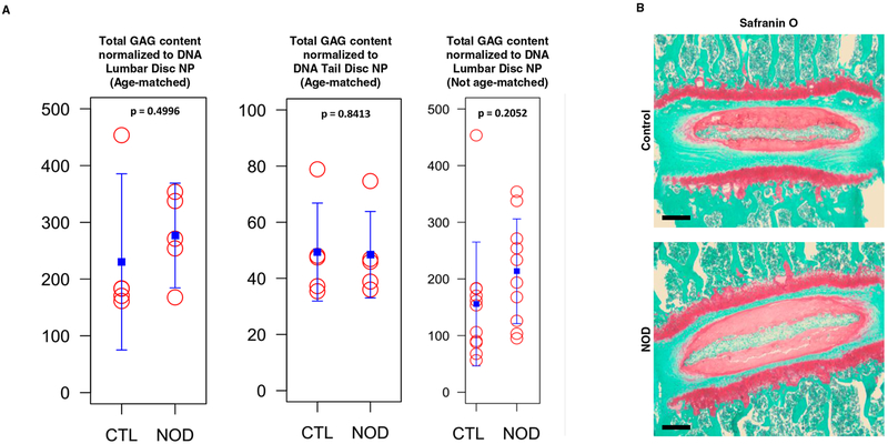
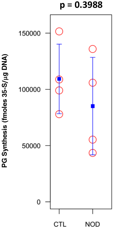
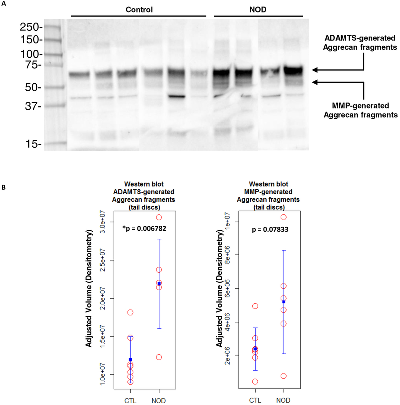
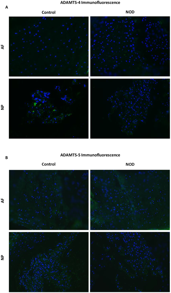
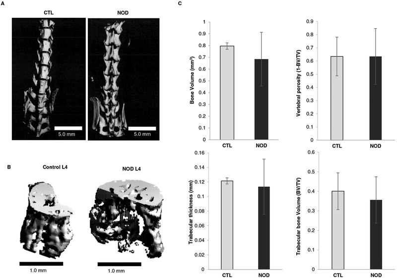
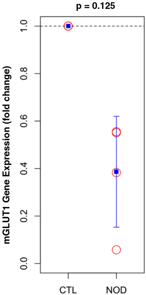
Methods: Total disc glycosaminoglycan content, proteoglycan synthesis, aggrecan fragmentation, glucose transporter gene expression, and apoptosis were assessed in NOD mice and wild-type euglycemic control mice. Spinal structural and molecular changes were analyzed by micro-computed tomography, histological staining (Safranin-O and fast green), and quantitative immunofluorescence (anti-ADAMTS-4 and -5 antibodies).

Results: Compared with euglycemic controls, NOD mice showed increased disc apoptosis and matrix aggrecan fragmentation. Disc glycosaminoglycan content and histological features of NOD mice did not significantly differ from those of euglycemic littermates.

Conclusion: These data demonstrate that DM may contribute to IDD by increasing aggrecan degradation and promoting cell apoptosis, which may represent early indicators of the involvement of DM in the pathogenesis of IDD.

Level of evidence: N/A.


Figures

Fig. 1
Fig. 1
Diabetic mice did not show a significant loss of PG and GAG in disc matrix. a DMMB assay for total GAG content normalized to DNA in NP tissue of control and NOD mice. b A representative Safranin O/fast green histological staining of thoracic disc for matrix PG in control and NOD mice. Red, proteoglycan staining.
Fig. 2
Fig. 2
PG synthesis was measured by 35S-sulfate incorporation in control and NOD mice IVDs and was not significantly affected.
Fig. 3
Fig. 3
T1DM increased aggrecan fragmentation within IVD matrix. a A representative immunoblot with corresponding quantitative analysis showing the levels of tail IVD aggrecan fragments generated by ADAMTS and MMP activity within the aggrecan interglobular domain. b Values of ADAMTS-mediated cleavage were higher and statistically significant in NOD mice (*p < 0.05) compared to euglycemic littermates. MMP-mediated fragmentation was increased in diabetic mice as well (p > 0.05).
Fig. 4
Fig. 4
ADAMTS quantitative immunofluorescence assay with anti-ADAMTS-4 (a) and −5 antibodies (b) did not show metalloproteinase upregulation within disc tissues.
Fig. 5
Fig. 5
TUNEL assay to identify apoptotic cells in disc tissue of control and NOD mice. a Histological sections including vertebral bodies, cartilaginous endplates and intervertebral disc of both wild-type and NOD mice were analyzed under 40x and 200x magnification. Blue, DAPI (nuclei); green, apoptotic cells. b TUNEL assay quantification was calculated as the percentage of TUNEL positive cells in control animals and NOD mice in nine random fields. Apoptosis was significantly enhanced (*p < 0.05) in NOD disc tissue under high magnification (200x).
Fig. 6
Fig. 6
Diabetes did not affect vertebral bone microarchitecture. a Representative 3D reconstruction of the micro-computed tomographical images of the lumbar spine and L4 vertebra (b) in both control and NOD mice. c Quantitative bone parameters did not significantly differ from wild type mice and diabetic littermates.
Fig. 7
Fig. 7
Diabetic mice showed a downregulation in mouse glucose transporter 1 (mGLUT1) gene expression compared to control littermates.

References

    1. Vadala G, Russo F, Ambrosio L, et al. Biotechnologies and Biomaterials in Spine Surgery. J Biol Regul Homeost Agents. 2015;29(4 Suppl):137–47. - PubMed
    1. Vadala G, Russo F, Di Martino A, et al. Intervertebral disc regeneration: from the degenerative cascade to molecular therapy and tissue engineering. J Tissue Eng Regen Med 2015;9(6):679–90. - PubMed
    1. Nasto LA, Ngo K, Leme AS, et al. Investigating the role of DNA damage in tobacco smoking-induced spine degeneration. Spine J 2014;14(3):416–23. - PMC - PubMed
    1. Russo F, Hartman RA, Bell KM, et al. Biomechanical Evaluation of Transpedicular Nucleotomy With Intact Annulus Fibrosus. Spine (Phila Pa 1976). 2017;42(4):E193–E201. - PubMed
    1. Vadala G, Russo F, Ambrosio L, et al. Stem cells sources for intervertebral disc regeneration. World J Stem Cells. 2016;8(5):185–201. - PMC - PubMed

MeSH terms