. 2019 Apr 1;60(5):1491-1500.
doi: 10.1167/iovs.18-25966.

The Retina in Alzheimer's Disease: Histomorphometric Analysis of an Ophthalmologic Biomarker

Affiliations

The Retina in Alzheimer's Disease: Histomorphometric Analysis of an Ophthalmologic Biomarker

Samuel Asanad et al. Invest Ophthalmol Vis Sci. .

Abstract

Purpose: To provide a histopathologic, morphometric analysis of the retina in Alzheimer's disease (AD).

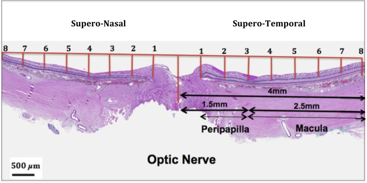
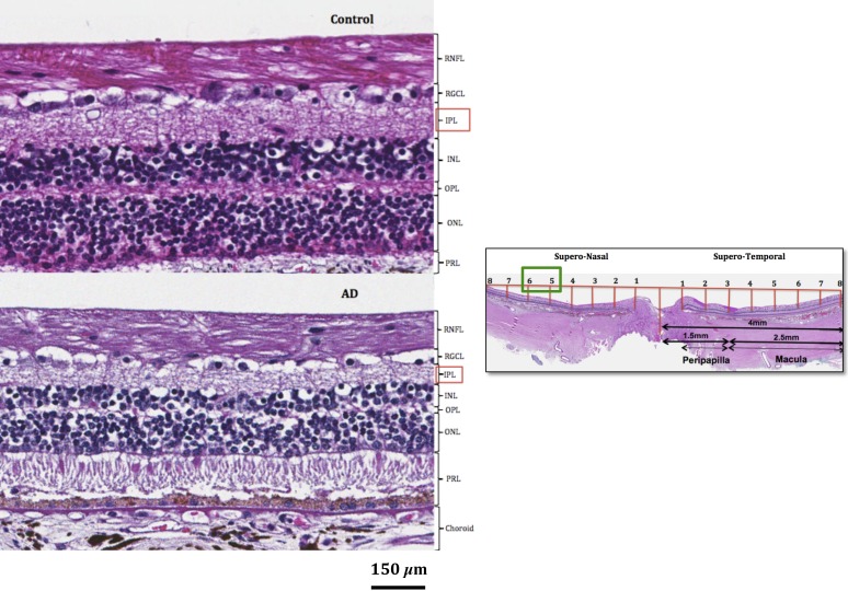
Methods: Human postmortem retinas from eight patients with AD (mean age: 80 ± 12.7 years) and from 11 age-matched controls (mean age: 78 ± 16.57 years) were analyzed. The retinas were sampled from the superior quadrant on both the temporal and nasal sides with respect to the optic nerve. Thickness of the inner and outer layers involving the retinal nerve fiber layer (RNFL), retinal ganglion cell layer (RGCL), inner plexiform layer (IPL), inner nuclear layer (INL), and outer nuclear layer (ONL) were measured and compared between controls and AD. A total of 16 measurements of retinal thickness were acquired for each layer.

Results: RNFL thinning supero-temporally was significant closest to the optic nerve (∼35% thickness reduction; P < 0.001). Supero-nasally, RNFL was thinner throughout all points (∼40% reduction; P < 0.001). Supero-temporally, RGCL thinning was pronounced toward the macula (∼35% thickness reduction; P < 0.001). Supero-nasally, RGCL showed uniform thinning throughout (∼35% reduction; P < 0.001). IPL thinning supero-temporally was statistically significant in the macula (∼15% reduction; P < 0.01). Supero-nasal IPL featured uniform thinning throughout (∼25% reduction; P < 0.001). Supero-temporally, INL and ONL thinning were pronounced toward the macula (∼25% reduction; P < 0.01). Supero-nasally, INL and ONL were thinner throughout (∼25% reduction; P < 0.01).

Conclusions: Our study revealed marked thinning in both the inner and outer layers of the retina. These quantified histopathologic findings provide a more comprehensive understanding of the retina in AD than previously reported.


Figures

Figure 1
Figure 1
Illustrates challenges of retinal layer identification on enhanced depth imaging optical coherence tomography (EDI-OCT) (A) relative to accurate identification and delineation of the corresponding retinal layers on histology in a representative control micrograph stained with hematoxylin and eosin (B) within the macula.
Figure 2
Figure 2
Morphometric analysis of the retinal layers. Thickness measurements were acquired along eight points beginning from the middle of the optic nerve to the macular region supero-temporally. The same distance was applied to the supero-nasal side, resulting in a total of 16 total thickness measurements for all five retinal layers.
Figure 3
Figure 3
Supero-temporal RNFL thickness (black arrows) in control (top) and AD postmortem tissue (bottom) on light microscopy. Depicts supero-temporal RNFL thinning most pronounced closest to the optic nerve. Low magnification view of the retina is depicted on the right with the corresponding retinal region marked (green box).
Figure 4
Figure 4
Qualitative assessment of the supero-temporal RGCL, INL, and ONL (marked by red boxes) in representative control (top) and AD (bottom) micrographs on light microscopy. Depicts supero-temporal RGCL, INL, and ONL thinning most pronounced in the macular region. Low magnification view of the retina is depicted on the right with the corresponding retinal region marked (green box).
Figure 5
Figure 5
Qualitative assessment of the IPL (marked by red box) in representative control (top) and AD (bottom) micrographs on light microscopy. Depicts IPL thinning most pronounced supero-nasally relative to the optic nerve. Low magnification view of the retina is depicted on the right with the corresponding retinal region marked (green box).
Figure 6
Figure 6
Average RNFL thickness comparison per sector between controls (blue) and AD (red) tissue samples (*P < 0.05; **P < 0.001). Error bars denote standard deviation.
Figure 7
Figure 7
Average RGCL thickness comparison per sector between controls (blue) and AD (red) tissue samples (*P < 0.05; **P < 0.001). Error bars denote standard deviation.
Figure 8
Figure 8
Average IPL thickness comparison per sector between controls (blue) and AD (red) tissue samples (*P < 0.05; **P < 0.01). Error bars denote standard deviation.
Figure 9
Figure 9
Average INL thickness comparison per sector between controls (blue) and AD (red) tissue samples (*P < 0.05). Error bars denote standard deviation.
Figure 10
Figure 10
Average ONL thickness comparison per sector between controls (blue) and AD (red) tissue samples (*P < 0.05). Error bars denote standard deviation.

References

    1. Tsai Y, Lu B, Ljubimov AV, et al. Ocular changes in TgF344-AD rat model of Alzheimer's disease. Invest Ophthalmol Vis Sci. 2014;55:523–534. - PMC - PubMed
    1. Batsch NL, Mittelman MS. World Alzheimer Report 2012: Overcoming the Stigma of Dementia. London: Alzheimer's Disease International; 2012. p. 75.
    1. Holtzman DM, Morris JC, Goate AM. Alzheimer's disease: the challenge of the second century. Sci Transl Med. 2011;3:77sr1. - PMC - PubMed
    1. Uhlmann RF, Larson EB, Koepsell TD, Rees TS, Duckert LG. Visual impairment and cognitive dysfunction in Alzheimer's disease. J Gen Intern Med. 1991;6:126–132. - PubMed
    1. Guo L, Salt TE, Maass A, et al. Assessment of neuroprotective effects of glutamate modulation on glaucoma-related retinal ganglion cell apoptosis in vivo. Invest Ophthalmol Vis Sci. 2006;47:626–633. - PMC - PubMed

Publication types