. 2019 Apr 11;5(10):e124460.
doi: 10.1172/jci.insight.124460.

Contractile and hemodynamic forces coordinate Notch1b-mediated outflow tract valve formation

Affiliations

Contractile and hemodynamic forces coordinate Notch1b-mediated outflow tract valve formation

Jeffrey J Hsu et al. JCI Insight. .

Abstract

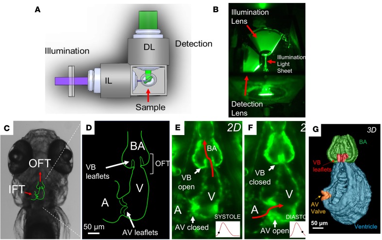
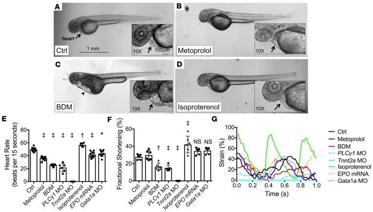
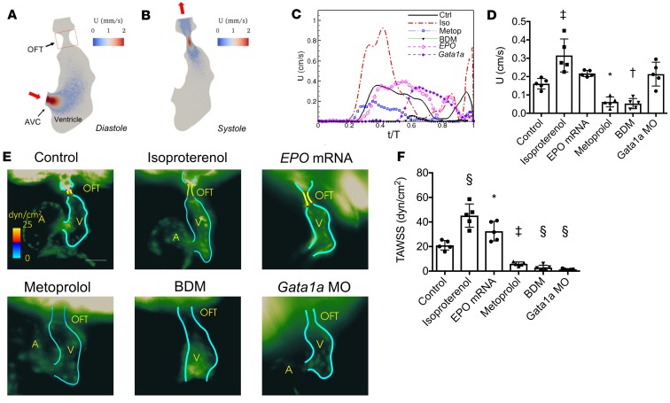
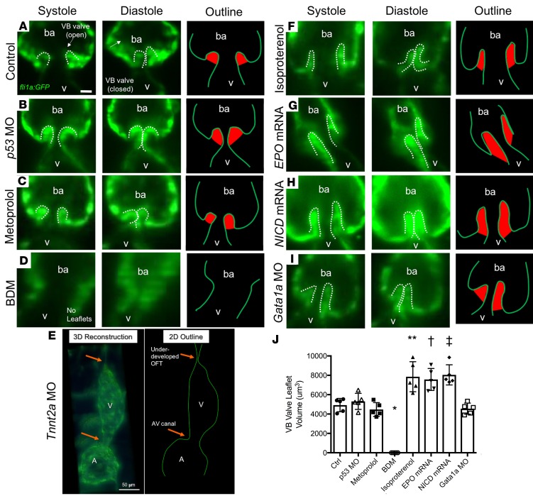
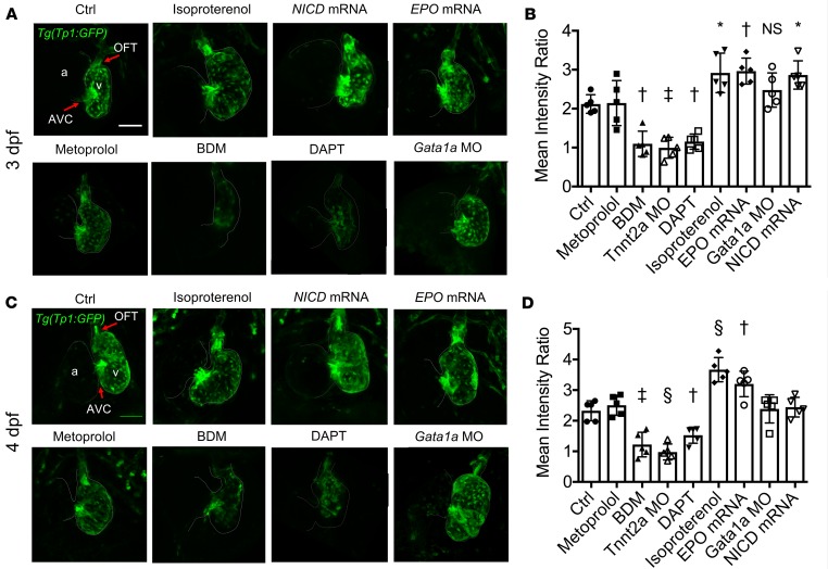
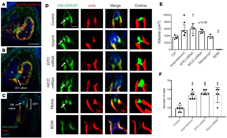
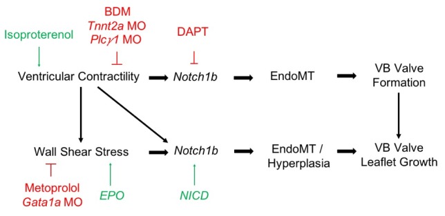
Biomechanical forces and endothelial-to-mesenchymal transition (EndoMT) are known to mediate valvulogenesis. However, the relative contributions of myocardial contractile and hemodynamic shear forces remain poorly understood. We integrated 4-D light-sheet imaging of transgenic zebrafish models with moving-domain computational fluid dynamics to determine effects of changes in contractile forces and fluid wall shear stress (WSS) on ventriculobulbar (VB) valve development. Augmentation of myocardial contractility with isoproterenol increased both WSS and Notch1b activity in the developing outflow tract (OFT) and resulted in VB valve hyperplasia. Increasing WSS in the OFT, achieved by increasing blood viscosity through EPO mRNA injection, also resulted in VB valve hyperplasia. Conversely, decreasing myocardial contractility by Tnnt2a morpholino oligonucleotide (MO) administration, 2,3-butanedione monoxime treatment, or Plcγ1 inhibition completely blocked VB valve formation, which could not be rescued by increasing WSS or activating Notch. Decreasing WSS in the OFT, achieved by slowing heart rate with metoprolol or reducing viscosity with Gata1a MO, did not affect VB valve formation. Immunofluorescent staining with the mesenchymal marker, DM-GRASP, revealed that biomechanical force-mediated Notch1b activity is implicated in EndoMT to modulate valve morphology. Altogether, increases in WSS result in Notch1b- EndoMT-mediated VB valve hyperplasia, whereas decreases in contractility result in reduced Notch1b activity, absence of EndoMT, and VB valve underdevelopment. Thus, we provide developmental mechanotransduction mechanisms underlying Notch1b-mediated EndoMT in the OFT.

Keywords: Cardiology; Cardiovascular disease; Development; Embryonic development.


Conflict of interest statement

Conflict of interest: The authors have declared that no conflict of interest exists.

Figures

Figure 1
Figure 1. 4D light-sheet imaging of zebrafish embryos for assessment of myocardial contractility and valvular morphology.
(A) Schematic diagram and (B) photograph of the orthogonal optical pathway for single-sided illumination and dual-channel detection of the sample. (C) Outline of cardiac anatomy superimposed on a bright-field image from a zebrafish embryo at 5 days after fertilization (dpf) indicating orientation of the inflow tracts (IFTs) and OFTs (red arrows). (D) Schematic diagram of the embryonic heart (coronal section) highlighting the atrioventricular (AV) and ventriculobulbar (VB) valves, atrium, ventricle, OFT, and bulbus arteriosus. (E and F) Light-sheet fluorescence microscopy image of the embryonic heart at 5 dpf, showing (E) the open VB valve during systole and (F) the open AV valve during diastole. (G) 3D reconstruction of the embryonic heart during systole with VB and AV valves shown in red and orange, respectively.
Figure 2
Figure 2. Effects of pharmacological interventions and genetic modification on cardiac hemodynamics in zebrafish embryos.
(AD) Bright-field microscopic images (original magnification, ×2) of Tg(fli1a:GFP) zebrafish embryos treated with (A) control vehicle, (B) metoprolol, (C) 2,3-butanedione monoxime (BDM), and (D) isoproterenol at 48 hpf. Precordial edema (arrowhead) with pooling of red blood cells in the sinus venosus is seen in the BDM-treated embryo (arrow). Scale bar: 1 mm. (E) HRs at 48 hpf and (F) FS measurements at 56 hpf in response to pharmacological interventions and genetic modifications (n = 10 for HR measurements; n = 6 for FS measurements, except n = 5 for PLC1 MO group). Data are presented as mean ± SD; *P < 0.01; P < 0.001; P < 0.0001; 1-way ANOVA with Dunnett’s multiple-comparisons test. (G) Representative strain measurements for the listed treatment groups of embryonic hearts at 56 hpf.
Figure 3
Figure 3. Moving-domain 2D CFD quantification of velocities and WSS in the developing OFT.
(A and B) Velocity (U) profiles during (A) diastole and (B) systole of a control embryo at 56 hpf. (C) Representative average velocities in the OFT of embryos at 56 hpf. (D) Average OFT velocities in the various treatment groups (n = 5 per group). (E) Representative endocardial border profiles of the time-averaged WSS (TAWSS) in the OFT of Tg(fli1a:GFP) embryos at 56 hpf, where (A) and (V) label the atria and ventricles. (F) Comparison of the TAWSS among the treatment groups (n = 5 per group). Data are represented as mean ± SD; *P < 0.05; P < 0.01; P < 0.001; §P < 0.0001; 1-way ANOVA with Dunnett’s multiple-comparisons test.
Figure 4
Figure 4. Effects of changes in hemodynamic shear force on VB valve leaflet formation.
Selective-plane illumination microscopy (SPIM) images of VB valves during ventricular systole and diastole at 5 dpf, with corresponding outline of the endocardium/endothelium (with valve leaflets highlighted in red), in Tg(fli1a:GFP) zebrafish embryos. (A) Control (untreated) embryo. (B) Vehicle control p53 MO–injected embryo showing similar size of VB valvular leaflets. (C) Metoprolol-treated embryo showing similar size of VB valvular leaflets despite reduction in HR. (D) BDM-treated embryo showing absence of VB valvular leaflets in response to significantly decreased HR and contractility. (E) Tnnt2a MO–injected embryo showing absence of VB leaflets in response to complete inhibition of myocardial contractile forces. 3D reconstruction at 5 dpf in a Tg(fli1a:GFP) zebrafish reveals an absence of cardiac looping accompanied by an underdeveloped OFT. The 2D schematic outline delineates the endocardial borders. (F) Isoproterenol-treated embryo showing prominence of VB valvular leaflets in response to increased HR and contractility. (G) EPO mRNA–injected embryo showing prominence of VB valve leaflets in response to increased blood viscosity with concomitant increase in shear stress. (H) NICD mRNA–injected embryo showing hyperplastic VB valve leaflets. (I) Gata1a MO–injected embryo showing normal leaflet morphology despite reductions in blood viscosity and endocardial shear stress. (J) Quantification of the VB valve leaflet volumes after 3D reconstruction (n = 5 per group). Scale bar: 10 μm in all except 50 μm in E. Data are presented as mean ± SD (*P < 0.0001, and **P = 0.0001 compared with Ctrl. P = 0.003, and P = 0.0003 compared with p53 MO. One-way ANOVA with Dunnett’s multiple-comparisons test with respective control groups). ba, bulbus arteriosus.
Figure 5
Figure 5. Effects of changes in hemodynamic shear force on Notch1b activity in the developing OFT.
Confocal microscopy images (maximum-intensity projections) of the OFT and AVC in the Tg(tp1:GFP) Notch1b reporter zebrafish line at (A) 3 dpf and (C) 4 dpf with the indicated treatments. Scale bar: 50 μm. Notch1b activity was prominent in both the OFT and AVC in control embryos. Notch1b activity was more prominent in the OFT in response to isoproterenol, as well as with increasing blood viscosity with EPO mRNA microinjection and with upregulation of Notch activity with NICD mRNA microinjection. Notch1b activity in the OFT was nearly absent in response to BDM and DAPT treatments. (B and D) 3D quantification of the mean intensity of Notch1b activity in the OFT (normalized to activity in the respective ventricle) at (B) 3 dpf and (D) 4 dpf was significantly higher with isoproterenol treatment and EPO mRNA injection, consistent with these groups having more prominent VB valvular leaflets. Additionally, Notch1b activity in the OFT was significantly lower with BDM and DAPT treatments (n = 5 per group). Data are presented as mean ± SD; *P < 0.05; P < 0.01; P < 0.001; §P < 0.0001; 1-way ANOVA with Dunnett’s multiple-comparisons test.
Figure 6
Figure 6. Effects of hemodynamic modulation on endocardial cell differentiation during VB valve formation in the OFT.
Immunostaining was performed against DM-GRASP, a cell surface adhesion protein expressed by differentiated valve-forming endocardial cells, in Tg(cmlc:mCherry) zebrafish embryos at 4 dpf. (AC) Confocal imaging (original magnification, ×20) demonstrates sections through the (A) ventricle, (B) AVC, and (C) OFT of a control embryo. Green fluorescent protein (GFP) signal is observed between the cmlc+ myocardial cells, as well as in the AV and VB valve leaflets, which lack a cmlc signal, suggesting these are differentiated endocardial/endothelial cells. Scale bar: 50 μm. (D) Confocal images through the OFT of embryos subjected to the indicated treatments. Arrows denote DM-GRASP+, cmlc valve leaflets. Scale bar: 25 μm. The outline denotes the DM-GRASP+, cmlc valve leaflets in green and the cmlc+ myocardium in red. (E) Quantification of the volumes of DM-GRASP+, cmlc valve leaflets in the indicated treatment groups (n = 4 per group). (F) Quantification of the cell counts in the DM-GRASP+, cmlc valve leaflets in the indicated treatment groups (n = 6 per group, except n = 5 for the NICD mRNA group). Data are presented as mean ± SD; *P < 0.05; P < 0.01; P < 0.001; §P < 0.0001; 1-way ANOVA with Dunnett’s multiple-comparisons test.
Figure 7
Figure 7. Schematic of proposed mechanism by which myocardial contractile and hemodynamic shear forces coordinate to promote Notch1b- and EndoMT-mediated valve formation in the developing OFT.

References

    1. Combs MD, Yutzey KE. Heart valve development: regulatory networks in development and disease. Circ Res. 2009;105(5):408–421. doi: 10.1161/CIRCRESAHA.109.201566. - DOI - PMC - PubMed
    1. Hoffman JI, Kaplan S. The incidence of congenital heart disease. J Am Coll Cardiol. 2002;39(12):1890–1900. doi: 10.1016/S0735-1097(02)01886-7. - DOI - PubMed
    1. Armstrong EJ, Bischoff J. Heart valve development: endothelial cell signaling and differentiation. Circ Res. 2004;95(5):459–470. doi: 10.1161/01.RES.0000141146.95728.da. - DOI - PMC - PubMed
    1. Hove JR, Köster RW, Forouhar AS, Acevedo-Bolton G, Fraser SE, Gharib M. Intracardiac fluid forces are an essential epigenetic factor for embryonic cardiogenesis. Nature. 2003;421(6919):172–177. doi: 10.1038/nature01282. - DOI - PubMed
    1. Bartman T, et al. Early myocardial function affects endocardial cushion development in zebrafish. PLoS Biol. 2004;2(5):E129. doi: 10.1371/journal.pbio.0020129. - DOI - PMC - PubMed

Publication types

MeSH terms