. 2019 Jun:24:101183.
doi: 10.1016/j.redox.2019.101183. Epub 2019 Apr 3.

The early response of αB-crystallin to a single bout of aerobic exercise in mouse skeletal muscles depends upon fiber oxidative features

Affiliations

The early response of αB-crystallin to a single bout of aerobic exercise in mouse skeletal muscles depends upon fiber oxidative features

Ivan Dimauro et al. Redox Biol. 2019 Jun.

Abstract

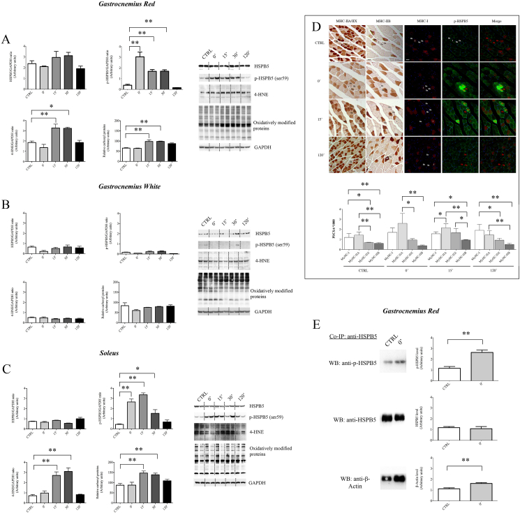
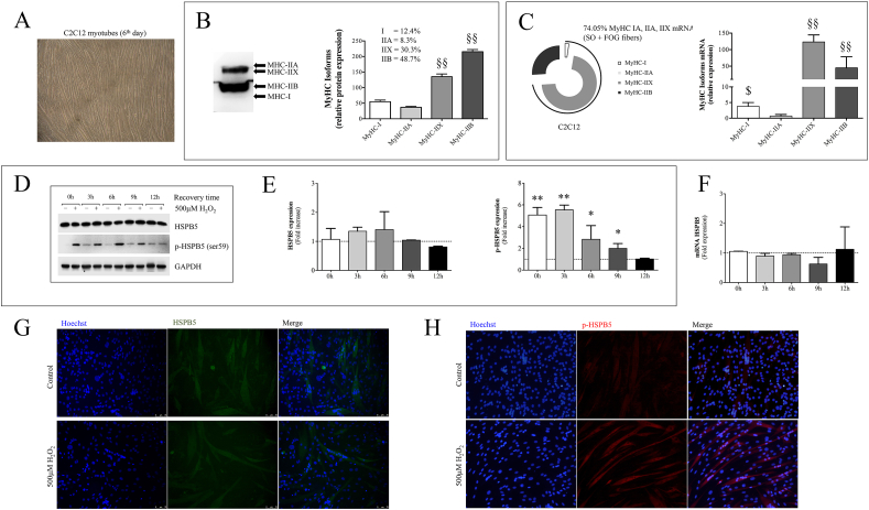
Besides its substantial role in eye lens, αB-crystallin (HSPB5) retains fundamental function in striated muscle during physiological or pathological modifications. In this study, we aimed to analyse the cellular and molecular factors driving the functional response of HSPB5 protein in different muscles from mice subjected to an acute bout of non-damaging endurance exercise or in C2C12 myocytes upon exposure to pro-oxidant environment, chosen as "in vivo" and "in vitro" models of a physiological stressing conditions, respectively. To this end, red (GR) and white gastrocnemius (GW), as sources of slow-oxidative and fast-glycolytic/oxidative fibers, as well as the soleus (SOL), mainly composed of slow-oxidative type fibers, were obtained from BALB/c mice, before (CTRL) and at different times (0', 15', 30' 120') following 1-h of running. Although the total level of HSPB5 protein was not affected by exercise, we found a significantly increase of phosphorylated HSPB5 (p-HSPB5) only in GR and SOL skeletal muscle with a higher amount of type I and IIA/X myofibers. The fiber-specific activation of HSPB5 was correlated to its interaction with the actin filaments, as well as to an increased level of lipid peroxidation and carbonylated proteins. The role of the pro-oxidant environment in HSPB5 response was investigated in terminally differentiated C2C12 myotubes, where most of HSPB5/pHSPB5 pool was present in the cytosolic compartment in standard culture conditions. As a result of exposure to pro-oxidizing, but not cytotoxic, H2O2 concentration, the p-38MAPK-mediated phosphorylation of HSPB5 resulted functional to promote its interaction with the myofibrillar components, such as β-actin, desmin and filamin 1. This study provides novel information on the molecular pathway underlying the HSPB5 physiological function in skeletal muscle, confirming the contribution of the pro-oxidant environment in HSPB5 activation and interaction with substrate/client myofibrillar proteins, offering new insights for the study of myofibrillar myopathies and cardiomyopathies.

Keywords: Endurance exercise; Oxidative stress; Skeletal muscle; αB-crystallin phosphorylation.


Figures

Fig. 1
Fig. 1
Analysis of the muscle type fibers in the posterior group of hindlimb muscles (i.e. Soleus, Gastrocnemius Red and White). (A) Images of mouse hindlimb muscles and a cross-section showing the cuts made during the dissection of the muscles. (B) Representative images used for quantitative analysis of serial cross-sections immunostained for MHC-I and MHC-IIA/X. Type IIB fibers are negative to antibodies anti-MHC-I and anti-MHC-IIA/X. The analysis was performed using 5 fields per section, 5 sections per mice (40 μm) between sections). An average of 400 fibers was analysed for each mouse. The percentage of the fibers was shown as histogram. (C) qRT-PCR analysis of MyHC genes levels. The exploded doughnut shows the percentage of MyHC isoforms expression in SOL, GR, GW and GS. Bar of each histogram shows the MyHC isoforms gene expression levels normalized for the reference gene (2−ΔCT). Intermediate hybrid fibers were not taken into consideration in these analyses because of their limited number. Data are presented as means ± SD. Statistical significance was determined using a one-way ANOVA with Bonferroni's post-hoc test. Φ p < 0.01 vs. all MyHC isoforms; Ψ p < 0.01 vs. MHC-IIX and MHC-IIB; **p < 0.01 vs. all other tissues; $ p < 0.05 vs. GW and GS; §p < 0.01 vs. SOL. SO, Slow Oxidative fibers; FOG, Fast Oxidative-Glycolytic fibers; GR, Gastrocnemius red; GW, Gastrocnemius white; SOL, Soleus; GS, Whole Gastrocnemius.
Fig. 2
Fig. 2
Densitometric analysis of Western blot related to HSPB5 modulation/activation, signal transduction and oxidative stress proteins in Gastrocnemius (A) Red, (B) White and (C) Soleus from control (CTRL) mice and during recovery period after an acute endurance exercise (0′-15′-30′-120′). (D) Immunohistochemistry and immunofluorescence for p-HSPB5, MHC-I and MHC-IIA/X in serial cross-sections of the whole gastrocnemius. Here are showed representative images used for densitometric analysis of the staining intensity. The analysis was performed using 5 fields per section, 5 sections per mice. An average of 435 fibers was analysed for each mouse. The staining intensity of the fibers was expressed as the mean pixel intensity (PI) normalized to the cross-sectional area (CSA) using ImageJ 1.50 h and Leica application suite advanced fluorescence's software. E) Western Blot analysis of co-immunoprecipitated proteins of Gastrocnemius Red from mice immediately after exercise (0′) showing a band corresponding to HSPB5 that co-immunoprecipitate with p-HSPB5 and β-Actin. Data are presented as the means ± SD. Statistical significance was determined using a one-way ANOVA with Bonferroni's post-hoc test or a Student's t-test. **p < 0.01; *p < 0.05.
Fig. 3
Fig. 3
(A) C2C12 myotubes at 6th day in differentiation medium. (B) A representative Western blot showing the protein levels of MyHC isoforms from C2C12 myotubes. (C) qRT-PCR analysis. The percentage of MyHC isoforms expression is shown as exploded doughnut. Bar diagram shows the relative expression of mRNA MyHC isoforms. In D and E whole-cell extracted by myotubes treated with 500 μM H2O2 for 1 h and collected at indicated recovery times were immunoblotted with antibodies against HSPB5, p-HSPB5 (Ser59). The quantification of each protein level was relative to GAPDH and represented as fold change. F) HSPB5 mRNA expression level was carried out at the same experimental points of protein expression analysis and represented as fold change (mean ± SD, n = 3). Myotubes treated with H2O2 and collected after 3 h were also fixed and immunoblotted with primary and secondary antibodies to detect total HSPB5 protein (G) and its phosphorylated form (p-HSPB5) (H). Hoechst nucleic acid stain was utilized to display nuclei. Scale bars = 75μM. Data are presented as the means ± SD (n = 3). Statistical significance was determined using either a one-way ANOVA with Bonferroni's post-hoc test or t-test analysis. §§p < 0.01 vs. MyHC-I and MyHC-IIA; $p < 0.05 vs. MyHC-IIA; **p < 0.01 vs. CTRL level; *p < 0.05 vs. CTRL level. SO, Slow Oxidative fibers; FOG, Fast Oxidative-Glycolytic fibers. (-------) CTRL level.
Fig. 4
Fig. 4
(A) Analysis of HSP1A1, 4-HNE, and oxidatively modified proteins content in C2C12 myotubes treated with H2O2. The quantification of each protein level was relative to GAPDH. Oxidatively modified proteins were determined by oxyblot analysis. Data are presented as the means ± SD (n = 3). (B) Densitometric analysis of Western blot related to p-38MAPK and p-p38MAPK in whole-cell extracted by myotubes treated with H2O2 and collected at indicated recovery times. Data were represented as fold change mean (n = 3). C) Fully differentiated myotubes were also treated with H2O2, in presence or not of both pharmacological inhibitors of p38MAPK (SB239063 and SB203580), to analyse the protein levels of p38MAPK, HSPB5 and their phosphorylated forms. D) Immunoprecipitation analysis of phospho-MAPAPK-2 in C2C12 myotubes exposed to pro-oxidant environment. (------) CTRL level. Unless different indicated, C2C12 cells were treated with H2O2 for 1 h followed from 3 h of recovery. Statistical significance was determined using either a one-way ANOVA with Bonferroni's post-hoc test or Student's t-test. **p < 0.01 vs. CTRL; *p < 0.05 vs. CTRL.
Fig. 5
Fig. 5
In (A) and (B) C2C12 myotubes were exposed to pro-oxidant and to p38MAPK inhibitors were fractionated into insoluble and soluble protein pools. Solubilized lysates were then separated by SDS-PAGE and analysed for HSPB5 and p-HSPB5 levels. C) Both whole and fractionated pools of proteins were obtained from myotubes exposed to cytochalasin D for 1 h and also followed a period of recovery. Soluble and insoluble fraction were analysed for HSPB5 and p-HSPB5 content. Whole proteins extracted from C2C12 myotubes treated with H2O2 in presence or not of both pharmacological inhibitors of p38MAPK were firstly co-immunoprecipitated for HSPB5 and then both D) visualized on gel staining the proteins with silver nitrate and E) analysed through SDS-PAGE for the indicated antibodies. Unless different indicated, C2C12 cells were treated with H2O2 for 1 h followed from 3 h of recovery.
Supplementary Fig. 1
Analysis of apoptotic markers in (A) red gastrocnemius (GR), (B) white gastrocnemius (GW); and (C) soleus (SOL) from control (CTRL) mice and at different times during recovery from acute endurance exercise (EX: 0′-15′-30′-120′); and in (D) C2C12 myotubes treated with H2O2 (500 μM for 1h followed by 3h of recovery). Representative SDS-PAGE showing the protein levels of selected proteins were reported. GAPDH served as loading control. The analysis was performed using 7 mice per group and in three independent experiments. Data are presented as the means ± SD. Statistical significance was determined using either a one-way ANOVA with Bonferroni's post-hoc test or Student's t-test.
Supplementary Fig. 2
Analysis of HSP1A1, NFkB-p65 and NFkB-p-p65 levels in (A) red gastrocnemius (GR), (B) white gastrocnemius (GW) and (C) soleus (SOL) from control (CTRL) mice and at different times during recovery from acute endurance exercise (0′-15′-30′-120′). D) Comparative analysis of the basal levels of HSPB5 and p-HSPB5 among all skeletal muscles examined. A representative SDS-PAGE of selected proteins was shown. GAPDH served as loading control. Data are presented as the means ± SD. Statistical significance was determined using either a one-way ANOVA with Bonferroni's post-hoc test or t-test analysis. **p < 0.01 vs. GW.
Supplementary Fig. 3
Representative immunofluorescence for HSPB5 in serial cross-sections of both skeletal muscles, red gastrocnemius and soleus, from control (CTRL) mice, immediately after an acute endurance exercise (0′) and at 120 min during recovery period. GR, Gastrocnemius Red; SOL, Soleus.
Supplementary Fig. 4
Densitometric analysis of Western blot related to p-38MAPK and p-p38MAPK in (A) Red-, (B) White-Gastrocnemius and (C) Soleus from control (CTRL) mice and during recovery period after an acute endurance exercise (0′-15′-30′-120′). Data are presented as the means ± SD. Statistical significance was determined using one-way ANOVA with Bonferroni's post-hoc test.

Similar articles

Cited by

References

    1. Hartl F.U., Bracher A., Hayer-Hartl M. Molecular chaperones in protein folding and proteostasis. Nature. 2011;475:324–332. - PubMed
    1. Bakthisaran R., Tangirala R., Rao C.M. Small heat shock proteins: role in cellular functions and pathology. Biochim. Biophys. Acta. 2015;1854(4):291–319. - PubMed
    1. Thornell E., Aquilina A. Regulation of αA- and αB-crystallins via phosphorylation in cellular homeostasis. Cell. Mol. Life Sci. 2015;72:4127–4137. - PMC - PubMed
    1. Benjamin I.J., Shelton J., Garry D.J., Richardson J.A. Temporospatial expression of the small HSP/alpha B-crystallin in cardiac and skeletal muscle during mouse development. Dev. Dynam. 1997;208:75–84. - PubMed
    1. Brady J.P., Garland D.L., Green D.E., Tamm E.R., Giblin F.J., Wawrousek E.F. AlphaB crystallin in lens development and muscle integrity: a gene knockout approach. Investig. Ophthalmol. Vis. Sci. 2001;42:2924–2934. - PubMed

Publication types