

Editorial
doi: 10.3390/molecules24071399.
Introduction to the Molecules Special Edition Entitled ' Heparan Sulfate and Heparin: Challenges and Controversies': Some Outstanding Questions in Heparan Sulfate and Heparin Research
Affiliations
- PMID: 30974725
- PMCID: PMC6479682
- DOI: 10.3390/molecules24071399
Item in Clipboard
Editorial
Introduction to the Molecules Special Edition Entitled ' Heparan Sulfate and Heparin: Challenges and Controversies': Some Outstanding Questions in Heparan Sulfate and Heparin Research
Molecules.
.
Abstract
The scope of this article is to provide a brief general introduction to heparan sulfate (HS) and heparin, and attempt to identify some of the central challenges regarding research into the chemistry and biology of glycosaminoglycans (GAGs), some of which are the subject of contributions to the special issue of Molecules (published in volume 23, 2018) entitled 'Heparan Sulfate and Heparin: Challenges and Controversies' [...].
Conflict of interest statement
The authors declare no conflict of interest.
Figures
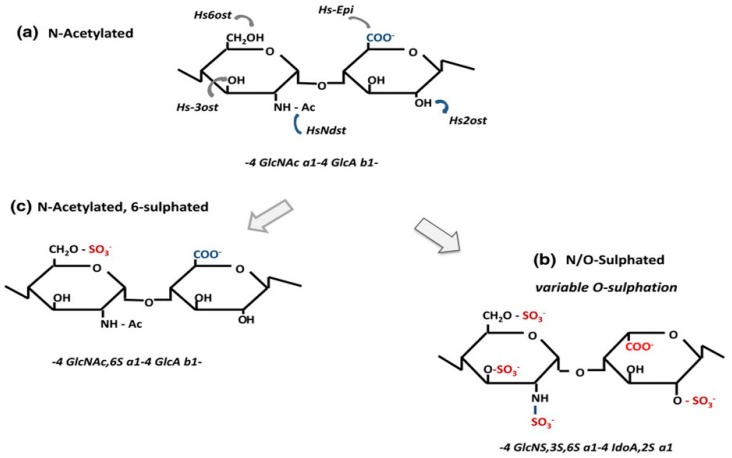
Enzymatic Modifications in the Biosynthesis of Heparin and Heparan Sulphate. Heparin and heparan sulphate (HS) are first synthesised as non-sulphated polymers (GlcNAc-GlcA repeat units) that are modified to different degrees by a combination of N- and O-sulphation and epimerisation. The initial modification step carried out by the Ndst enzymes is the conversion of GlcNAc to GlcNS; in heparin, about 90% of GlcNAc residues are converted to the N-sulphated derivative whereas in HS the level of conversion is normally in the range of 40 to 50%. In a mature heparin polymer, the main disaccharide is a trisulphated unit (GlcNS6S-IdoA2S), whereas in HS the sulphated and epimerised disaccharides occur in clusters and with a low frequency (normally below 10%) of trisulphated units. In both polymers, 3-O-sulphation, though rare, is a key functional group in the high-affinity antithrombin binding sequence, but can also be found in non-anticoagulant sequences. Key to enzymes: HsNdst: N-deacetylase/N-sulphotransferase, Hsepi: C5-epimerase, Hs2ost: 2-O-sulphotransferase, Hs6ost: 6-O-sulphotransferase, Hs3ost 3-O-sulphotransferase. This diagram was originally published in [6].
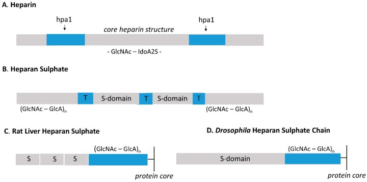
Differences in molecular design between characteristic regions of heparin (A) and heparan sulphate (B). The structure of the highly sulphated heparin is dominated by the trisulphated GlcNS6S—IdoA2S unit; however, there are less sulphated sequences that can be cleaved by an endoheparanase, hpa1, distributed throughout the chain at approximately 15 kDa intervals. The molecular design of HS is quite distinct from that of heparin. The sulphated regions composed of S- and NA/NS-domains (or T-zones) are arranged in a regular manner along the glycosaminoglycan (GAG) chain separated by extensive areas that lack any enzymatic modification (NA domains). A long NA sequence of nine to 10 disaccharides is present in the inner region of HS proximal to the protein core (shown in (C,D)), depicting, respectively, the asymmetric rat liver HS and the two-domain Drosophila HS). A comparable sequence is not present in heparin. The “composite regions” of sulphation in HS illustrated above are ~ 7 kDa in size and can be excised by K5- heparan lyase that acts specifically on NA sequences. This enzyme is inhibited by the presence of the N-sulphate group. Hpa1 susceptible sequences are present in HS probably at the junction of NA/NS and S-domains.
References
Publication types
MeSH terms
Substances
LinkOut - more resources
Full Text Sources
Medical
