. 2019 Apr 10;20(7):1761.
doi: 10.3390/ijms20071761.

Fibrin Sealant Derived from Human Plasma as a Scaffold for Bone Grafts Associated with Photobiomodulation Therapy

Affiliations

Fibrin Sealant Derived from Human Plasma as a Scaffold for Bone Grafts Associated with Photobiomodulation Therapy

Karina Torres Pomini et al. Int J Mol Sci. .

Abstract

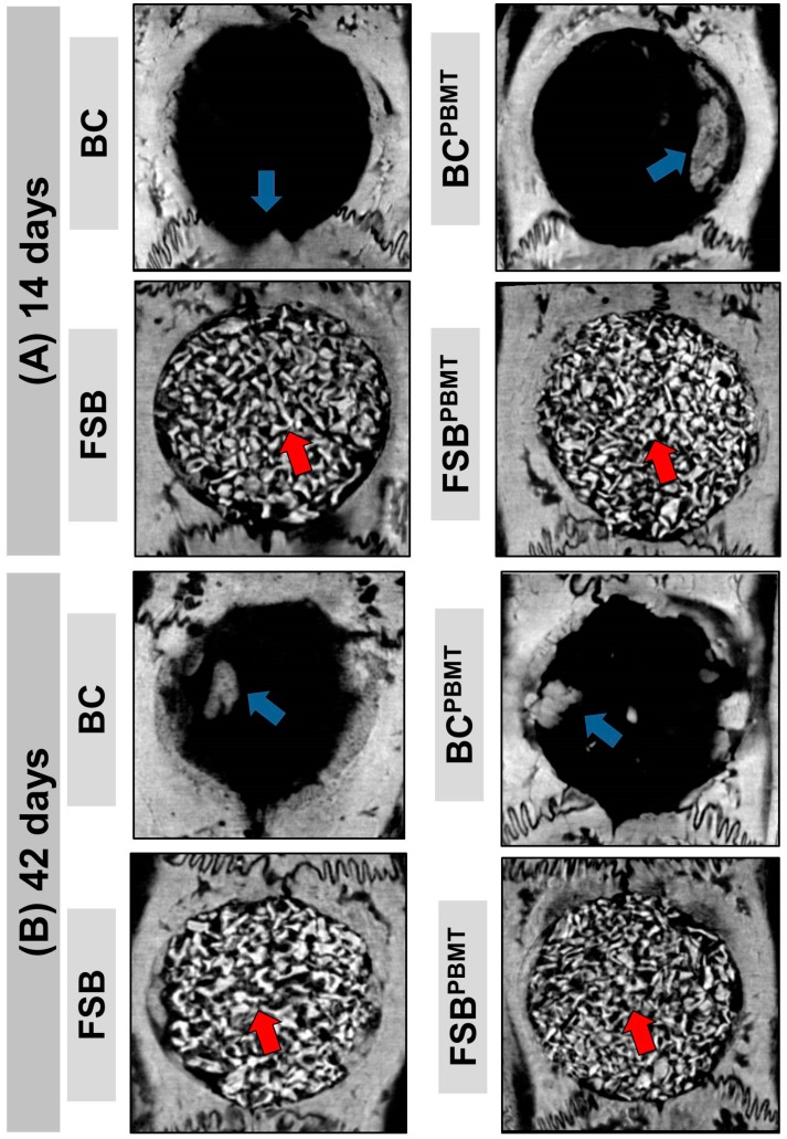
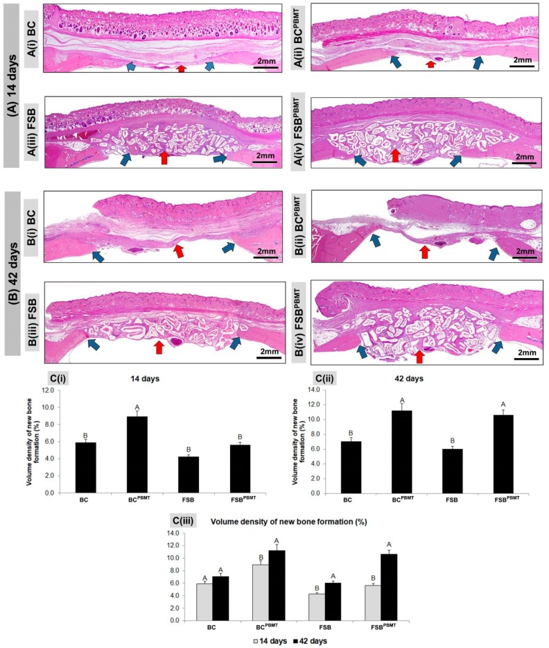
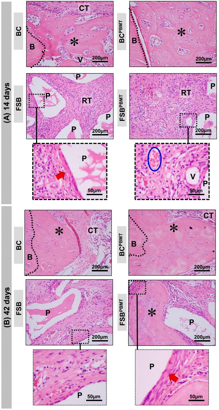
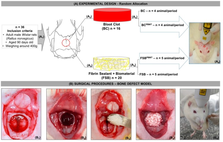
Fibrin sealants derived from human blood can be used in tissue engineering to assist in the repair of bone defects. The objective of this study was to evaluate the support system formed by a xenograft fibrin sealant associated with photobiomodulation therapy of critical defects in rat calvaria. Thirty-six rats were divided into four groups: BC (n = 8), defect filled with blood clot; FSB (n = 10), filled with fibrin sealant and xenograft; BCPBMT (n = 8), blood clot and photobiomodulation; FSBPBMT (n = 10), fibrin sealant, xenograft, and photobiomodulation. The animals were killed after 14 and 42 days. In the histological and microtomographic analysis, new bone formation was observed in all groups, limited to the defect margins, and without complete wound closure. In the FSB group, bone formation increased between periods (4.3 ± 0.46 to 6.01 ± 0.32), yet with lower volume density when compared to the FSBPBMT (5.6 ± 0.45 to 10.64 ± 0.97) group. It was concluded that the support system formed by the xenograft fibrin sealant associated with the photobiomodulation therapy protocol had a positive effect on the bone repair process.

Keywords: biomaterial; bone regeneration; bone repair; fibrin sealant; low-level laser therapy; photobiomodulation therapy.


Conflict of interest statement

The authors declare no conflict of interest.

Figures

Figure 1
Figure 1
Microtomographic images showing the evolution of the repair of defects filled with clot and fibrin sealant plus xenograft (biomaterial) with or without low-level laser biostimulation therapy. Biomaterial particles (red arrow) and newly formed bone tissue (blue arrow). Two-dimensional trans-axial cuts at (A) 14 days; and (B) 42 days, respectively.
Figure 2
Figure 2
Panoramic histological views at (A) 14 days; and (B) 42 days, respectively; (C) graphs of volume density of newly formed bone in skull defects filled with a blood clot or fibrin sealant plus xenograft and with or without laser photobiomodulation therapy. (A) A(i)–A(ii) bone formation (blue arrows) occurring at the defect border and under the dura mater surface. A(iii)–A(iv): the defect showed trabecular bone formation (blue arrows) adjacent to the defect border, in a more advanced stage of bone maturation. (B) B(i)–B(ii) both groups showed similar bone formation limited to the defect border and a large region filled with fibrous connective tissue (red arrows); B(iii)–B(iv) a large part of the defect was filled by connective tissue and biomaterials (red arrows), but in the FSBPBMT group, greater bone formation defect could be observed compared to the FSB group; (C) Graphs of newly formed bone showed smaller bone formation in the non-biostimulated group (BC and FSB) than the biostimulated group (BCPBMT and FSBPBMT). (BC and BCPBMT: N = 4/group and periods), (FSB and FSBPBMT: N = 5/group and periods). C(i) and C(ii) where different letters (A≠B) indicate a statistically significant difference between groups in the same period and C(iii) where the different letters (A≠B) indicate a statistically significant difference in the same group in the two periods analysed (p < 0.05). (HE; original magnification × 4; bar = 2 mm).
Figure 3
Figure 3
Details of the evolution of the bone healing of the skull defects filled with a blood clot or fibrin sealant plus xenograft (biomaterial) with or without low-level laser biostimulation therapy. (A) At 14 days, BC and BCPBMT: the defect shows the trabecular bone formation (asterisks) adjacent to the defect border and spaces between the trabeculae filled by connective tissue. FSB: the defect was filled by particles of the biomaterial (P) surrounded by connective tissue with some inflammatory cells (RT—reactional tissue). Collagen fibres surrounding the xenograft particles (red arrow). FSBPBMT: the defect was filled by particles of the biomaterial (P) surrounded by connective tissue with some inflammatory cells (RT—reactional tissue). Presence of many red blood cells (inside the blue lined area) and blood vessels (V) permeating connective tissue; (B) At 42 days, BC and BCPBMT: the new bone shows a gradual increase in thickness of the trabeculae leading to a compact structure. FSB: the new bone formation increases, becoming compact, there is a presence of xenograft particles and a decrease in inflammatory response. In FSBPBMT, the collagen fibres are arranged in more layers surrounding the particles. (HE; original magnification × 40; bar = 200 µm; and Insets, magnified images × 100; bar = 50 µm).
Figure 4
Figure 4
(A) Experimental design (A1) Random allocation: Thirty-six rats were divided into two groups; (A2) BC (n = 16)—Blood Clot and FSB (n = 20)—Fibrin Sealant + Biomaterial; (A3) After surgical procedures, two subgroups were preformatted according to treatment: BC, n = 8 (Blood Clot, the defect was filled with blood clot and without photobiomodulation), BCPBMT, n = 8 (Blood Clot, the defect was filled with blood clot and photobiomodulation), FSB, n = 10 (the defect was filled with a mixture of biomaterial and fibrin sealant and without photobiomodulation), and FSBPBMT, n = 10 (the defect was filled with a mixture of biomaterial and fibrin sealant and photobiomodulation); (A4) illustration of the four points of cross-application of the low-level laser on rat calvarium; (B) Surgical procedures—bone defect model; (B1) Osteotomy using an 8 mm trephine bur with exposure of the fragment removed from the parietal bones; (B2) defect filled with blood clot; (B3) deposition of the mixture fibrin sealant + biomaterial in the defect; (B4) defect filled with mixture; (B5) low-level laser therapy (photobiostimulation).

References

    1. Nee E.A., Chrzanowski W., Salih V., Kim H., Knowles J. Tissue engineering in dentistry. J. Dent. 2014;42:915–928. doi: 10.1016/j.jdent.2014.05.008. - DOI - PubMed
    1. Buchaim D.V., Bueno P.C.D.S., Andreo J.C., Roque D.D., Roque J.S., Zilio M.G., Salatin J.A., Kawano N., Furlanette G., Buchaim R.L. Action of a deproteinized xenogenic biomaterial in the process of bone repair in rats submitted to inhalation of cigarette smoke. Acta Cir. Bras. 2018;33:324–332. doi: 10.1590/s0102-865020180040000004. - DOI - PubMed
    1. Pomini K., Cestari M., German I., Rosso M., Gonçalves J., Buchaim D., Pereira M., Andreo J., Júnior G.M.R., della Coletta B., et al. Influence of experimental alcoholism on the repair process of bone defects filled with beta-tricalcium phosphate. Drug Alcohol Depend. 2019;197:315–325. doi: 10.1016/j.drugalcdep.2018.12.031. - DOI - PubMed
    1. Campana V., Milano G., Pagano E., Barba M., Cicione C., Salonna G., Lattanzi W., Logroscino G. Bone substitutes in orthopaedic surgery: From basic science to clinical practice. J. Mater. Sci. Mater. Med. 2014;25:2445–2461. doi: 10.1007/s10856-014-5240-2. - DOI - PMC - PubMed
    1. Wang W., Yeung K.W.K. Bioactive Materials Bone grafts and biomaterials substitutes for bone defect repair: A review. Bioact. Mater. 2017;2:224–247. doi: 10.1016/j.bioactmat.2017.05.007. - DOI - PMC - PubMed

Substances