. 2019 Apr 11;17(1):33.
doi: 10.1186/s12915-019-0652-6.

Derivation of adult canine intestinal organoids for translational research in gastroenterology

Affiliations

Derivation of adult canine intestinal organoids for translational research in gastroenterology

Lawrance Chandra et al. BMC Biol. .

Abstract

Background: Large animal models, such as the dog, are increasingly being used for studying diseases including gastrointestinal (GI) disorders. Dogs share similar environmental, genomic, anatomical, and intestinal physiologic features with humans. To bridge the gap between commonly used animal models, such as rodents, and humans, and expand the translational potential of the dog model, we developed a three-dimensional (3D) canine GI organoid (enteroid and colonoid) system. Organoids have recently gained interest in translational research as this model system better recapitulates the physiological and molecular features of the tissue environment in comparison with two-dimensional cultures.

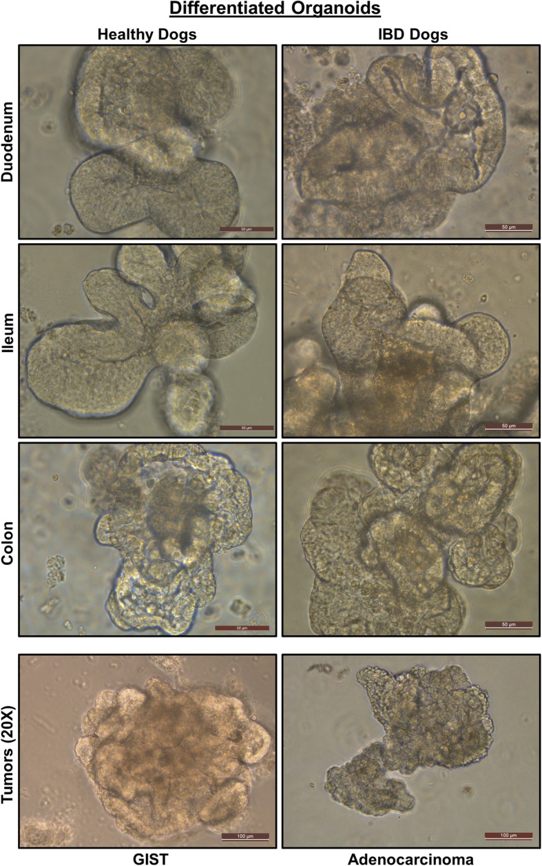
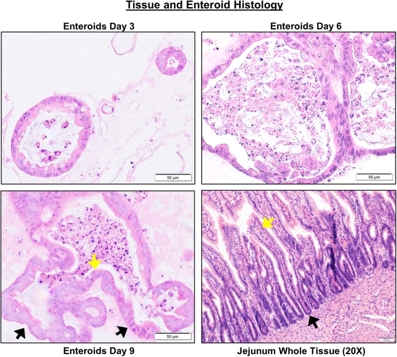
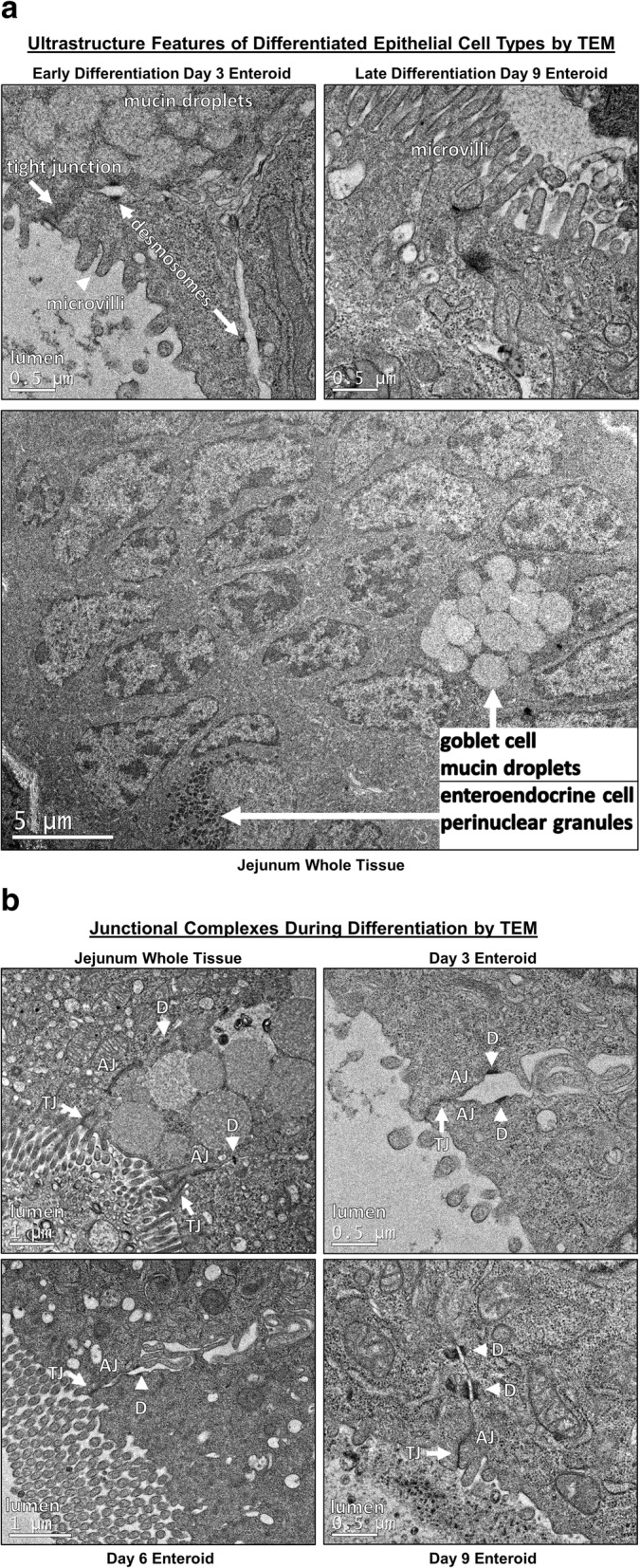
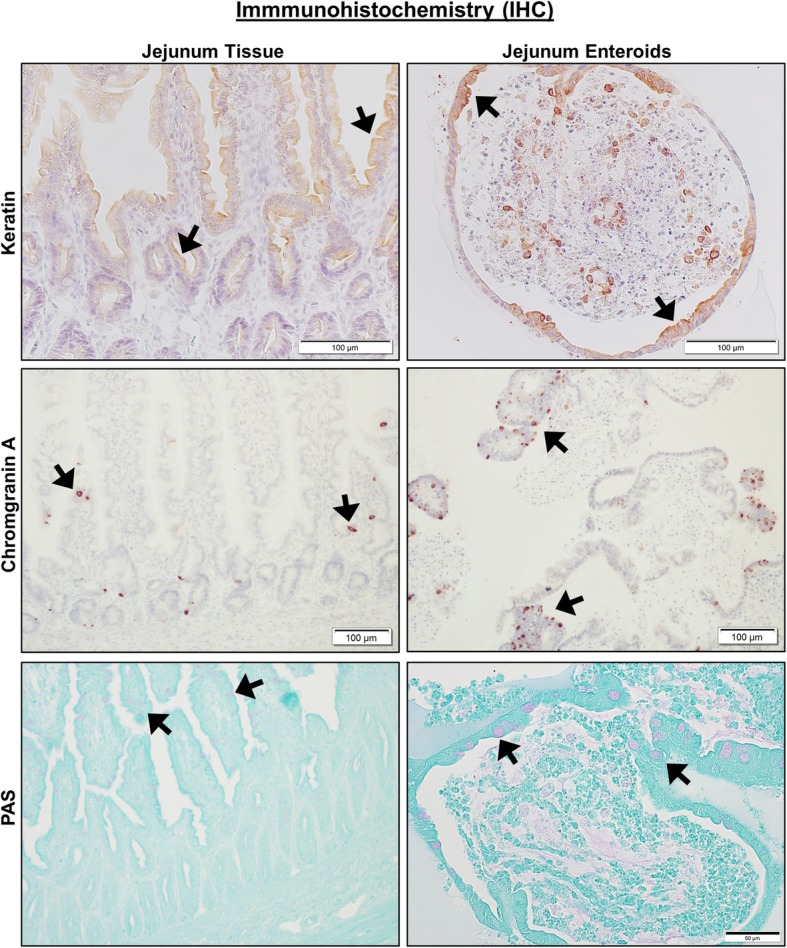
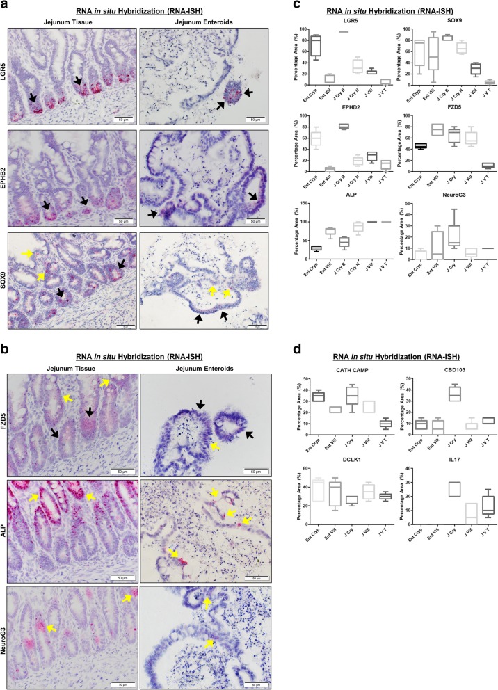
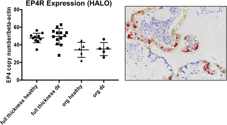
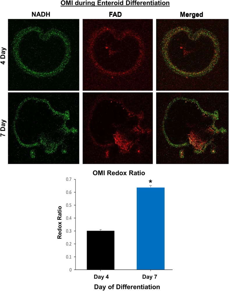
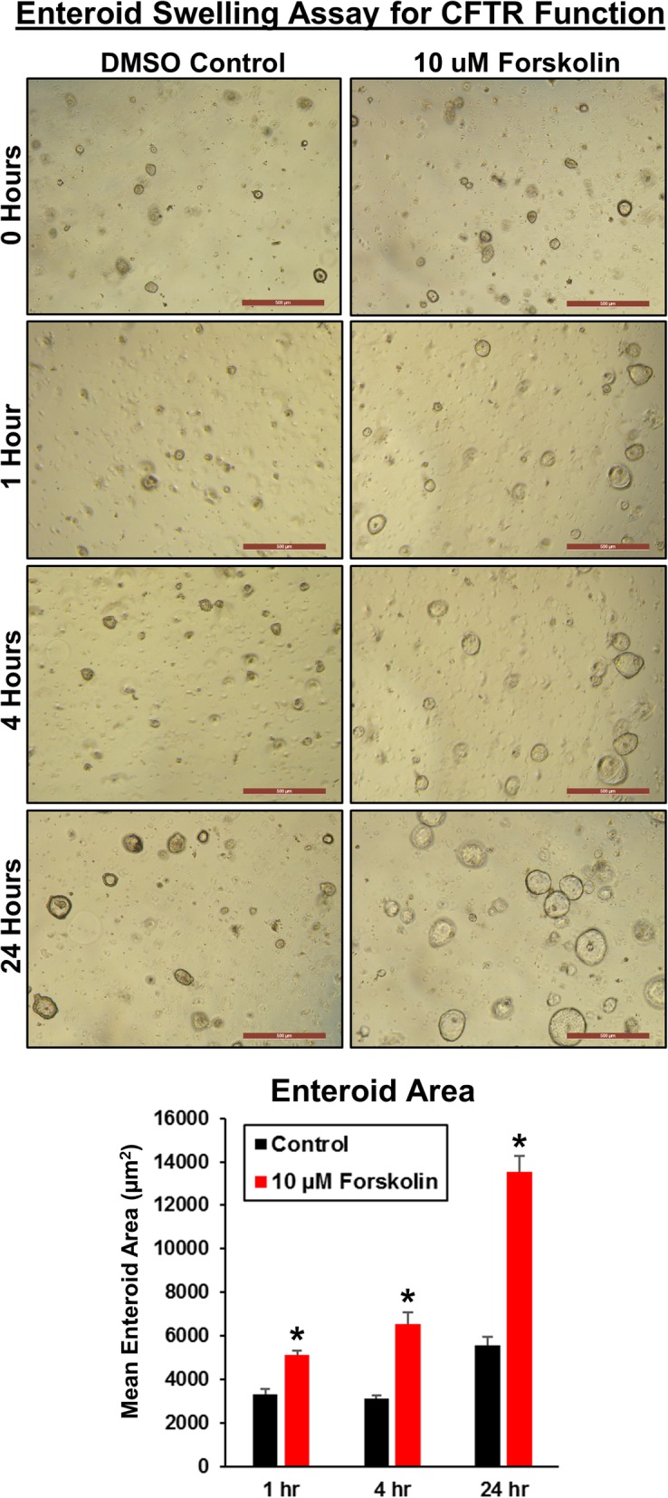
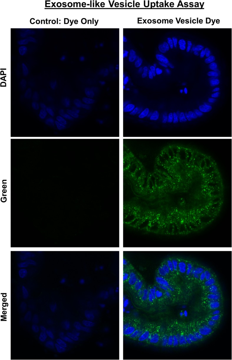
Results: Organoids were derived from tissue of more than 40 healthy dogs and dogs with GI conditions, including inflammatory bowel disease (IBD) and intestinal carcinomas. Adult intestinal stem cells (ISC) were isolated from whole jejunal tissue as well as endoscopically obtained duodenal, ileal, and colonic biopsy samples using an optimized culture protocol. Intestinal organoids were comprehensively characterized using histology, immunohistochemistry, RNA in situ hybridization, and transmission electron microscopy, to determine the extent to which they recapitulated the in vivo tissue characteristics. Physiological relevance of the enteroid system was defined using functional assays such as optical metabolic imaging (OMI), the cystic fibrosis transmembrane conductance regulator (CFTR) function assay, and Exosome-Like Vesicles (EV) uptake assay, as a basis for wider applications of this technology in basic, preclinical and translational GI research. We have furthermore created a collection of cryopreserved organoids to facilitate future research.

Conclusions: We establish the canine GI organoid systems as a model to study naturally occurring intestinal diseases in dogs and humans, and that can be used for toxicology studies, for analysis of host-pathogen interactions, and for other translational applications.

Keywords: Canine; Enteroid; GI diseases; Intestinal stem cell; Organoid model; Translational research.


Conflict of interest statement

Authors’ information

Not applicable

Ethics approval and consent to participate

All animal studies were reviewed and approved by Iowa State University IACUC, as detailed in the Methods.

Consent for publication

Not applicable.

Competing interests

JPM, KA, and AJ would like to disclose a competing financial interest and management role in 3D Health Solutions, Inc., an entity that provides canine 3D organoid testing services. The other authors declare that they have no competing interests.

Publisher’s Note

Springer Nature remains neutral with regard to jurisdictional claims in published maps and institutional affiliations.

Figures

Fig. 1
Fig. 1
Canine intestinal 3D organoids (enteroids and colonoids) from healthy and inflammatory bowel disease (IBD) dogs. Representative phase contrast images at × 40 magnification of fully differentiated 5–7-day-old duodenal and ileal enteroids and colonoids from healthy dogs and dogs with IBD, as well as gastrointestinal stromal tumor (GIST) and colorectal adenocarcinoma tumor (tumors at × 20 magnification). The structures of enteroids and colonoids from healthy dogs and dogs with IBD appeared comparable. Representative photomicrographs of at least n = 15 (healthy dogs), n = 7 (IBD dogs), and n = 4 (tumors) images of enteroids/colonoids are shown for each location as indicated
Fig. 2
Fig. 2
Characterization of canine jejunal tissue and jejunal enteroid histology shows similarities of epithelial structure. Histological images of hematoxylin and eosin (H&E) staining show the development and differentiation of canine enteroids at 3, 6, and 9 days after isolation or passaging. Spheroid-like epithelial structures are visualized in 3-day enteroids compared to crypt-villi epithelial structures on the sixth and ninth days. In the whole jejunal tissue, there are crypt-villi epithelial structures as well as non-epithelial cell types. Arrows indicate examples of crypts (black arrows) and villi (yellow arrows). Representative images from at least n = 15 enteroids per condition
Fig. 3
Fig. 3
Ultrastructural features of differentiated enteroids mimic intact small intestinal tissue. a Canine jejunum enteroids, during early (day 3) and late (day 9) stages of differentiation, using whole tissue showing features of cellular differentiation, such as apical microvilli, electron-lucent cytoplasmic vacuoles (i.e., mucus), and electron-dense perinuclear granules (i.e., neurosecretory granules), consistent with development of absorptive enterocytes, goblet cells, and enteroendocrine cells, respectively. Ultrastructure features are visualized by representative transmission electron micrographs (TEM). b TEM of canine organoids show the progressive development (days 3–9 of differentiation) of intercellular structures important for intestinal barrier function. Adherens junction (AJ), tight junction (TJ), and desmosomes (D) structures are seen in both canine enteroids and native jejunum. On day 3, the developing tight junction had dilated paracellular space adjacent to tight junctions; however, the paracellular spaces were smaller on day 6, and no longer apparent by day 9. Representative images from at least n = 10 enteroids per condition
Fig. 4
Fig. 4
Canine jejunal tissues and enteroids both express markers of epithelial cell lineage. Representative immunohistochemistry (IHC) images comparing staining for marker proteins of epithelial cells and their lineage, including Keratin (epithelial cells, upper panels), Chromogranin A (enteroendocrine cells, middle panels), and PAS (goblet cells, lower panels) on both intact whole jejunal tissues and jejunal enteroids. Black arrows indicate representative positive staining for epithelial, enteroendocrine, or goblet cells. Tissue and enteroids were counterstained with Hematoxylin (upper and middle panels) or Alcian Blue (lower panels). Representative images from n = 20 or more enteroids per condition
Fig. 5
Fig. 5
Both canine jejunal tissues and enteroids express mRNA for markers of various epithelial cell lineages. a Representative RNA in situ hybridization (RNA-ISH) images reveal expression of stem cell markers LGR5, SOX9, and EPHB2 on both intact jejunal tissues and enteroids. SOX9 is also a marker of enteroendocrine and tuft cells, while EPHB2 is also a marker of Paneth-like cells. Arrows indicate representative positive red areas in crypts (black arrows) or villi (yellow arrows). Representative images from at least n = 15 organoids per condition. b Representative images of gene expression for markers of epithelial lineage, including FZD5 (Paneth-like cell), ALP (absorptive epithelium), and Neuro G3 (Enteroendocrine cells) in both intact jejunal tissues and enteroids, as determined by RNA-ISH. Representative images from at least n = 15 organoids per condition. c Semi-quantitative scoring of RNA-ISH staining (box and whisker plots) for expression of stem cell markers (LGR5, SOX9), Paneth-like cell markers (FZD5, EPHB2), absorptive epithelial markers (ALP), and enteroendocrine cells (Neuro G3) in both intact jejunal tissues and enteroids. Specific sites include Enteroid Crypt (Ent Cryp), Enteroid Villus (Ent Vill), whole tissue Jejunum Crypt Base (J Cry B), Jejunum Crypt Neck (J Cry N), Jejunum Villus (J Vill), and Jejunum Villus Tip (J V T). Scoring of at least n = 6 images per condition. d Semi-quantitative expression of Paneth cell markers IL-17, CBD 103, and CATH as well as tuft cell marker Dclk1, in both intact jejunal tissues and enteroids, in specific sites as above. Cells and tissue were counterstained with hematoxylin. Scoring of at least n = 6 images per condition
Fig. 6
Fig. 6
Prostaglandin E2 receptor-4 expression is similar between tissue and enteroids from healthy and IBD dogs. Representative RNA-ISH image illustrates how the Prostaglandin E2 receptor-4 (EP4R) staining was marked for quantification using Halo software. Box and whisker plot compares the EP4R expression among biopsy tissues (full thickness) and enteroids (org) obtained from both healthy and IBD (dz) dogs. Scoring of at least n = 5 images per condition
Fig. 7
Fig. 7
Optical metabolic imaging (OMI) reveals metabolic differences during differentiation of canine enteroids. Representative fluorescent images from 4- and 7-day-old enteroids are shown. The green image indicates the NADH measurement whereas the red image indicates the FAD, with the merged images on the right. The graph shows the optical redox ratio of 4-day-old versus 7-day-old enteroids (0.303 + 0.008 versus 0.637 + 0.013, respectively, for mean + SEM). n = 15 organoids per condition
Fig. 8
Fig. 8
Forskolin induces swelling of canine enteroids in a time-dependent manner, indicating presence of functional CFTR. Enteroids were passaged and seeded in Matrigel into 24-well plates. After 2 days, enteroids were incubated in media containing vehicle control (DMSO) or 10 μM forskolin. Representative images of enteroids were taken after 0, 1, 4, and 24 h, at × 5 magnification. Graph of mean area of enteroids (15–25 per field) from n = 12 fields per condition as determined by ImageJ (mean + SEM; p < 0.05 vs. control for each time)
Fig. 9
Fig. 9
Canine enteroids uptake exosome-like vesicles secreted from the parasite Ascaris suum. Representative confocal microscope images taken after 24-h incubation with control fluorophore alone (green fluorescent PKH67) or exosome-like vesicles (EV) labeled with PKH67 green fluorescent dye. Enteroids were counterstained with the nuclear marker DAPI (blue fluorescence). Only exosome+ PKH67 group demonstrated green fluorescence within epithelial cells and within the organoid lumen. The merged image shows the intracytoplasmic localization of the PKH67 green fluorescent dye labeled EV obtained from Ascaris suum

References

    1. Perlman RL. Mouse models of human disease: an evolutionary perspective. Evol Med Public Health. 2016;1:170–176. - PMC - PubMed
    1. Ziegler A, Gonzalez L, Blikslager A. Large animal models: the key to translational discovery in digestive disease research. Cell Mol Gastr Hepatol. 2016;2(6):716–712. doi: 10.1016/j.jcmgh.2016.09.003. - DOI - PMC - PubMed
    1. Wong SK, Chin KY, Suhaimi FH, Fairus A, Ima-Nirwana S. Animal models of metabolic syndrome: a review. Nutr Metab. 2016;13:65. doi: 10.1186/s12986-016-0123-9. - DOI - PMC - PubMed
    1. Mochel JP, Jergens AE, Kingsbury D, Kim HJ, Martín MG, Allenspach K. Intestinal stem cells to advance drug development, precision, and regenerative medicine: a paradigm shift in translational research. AAPS J. 2017;20(1):17. doi: 10.1208/s12248-017-0178-1. - DOI - PMC - PubMed
    1. Ericsson AC, Crim MJ, Franklin CL. A brief history of animal modeling. Mo Med. 2013;110(3):201–205. - PMC - PubMed

Publication types