. 2019 Apr 11;16(1):82.
doi: 10.1186/s12974-019-1458-8.

GPR34 in spinal microglia exacerbates neuropathic pain in mice

Affiliations

GPR34 in spinal microglia exacerbates neuropathic pain in mice

Akira Sayo et al. J Neuroinflammation. .

Abstract

Background: Neuropathic pain is caused by sensory nerve injury, but effective treatments are currently lacking. Microglia are activated in the spinal dorsal horn after sensory nerve injury and contribute to neuropathic pain. Accordingly, molecules expressed by these cells are considered potential targets for therapeutic strategies. Our previous gene screening study using a mouse model of motor nerve injury showed that the G-protein-coupled receptor 34 gene (GPR34) is induced by nerve injury. Because GPR34 is now considered a microglia-enriched gene, we explored the possibility that it might be involved in microglial activation in the dorsal horn in a mouse model of neuropathic pain.

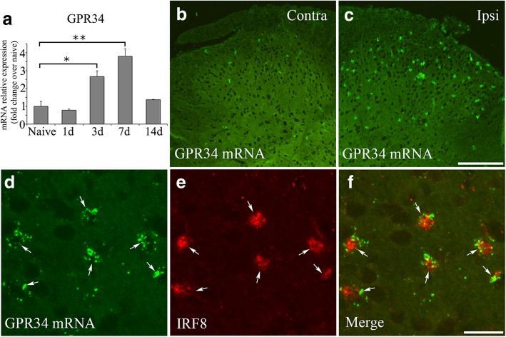
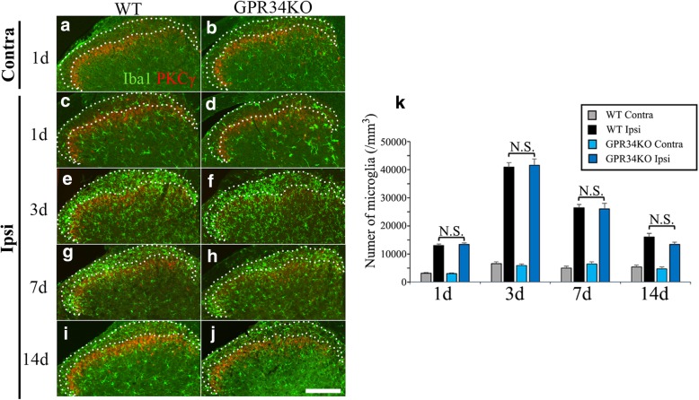
Methods: mRNA expression of GPR34 and pro-inflammatory molecules was determined by quantitative real-time PCR in wild-type and GPR34-deficient mice with L4 spinal nerve injury. In situ hybridization was used to identify GPR34 expression in microglia, and immunohistochemistry with the microglial marker Iba1 was performed to examine microglial numbers and morphology. Mechanical sensitivity was evaluated by the von Frey hair test. Liquid chromatography-tandem mass spectrometry quantified expression of the ligand for GPR34, lysophosphatidylserine (LysoPS), in the dorsal horn, and a GPR34 antagonist was intrathecally administrated to examine the effect of inhibiting LysoPS-GPR34 signaling on mechanical sensitivity.

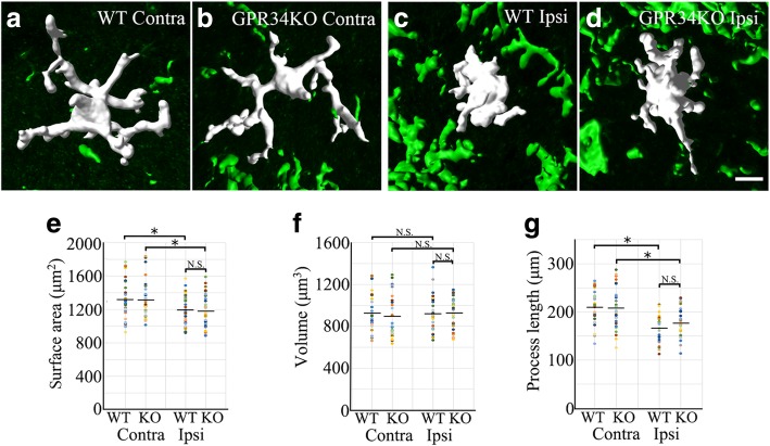
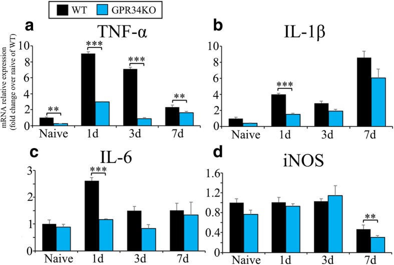
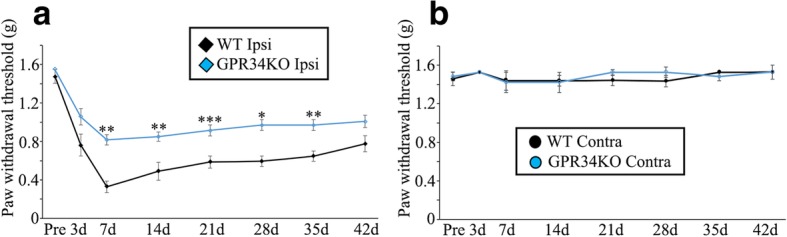
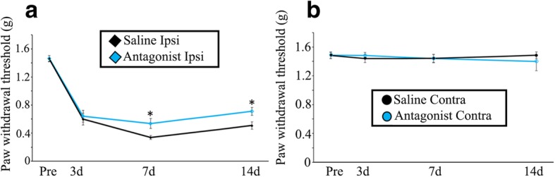
Results: GPR34 was predominantly expressed by microglia in the dorsal horn after L4 nerve injury. There were no histological differences in microglial numbers or morphology between WT and GPR34-deficient mice. However, nerve injury-induced pro-inflammatory cytokine expression levels in microglia and pain behaviors were significantly attenuated in GPR34-deficient mice. Furthermore, the intrathecal administration of the GPR34 antagonist reduced neuropathic pain.

Conclusions: Inhibition of GPR34-mediated signal by GPR34 gene deletion reduced nerve injury-induced neuropathic pain by suppressing pro-inflammatory responses of microglia without affecting their morphology. Therefore, the suppression of GPR34 activity may have therapeutic potential for alleviating neuropathic pain.

Keywords: Allodynia; GPCR; GPR34; LysoPS; Microglia; Neuroinflammation; Pain.


Conflict of interest statement

Ethics approval and consent to participate

This study was approved by the local animal ethics committee in accordance with the regulations for animal experiments at Nagoya University (permission No. 27204, 28303, 29281, and 30178).

Consent for publication

Not applicable.

Competing interests

The authors declare that they have no competing interests.

Publisher’s Note

Springer Nature remains neutral with regard to jurisdictional claims in published maps and institutional affiliations.

Figures

Fig. 1
Fig. 1
GPR34 expression is induced in microglia in the spinal dorsal horn after L4 nerve injury. a qRT-PCR analysis of GPR34 mRNA in the ipsilateral L4 dorsal horn of WT mice before (Naive) and after L4 nerve injury. Results are normalized to the housekeeping gene GAPDH (n = 4 for each time point). Values are mean ± SEM. Data are shown as fold change over naive sample. *p < 0.05, **p < 0.01 (one-way ANOVA with post hoc Tukey’s test). b, c In situ hybridization shows increased GPR34 mRNA expression in the spinal dorsal horn 7 days after L4 nerve injury (low-magnification images). Contra contralateral side, Ipsi ipsilateral side. Scale bar = 200 μm. df Localization of GPR34 mRNA in microglia in the ipsilateral dorsal horn (high-magnification images). Hybridized cells (d) display IRF8 immunoreactivity (e) in their nuclei (arrows). Scale bar = 30 μm
Fig. 2
Fig. 2
Microglial numbers are not affected by GPR34 deficiency. a, b Contralateral dorsal horn in WT (a) and GPR34-deficient (b) mice at 1 day after L4 nerve injury. cj Ipsilateral dorsal horn in WT (c, e, g, i) and GPR34-deficient (d, f, h, j) mice at 1 (c, d), 3 (e, f), 7 (g, h), and 14 (ij) days after L4 nerve injury. Sections were stained by anti-Iba1 antibody (green) and anti-PKCγ antibody (red) for visualization of microglia and inner lamina II, respectively. Iba1-positive microglia within lamina I/IIo, identified by a dotted line, were counted. Scale bar = 200 μm. k Quantification of microglial numbers in lamina I/IIo of the ipsilateral and contralateral L4 dorsal horn in WT and GPR34-deficient mice after L4 nerve injury (n = 4 for each time point). Values are mean ± SEM. There was no significant difference between WT and GPR34-deficient mice at any time points (one-way ANOVA with post-hoc Tukey’s test)
Fig. 3
Fig. 3
Microglial morphology is not affected by GPR34 deficiency. ad Representative 3D reconstructions of microglia in the contralateral (Contra) and ipsilateral (Ipsi) dorsal horn in WT and GPR34-deficient mice. Scale bar = 10 μm. eg Quantitative morphometric analysis of microglial surface area (e), volume (f), and process length (g) (n = 32 cells from four animals). Values are mean ± SEM. *p < 0.05 (one-way ANOVA with post-hoc Tukey’s test). There was no significant difference in any parameter between WT and GPR34-deficient mice
Fig. 4
Fig. 4
Pro-inflammatory microglial molecules are reduced in GPR34-deficient mice. The ipsilateral L4 dorsal horn was obtained from WT and GPR34-deficient mice before (Naive) and after L4 nerve injury (n = 4 for each time point), and mRNA expression levels of neuropathic pain-related molecules were quantified by qRT-PCR. a TNF-α, b IL-1β, c IL-6, and d iNOS. Results are normalized to GAPDH. Data are shown as fold change over naive sample of WT mice. Values are mean ± SEM. **p < 0.01, ***p < 0.001 (two-way ANOVA with post-hoc Tukey’s test)
Fig. 5
Fig. 5
Nerve injury-induced mechanical allodynia is reduced in GPR34-deficient mice. PWT in WT and GPR34-deficient mice before (pre) and after L4 nerve injury (n = 6). PWT in the ipsilateral (Ipsi) (a) and contralateral (Contra) (b) side. *p < 0.05, **p < 0.01, ***p < 0.001 (one-way RM ANOVA with Bonferroni adjustment or the nonparametric RM ANOVA on ranks with Dunnett’s adjustment). On the ipsilateral side, the PWT was higher in GPR34-deficient mice than in WT mice from 7 to 35 days
Fig. 6
Fig. 6
Neuropathic pain is reduced by GPR34 antagonism. The PWT in the ipsilateral (Ipsi) (a) and contralateral (Contra) (b) side of WT mice with L4 nerve injury. Mice were intrathecally administered the GPR34 antagonist once a day for 14 days after nerve injury (n = 8), and the von Frey test was performed on days 3, 7, and 14. Values are mean ± SEM. *p < 0.05 (one-way RM ANOVA with Bonferroni adjustment or the nonparametric RM ANOVA on ranks with Dunnett’s adjustment)

References

    1. Inoue K. ATP receptors of microglia involved in pain. Novartis Found Symp. 2006;276:263–272. - PubMed
    1. Inoue K, Tsuda M. Microglia and neuropathic pain. Glia. 2009;57:1469–1479. doi: 10.1002/glia.20871. - DOI - PubMed
    1. Yasui M, Yoshimura T, Takeuchi S, Tokizane K, Tsuda M, Inoue K, Kiyama H. A chronic fatigue syndrome model demonstrates mechanical allodynia and muscular hyperalgesia via spinal microglial activation. Glia. 2014;62:1407–1417. doi: 10.1002/glia.22687. - DOI - PubMed
    1. Grace PM, Hutchinson MR, Maier SF, Watkins LR. Pathological pain and the neuroimmune interface. Nat Rev Immunol. 2014;14:217–231. doi: 10.1038/nri3621. - DOI - PMC - PubMed
    1. Maeda M, Tsuda M, Tozaki-Saitoh H, Inoue K, Kiyama H. Nerve injury-activated microglia engulf myelinated axons in a P2Y12 signaling-dependent manner in the dorsal horn. Glia. 2010;58:1838–1846. doi: 10.1002/glia.21053. - DOI - PubMed

MeSH terms

LinkOut - more resources