. 2019 Jun 6;74(5):936-950.e5.
doi: 10.1016/j.molcel.2019.03.014. Epub 2019 Apr 8.

Introducing a Spectrum of Long-Range Genomic Deletions in Human Embryonic Stem Cells Using Type I CRISPR-Cas

Affiliations

Introducing a Spectrum of Long-Range Genomic Deletions in Human Embryonic Stem Cells Using Type I CRISPR-Cas

Adam E Dolan et al. Mol Cell. .

Abstract

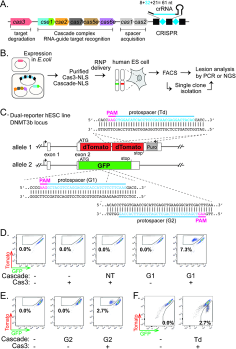
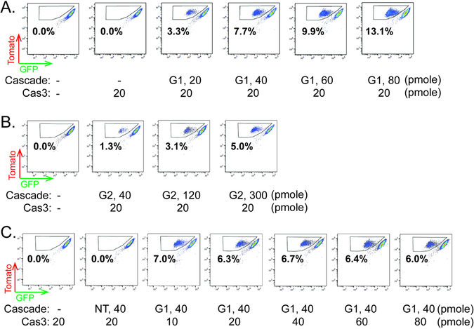
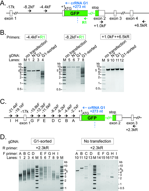
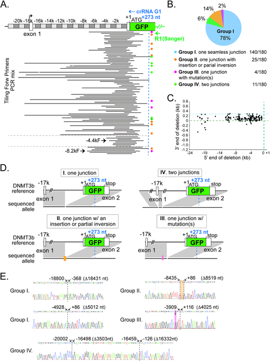
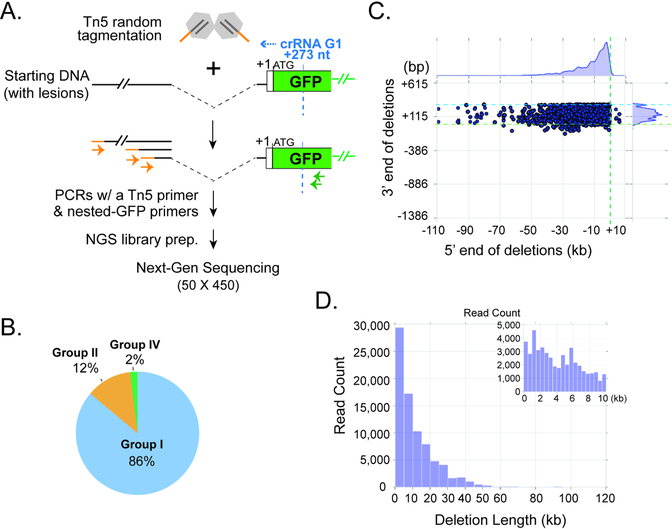
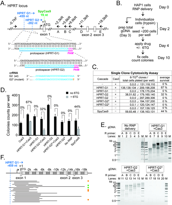
CRISPR-Cas systems enable microbial adaptive immunity and provide eukaryotic genome editing tools. These tools employ a single effector enzyme of type II or V CRISPR to generate RNA-guided, precise genome breaks. Here we demonstrate the feasibility of using type I CRISPR-Cas to effectively introduce a spectrum of long-range chromosomal deletions with a single RNA guide in human embryonic stem cells and HAP1 cells. Type I CRISPR systems rely on the multi-subunit ribonucleoprotein (RNP) complex Cascade to identify DNA targets and on the helicase-nuclease enzyme Cas3 to degrade DNA processively. With RNP delivery of T. fusca Cascade and Cas3, we obtained 13%-60% editing efficiency. Long-range PCR-based and high-throughput-sequencing-based lesion analyses reveal that a variety of deletions, ranging from a few hundred base pairs to 100 kilobases, are created upstream of the target site. These results highlight the potential utility of type I CRISPR-Cas for long-range genome manipulations and deletion screens in eukaryotes.

Keywords: CRISPR-Cas; Cas3; Cascade; RNA-guided; chromosome; embryonic stem cell; genome editing; large genome deletion; long-range; type I CRISPR.


Conflict of interest statement

Declaration of Interests

A patent application has been filed describing the invention reported herein.

Figures

Figure 1.
Figure 1.. Type I CRISPR-Cas can enable RNA-guided genome editing in human ES cells.
(A) Schematic diagram of the T. fusca Type I-E CRISPR-cas locus. Black rectangles and blue diamonds represent CRISPR repeats and spacers; colored boxes, cas genes. (B) Procedure of the genome editing experiments in hESCs. (C) Schematic of the hESC dual-reporter line bearing EGFP and tdTomato at the DNMT3B locus. Protospacers for the three reporter-targeting crRNAs are indicated in blue, and corresponding PAMs in magenta. (D-F) Flow cytometry analysis of the dual-reporter hESC line 4–5 days after RNP delivery. Percentages of EGFP-negative/tdTomato-positive cells (boxed) are indicated in (D) (E), and percentages of EGPF-positive/tdTomato-negative cells (boxed) are indicated in (F).
Figure 2.
Figure 2.. Optimization of genome editing efficiency.
(A-C) Flow cytometry analysis of dual-reporter hESC line 4 days after RNP delivery. Increasing amount of TfuCascade-G1 or TfuCascade-G2 was used in conjunction with constant TfuCas3 in (A) and (B). Increasing amount of TfuCas3 was used in conjunction with constant amount of TfuCascade-G1 in (C). Percentages of EGFP-negative/tdTomato-positive cells (boxed) are indicated. Amounts of Cascade or Cas3 delivered via 10 μL electroporation reactions were indicated.
Figure 3.
Figure 3.. Long-range PCR based characterization of genomic lesions induced by Type I CRISPR-Cas.
(A) (C) Schematic of the DNMT3B-EGFP locus and annealing sites for five PCR primers used in (B) or ten PCR primers used in (D). Positions relative to the EGFP translation start site (+1) are indicated. Recognition sites (2nd nt of the PAM) for Cascade- G1 are marked by the dashed blue line. Blue arrowhead, direction of Cas3 translocation. (B) Characterization of genomic lesions by long-range PCRs. A collection of DNA lesions was introduced upstream of GFP by a GFP-targeting Cascade-G1 and Cas3, in the sorted EGFP-negative/tdTomato-positive population, as well as in unsorted total cells. NT, total cells treated with a non-targeting Cascade and Cas3. PCR primers used are indicated and their annealing sites depicted in (A). (D) Heterogeneous large genomic deletions were introduced by Cascade-G1/Cas3 in the ~20 kb region upstream of GFP, revealed by serial long-range PCRs with tiling forward primers. NT, non-targeting Cascade. PCR primers used are indicated and their annealing sites depicted in (C). M, DNA size markers.
Figure 4.
Figure 4.. Type I CRISPR induced deletion/repair junctions are revealed at single nucleotide resolution by sanger-sequencing.
(A) Representative deletion locations at the DNMT3B-EGFP locus, revealed by cloning of pooled tiling PCRs in Fig. 3D and Sanger sequencing. The entire PCR in lanes 4 or 8 of Fig. 3B was also analyzed, the detected lesions are shown at the bottom. R1, reverse primer used for Sanger sequencing. Black lines, deleted genomic regions. Orange, magenta, green, and the lack of dots on the right indicate Groups II, III, IV, and I deletion junctions as described in (B). (B) A pie chart of the four groups of deletion junctions identified. Group I (light blue), one seamless junction; Group II (orange), one deletion with an small insertion or partial inversion; Group III (pink), one deletion with point mutations); Group IV (green), two deletions. (C) Scatter plot for the 180 unique junctions, showing the upstream (5’, X-axis) and downstream (3’, Y-axis) end points of the chromosomal deletions, relative to the EGFP translation start site (+1, marked by the dashed green line) at the DNMT3B locus. Dashed blue line, recognition site for Cascade-G1. (D) Schematics of the gene structure of representative groups I, II, III, and IV lesion junctions. Black horizontal line, reference and sequenced alleles; gray shadow; direct match between the reference and sequenced alleles. Lack of grey shadow at the reference locus represents a deletion in the sequenced allele. Orange bar, an insertion or partial inversion; pink line, point mutation(s). Cascade-G1 recognition site (+273, relative to the beginning of EGFP ORF +1) is marked by the dashed blue line in (A) and (C). (E) Example Sanger sequencing chromatograms showing representative lesion events. Dashed black line, deletion/repair junctions. Genomic positions of the flanking nucleotides, relative to the EGFP translation start site (+1), are indicated on the top. Sizes of the deletions are shown in parentheses. Orange, a 2 bp insertion; pink, a T to G point mutation.
Figure 5.
Figure 5.. Tn5- and NGS- based characterization of Type I CRISPR-induced genome deletions.
(A) Schematic of Tn5 tagmentation procedure and NGS library construction. (B) Pie chart of three main types of lesion-containing reads identified by Miseq analysis. Light blue, Group I (one deletion junction); orange, Group II (a deletion with a large [>=9 bp] insertion, including partial inversion); green, Group IV (two deletions). The coloring scheme and categorization for deletion junctions are consistent with Fig. 4B. (C) Scatter plot for all Group I lesion-containing reads, showing the upstream (5’, X-axis) and downstream (3’, Y-axis) end points of the chromosomal deletions, relative to the EGFP translation start site (+1) at the DNMT3B locus. Dashed green line, and light blue lines, the (+1) start of the EGFP ORF; dashed blue line, recognition site for Cascade-G1. Kernel density estimates for the marginal distributions are shown along the axes, revealing the narrow range of downstream deletion endpoints and very long-tailed distribution of upstream deletion endpoints. (D) Histogram showing the distribution of deletion lengths observed for all the Group I reads in (B). The inset view on the top right corner is a zoom-in on deletions smaller than 10 kb.
Figure 6.
Figure 6.. Efficient Editing at the Endogenous HPRT1 Locus in HAP1 Cells
(A) Schematic of the HPRT locus and annealing sites for six PCR primers used in (E). Protospacers for the two crRNAs are indicated in blue, and corresponding PAMs in magenta. Positions relative to HPRT translation start site (+1) are indicated. Recognition sites (2nd nt of the PAM) for HPRT G1 and G2 are marked by the dashed blue line. Blue arrowhead, direction of Cas3 translocation. Recognition site (1st G in PAM) for the SpyCas9 is marked by the dashed green line. The G2 guide sequence is shown at the bottom, with the single nt mutation in HPRTG2* in orange. (B) Procedure of the genome editing experiments in HAP1 cells. (C) Estimate HPRT targeting efficiency by single clone 6-TG cytotoxicity assay. (D) Bar graph plotting the HAP1 colony counts obtained in (C). The average 6-TG survival rates from three independent experiments are indicated. Error bar, standard deviation. (E) A collection of DNA lesions was induced in the HAP1 cells treated with Cas3 and Cascades HPRT-G1 or HPRT-G2, but not the HPRT-G2* mutant. PCR primers used are indicated and their annealing sites depicted in (A). M, DNA size markers. Lanes from two different agarose gels are separated by the dashed black line. (F) Locations of the HPRT genomic deletions. Black lines, deleted genomic regions. Orange, green, and the lack of dots on the right indicate Groups II, IV, and I deletion junctions as described in Fig. 4B.

Comment in

References

    1. Barrangou R, Fremaux C, Deveau H, Richards M, Boyaval P, Moineau S, Romero DA, and Horvath P (2007). CRISPR provides acquired resistance against viruses in prokaryotes. Science 315, 1709–1712. - PubMed
    1. Blosser TR, Loeff L, Westra ER, Vlot M, Kunne T, Sobota M, Dekker C, Brouns SJJ, and Joo C (2015). Two distinct DNA binding modes guide dual roles of a CRISPR-Cas protein complex. Mol Cell 58, 60–70. - PMC - PubMed
    1. Bolger AM, Lohse M, and Usadel B (2014). Trimmomatic: a flexible trimmer for Illumina sequence data. Bioinformatics 30, 2114–2120. - PMC - PubMed
    1. Bolotin A, Quinquis B, Sorokin A, and Ehrlich SD (2005). Clustered regularly interspaced short palindrome repeats (CRISPRs) have spacers of extrachromosomal origin. Microbiology 151, 2551–2561. - PubMed
    1. Boyle EA, Andreasson JOL, Chircus LM, Sternberg SH, Wu MJ, Guegler CK, Doudna JA, and Greenleaf WJ (2017). High-throughput biochemical profiling reveals sequence determinants of dCas9 off-target binding and unbinding. Proceedings of the National Academy of Sciences of the United States of America 114, 5461–5466. - PMC - PubMed

Publication types