. 2019 Jun 5;9(6):1869-1880.
doi: 10.1534/g3.119.400197.

An Interaction Network of the Human SEPT9 Established by Quantitative Mass Spectrometry

Affiliations

An Interaction Network of the Human SEPT9 Established by Quantitative Mass Spectrometry

Matthias Hecht et al. G3 (Bethesda). .

Abstract

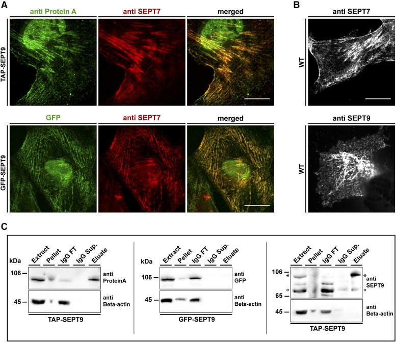
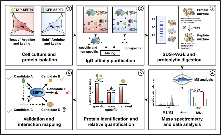
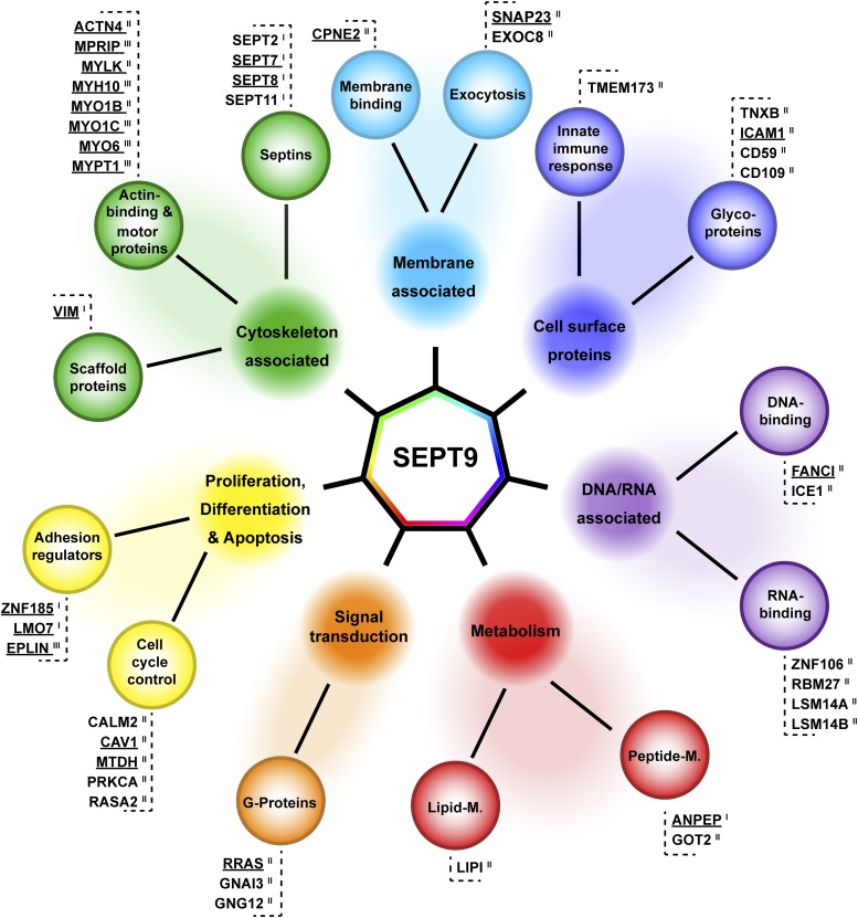
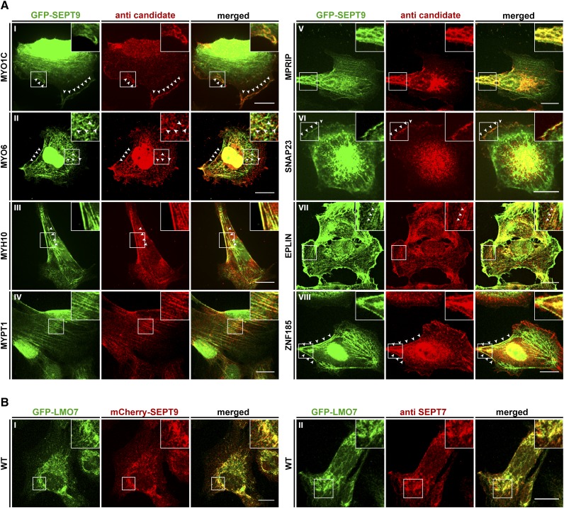
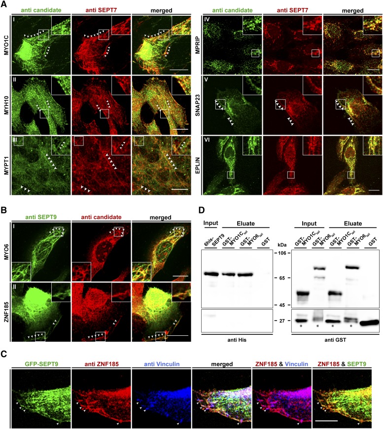
Septins regulate the organization of the actin cytoskeleton, vesicle transport and fusion, chromosome alignment and segregation, and cytokinesis in mammalian cells. SEPT9 is part of the core septin hetero-octamer in human cells which is composed of SEPT2, SEPT6, SEPT7, and SEPT9. SEPT9 has been linked to a variety of intracellular functions as well as to diseases and diverse types of cancer. A targeted high-throughput approach to systematically identify the interaction partners of SEPT9 has not yet been performed. We applied a quantitative proteomics approach to establish an interactome of SEPT9 in human fibroblast cells. Among the newly identified interaction partners were members of the myosin family and LIM domain containing proteins. Fluorescence microscopy of SEPT9 and its interaction partners provides additional evidence that SEPT9 might participate in vesicle transport from and to the plasma membrane as well as in the attachment of actin stress fibers to cellular adhesions.

Keywords: interaction map; proteomics; quantitative mass spectrometry; septins.


Figures

Figure 1
Figure 1
Cell lines and workflow for AP-MS of SEPT9 complexes. A) Characterization of the used cell lines. The colocalization of GFP-SEPT9 (upper panel) and TAP-SEPT9 (lower panel) with the endogenous septin cytoskeleton was probed by immunostaining of SEPT7. TAP-SEPT9 was visualized via an anti-ProteinA antibody. B) 1306 wild type fibroblasts were stained with anti SEPT9 and anti-SEPT7 antibodies. Scale bars represent 20 µm. C) Western blot analysis of the consecutive steps of the AP. Samples of the cell extract, pellet, supernatant of the beads after coupling (IgG sup.), washing step (IgG wash) and the eluate were separated by SDS-PAGE and Western blot was performed using an anti-ProteinA, anti-GFP or anti-SEPT9 antibody, respectively. The band marked with an asterisk represents TAP-SEPT9. The band marked with a circle represents the endogenous SEPT9.
Figure 2
Figure 2
Workflow of the performed “mixing after purification” AP approach with subsequent MS analysis.
Figure 3
Figure 3
Interaction map of SEPT9 in 1306 fibroblast cells. Class I, II and III SEPT9 interactors identified by SILAC based AP-MS were categorized into seven functional groups, (i) cytoskeleton associated proteins, (ii) proteins involved in proliferation, differentiation and apoptosis, (iii) cell surface proteins, (iv) metabolic proteins, (v) signal transduction proteins, (vi) nucleic acid associated proteins and (vii) proteins associated with the plasma membrane. Candidates validated by IF are underlined.
Figure 4
Figure 4
Colocalization of interaction partners with SEPT9 in GFP-SEPT9 expressing cells. A) The indicated candidate proteins were immunostained with a suitable primary antibody, followed by an Alexa555 coupled secondary antibody. GFP-SEPT9 was observed directly via its GFP. White arrowheads mark colocalizing structures. B) Colocalization of GFP-LMO7 with mCherry-SEPT9 in transiently transfected 1306 cells (left panel) and colocalization with the endogenous septin cytoskeleton by IF via an anti-SEPT7 antibody (right panel). Images were assembled from z-projections. The scale bars represent 20 µM.
Figure 5
Figure 5
Colocalization of interaction partners with the septin cytoskeleton in 1306 fibroblast cells and pulldown analysis showing a direct interaction of SEPT9 with myosin motors. A) The indicated candidate proteins were immunostained with a suitable primary antibody, followed by an Alexa488 coupled secondary antibody. The endogenous septin cytoskeleton was immunostained by an anti-SEPT7 primary antibody followed by an Alexa555 coupled secondary antibody. White arrowheads mark colocalizing structures. B) Colocalization of MYO6 and ZNF185 with the endogenous septin cytoskeleton by immunostaining of SEPT9 with an anti-SEPT9 primary antibody followed by an Atto488 coupled secondary antibody. Due to species incompatibility of the available primary antibodies against MYO6 and ZNF185, these two candidates had to be visualized via an Alexa555 coupled secondary antibody. White arrowheads mark colocalizing structures. C) Colocalization of ZNF185, Vinculin and SEPT9 in GFP-SEPT9 expressing cells. ZNF185 was visualized as in figure part B and Vinculin was visualized by an antibody directly coupled to Alexa647. White arrowheads mark sites of focal adhesions. Images in parts A-C were assembled from z-projections. The scale bars represent 20 µM. D) In vitro pulldown experiment showing direct binding of 6his-SEPT9 to tail fragments of myosin motors. GST-MYO6835-1294, GST-MYO1C809-1063 and free GST as control were immobilized on Glutathione sepharose and incubated with purified 6his-SEPT9. Samples of the SEPT9 input, the GST-fusion protein input and the eluates from the beads were subjected to SDS-PAGE and subsequent Western blot analysis using anti-his or anti-GST primary antibodies, respectively. Bands marked with an asterisk represent a GST containing degradation product of the GST-myosin fusion proteins.

References

    1. Abe K., Takeichi M., 2008. EPLIN Mediates Linkage of the Cadherin Catenin Complex to F-Actin and Stabilizes the Circumferential Actin Belt. Proc. Natl. Acad. Sci. USA 105: 13–19. 10.1073/pnas.0710504105 - DOI - PMC - PubMed
    1. Arden S. D., Puri C., Sui J., Au Y., Kendrick-Jones J., et al. , 2007. Myosin VI Is Required for Targeted Membrane Transport during Cytokinesis. Mol. Biol. Cell 18: 4750–4761. 10.1091/mbc.e07-02-0127 - DOI - PMC - PubMed
    1. Aschenbrenner L., Lee T., Hasson T., 2003. Myo6 Facilitates the Translocation of Endocytic Vesicles from Cell Peripheries. Mol. Biol. Cell 14: 2728–2743. 10.1091/mbc.e02-11-0767 - DOI - PMC - PubMed
    1. Bai X., Bowen J. R., Knox T. K., Zhou K., Pendziwiat M., et al. , 2013. Novel Septin 9 Repeat Motifs Altered in Neuralgic Amyotrophy Bind and Bundle Microtubules. J. Cell Biol. 203: 895–905. 10.1083/jcb.201308068 - DOI - PMC - PubMed
    1. Beites C. L., Campbell K. A., Trimble W. S., 2005. The Septin Sept5/CDCrel-1 Competes with α-SNAP for Binding to the SNARE Complex. Biochem. J. 385: 347–353. 10.1042/BJ20041090 - DOI - PMC - PubMed

Publication types

LinkOut - more resources