. 2019 Apr 12;364(6436):148-153.
doi: 10.1126/science.aav7942.

Structure and dynamics of the active human parathyroid hormone receptor-1

Affiliations

Structure and dynamics of the active human parathyroid hormone receptor-1

Li-Hua Zhao et al. Science. .

Abstract

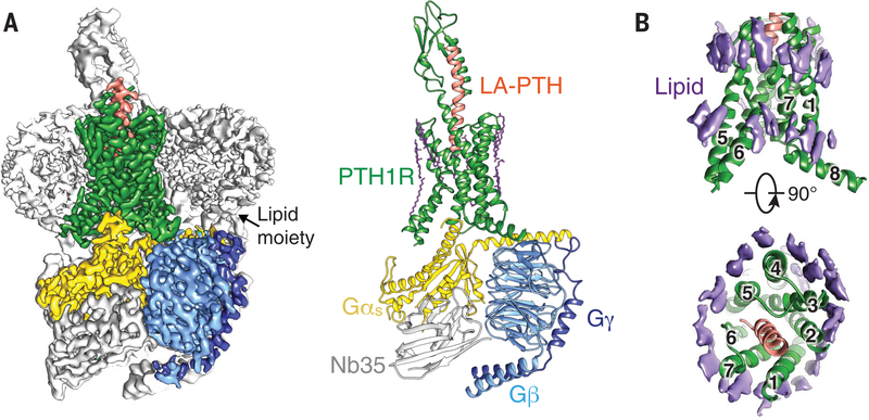
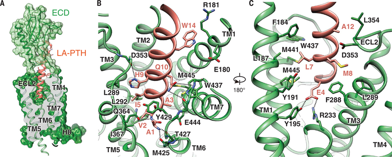
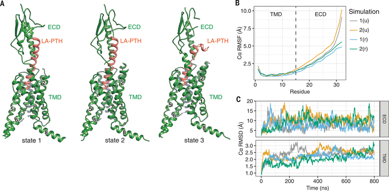
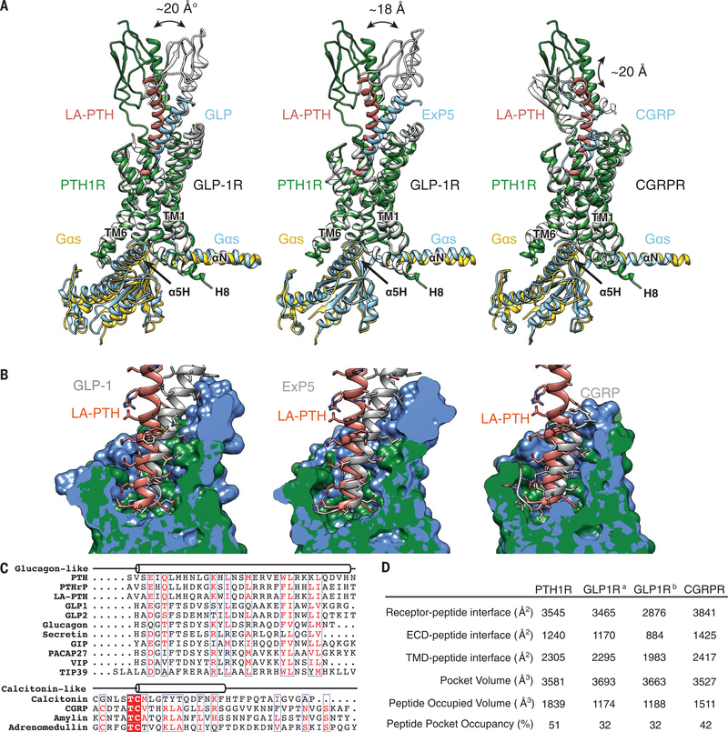
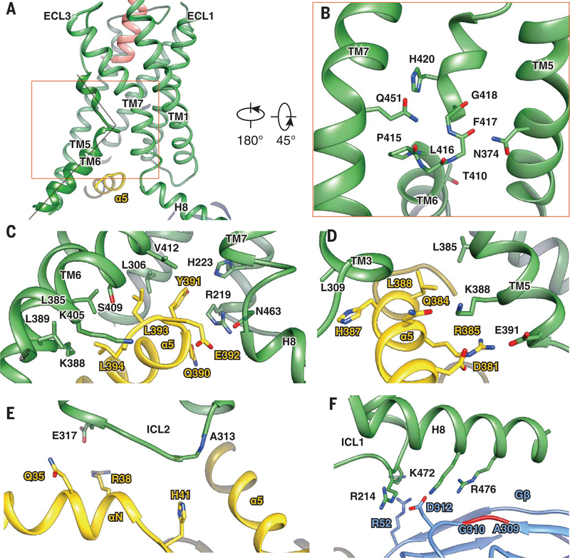
The parathyroid hormone receptor-1 (PTH1R) is a class B G protein-coupled receptor central to calcium homeostasis and a therapeutic target for osteoporosis and hypoparathyroidism. Here we report the cryo-electron microscopy structure of human PTH1R bound to a long-acting PTH analog and the stimulatory G protein. The bound peptide adopts an extended helix with its amino terminus inserted deeply into the receptor transmembrane domain (TMD), which leads to partial unwinding of the carboxyl terminus of transmembrane helix 6 and induces a sharp kink at the middle of this helix to allow the receptor to couple with G protein. In contrast to a single TMD structure state, the extracellular domain adopts multiple conformations. These results provide insights into the structural basis and dynamics of PTH binding and receptor activation.


Figures

Fig. 1.
Fig. 1.. Cryo-EM structure of LA-PTH–bound human PTH1R in complex with Gs.
(A) (Left) Cut-through view of cryo-EM density map that illustrates the LA-PTH–PTH1R–Gs complex and the disc-shaped micelle. The unsharpened cryo-EM density map at the 0.01 threshold shown as light gray surface indicates a micelle diameter of 12 nm. The colored cryo-EM density map is shown at 0.026 threshold. (Right) Cartoon representation of the LA-PTH–PTH1R–Gs complex is shown with annular lipids in purple stick representation. Green, PTH1R; orange, LA-PTH; gold, Gs Ras-like domain; light blue, Gβ; medium blue, Gγ; gray, Nb35. (B) Cryo-EM density of the ordered annular lipid layer around the receptor TMD shown in purple; numbers 1 through 7 represent TM1 to TM7; receptor ECD and G protein are omitted.
Fig. 2.
Fig. 2.. Molecular recognition of LA-PTH by PTH1R.
(A) The binding mode of LA-PTH with PTH1R, showing that LA-PTHN (ribbon and stick representation, orange) penetrates into a pocket formed by all TM helices except TM4, and by ECL2 and ECL3 whereas its C-terminal half is recognized by the ECD (ribbon in transparent surface, green). Single-letter abbreviations for the amino acid residues are as follows: A, Ala; C, Cys; D, Asp; E, Glu; F, Phe; G, Gly; H, His; I, Ile; K, Lys; L, Leu; M, Met; N, Asn; P, Pro; Q, Gln; R, Arg; S, Ser; T, Thr; V, Val; W, Trp; and Y, Tyr. (B and C) Detailed interactions of LA-PTH with the PTH1R TMD pocket with hydrogen bonds shown as dotted lines.
Fig. 3.
Fig. 3.. Conformational dynamics of the LA-PTH–PTH1R complex.
(A) Three conformational states of the LA-PTH–PTH1R complex. (B and C) The relative movement of the Ca positions of LA-PTH (B) and the PTH1R ECD and TMD in four independent MD simulations (C). The u and r in parentheses indicate that the Gs-binding interface is unrestrained or restrained, respectively, during simulation. RMSF, root mean square fluctuation; RMSD, root mean square deviation.
Fig. 4.
Fig. 4.. Basis of peptide hormone specificity.
(A) Pairwise comparison of the LA-PTH bound to PTH1R with GLP-1, Exendin-P5 (ExP5), and CGRP in complex with their corresponding receptors, showing the relative positions of peptide ligands and their receptor ECDs. The RAMP1 component was omitted from the CGRPR figure for clarity. (B) Pairwise comparison of the PTH1R structure with GLP-1R bound to GLP-1, ExP5, and CGRPR, showing the pocket shapes accommodating respective peptide ligands. (C) Sequence alignment of class B GPCR peptide ligands. (D) Pocket size and occupancy of peptide ligands in class B GPCR structures. GLP-1Ra is a GLP-1–bound GLP-1R structure, and GLP-1Rb is an ExP5–bound GLP-1R structure.
Fig. 5.
Fig. 5.. Common mechanism of class B GPCR activation and Gs coupling.
(A) The relative position of the LA-PTH N terminus (orange) toward the sharp kink of TM6 (green). (B) The kink and helix capping at TM6 are stabilized by conserved interactions with residues from TM5 (N3745.50b) and TM7 (Q4517.49b). (C to F) The PTH1R–Gs interface, including Gαs α5-PTH1R helix 8 interface (C); Gαs α5-PTH1R TM3/TM5 interface (D); PTH1R ICL2-Gαs αN helix interface (E); and helix 8–Gβ interface (F). PTH1R, green; Gαs, yellow; and Gβ, cyan.

References

    1. Gardella TJ, Vilardaga JP, Pharmacol. Rev 67, 310–337 (2015). - PMC - PubMed
    1. Kir S. et al., Nature 513, 100–104 (2014). - PMC - PubMed
    1. Pal K, Melcher K, Xu HE, Acta Pharmacol. Sin 33, 300–311 (2012). - PMC - PubMed
    1. Pioszak AA, Parker NR, Gardella TJ, Xu HE, J. Biol. Chem 284, 28382–28391 (2009). - PMC - PubMed
    1. Pioszak AA, Xu HE, Proc. Natl. Acad. Sci. U.S.A 105, 5034–5039 (2008). - PMC - PubMed

Publication types

MeSH terms

Substances