. 2019 Jun 3;216(6):1301-1310.
doi: 10.1084/jem.20190287. Epub 2019 Apr 11.

HIV-specific humoral immune responses by CRISPR/Cas9-edited B cells

Affiliations

HIV-specific humoral immune responses by CRISPR/Cas9-edited B cells

Harald Hartweger et al. J Exp Med. .

Abstract

A small number of HIV-1-infected individuals develop broadly neutralizing antibodies to the virus (bNAbs). These antibodies are protective against infection in animal models. However, they only emerge 1-3 yr after infection, and show a number of highly unusual features including exceedingly high levels of somatic mutations. It is therefore not surprising that elicitation of protective immunity to HIV-1 has not yet been possible. Here we show that mature, primary mouse and human B cells can be edited in vitro using CRISPR/Cas9 to express mature bNAbs from the endogenous Igh locus. Moreover, edited B cells retain the ability to participate in humoral immune responses. Immunization with cognate antigen in wild-type mouse recipients of edited B cells elicits bNAb titers that neutralize HIV-1 at levels associated with protection against infection. This approach enables humoral immune responses that may be difficult to elicit by traditional immunization.


Figures

Figure 1.
Figure 1.
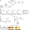
Efficient generation of indels in primary mouse B cells by CRISPR/Cas9. (A) Targeting scheme for Igh (crIgH) and Igk crRNA guides (crIgK1, crIgK2). (B) Experimental setup for C–E. Primary mouse B cells were cultured for 24 h in the presence of anti-RP105 antibody and then transfected with Cas9 RNPs and analyzed at the indicated time points. gDNA, genomic DNA. (C) Flow-cytometric plots of cultured B cells at the indicated time points after transfection. Control uses an irrelevant crRNA targeting the HPRT gene. (D) Quantification of C, percentage of Igκ Igλ B cells by flow cytometry (right y axis), and percentage of cells containing indels in the Igkc exon by TIDE analysis (left y axis). Control bars include irrelevant HPRT-targeting crRNAs or a scramble crRNA without known targets in the mouse genome. (E) Percentage of cells containing indels in the JH4 intron by TIDE analysis after targeting with crIgH or control. Bars indicate mean ± SEM in two (TIDE) or four (flow cytometry) independent experiments.
Figure 2.
Figure 2.
Engineering bNAb-expressing primary mouse B cells. (A) Schematic representation of the targeting strategy to create bNAb-expressing primary mouse B cells. ssDNA HDRT contained 110 nt 5′ and 790 nt 3′ homology arms flanking an expression cassette. The 5′ homology arm is followed by the 111 nt long splice acceptor site and the first two codons of Cμ exon 1, a stop codon, and a SV40 polyadenylation signal (CμSA SV40 pA). Then the mouse Ighv4-9 gene promoter, the leader, and variable and joining regions (VJ) of the respective antibody light chain and mouse κ constant region (Cκ) are followed by a furin-cleavage site, a glycine-serine-glycine (GSG)–linker, and a P2A self-cleaving oligopeptide sequence, the leader, VDJ of the respective antibody heavy chain, and 45 nt of the mouse JH1 intron splice donor site to splice into downstream constant regions. (B) Experimental setup for C. (C) Flow-cytometric plots of primary, mouse B cells, activated and transfected with RNPs targeting the Ighj4 intron and Igkc exon with or without ssDNA HDRTs encoding the 3BNC60SI, 3BNC117, or 10-1074 antibody. Non-transfected, antigen-binding B cells from 3BNC60SI knock-in mice cultured the same way are used as control for gating. (D) Quantification of C. Each dot represents one transfection. Data from seven independent experiments (B–D). (E) Experimental setup for F–H. (F) Flow-cytometric plots of primary, mouse B cells, activated and transfected using ssDNA HDRT encoding the antibodies 3BNC60SI, 3BNC117, PGT121, or 10-1074. B cells were expanded on feeder cells for 3 d. Cultured, nontransfected, antigen-binding B cells from PGT121 knock-in mice are shown for gating. (G) Quantification of F. (H) Total number of antigen-binding B cells before (24 h) or after 3 d (day 4) of feeder culture. Bars indicate mean ± SEM. Combined data from two independent experiments for E–H.
Figure 3.
Figure 3.
Engineering bNAb-expressing primary human B cells. (A) Schematic representation of the targeting strategy to create bNAb-expressing primary human B cells. The ssDNA HDRT is flanked by 179 nt and 521 nt homology arms. The central expression cassette contains 112 nt of the human splice acceptor site and the first two codons of Cμ exon 1, a stop codon and a SV40 polyadenylation signal (CμSA SV40 pA). Then the human IGHV1-69 gene promoter, the leader, variable and joining regions (VJ) of the respective antibody light chain, and human Cκ are followed by a furin-cleavage site, a GSG-linker, and a P2A self-cleaving oligopeptide sequence, the leader, VDJ of the respective antibody heavy chain, and 50 nt of the human JH4 intron splice donor site to splice into downstream constant regions. (B) Experimental setup for C and D. Primary human B cells were cultured for 24 h in the presence of anti-RP105 antibody and then transfected with RNPs ± HDRT. (C) Flow-cytometric plots of primary human B cells 48 h after transfection with RNPs containing crRNAs without target (scramble) or targeting the IGHJ6 intron or the IGKC exon. (D) Quantification of C. Bars indicate mean ± SEM. Combined data from three independent experiments are shown (B–D). (E) Flow-cytometric plots of antigen binding by Igλ primary human B cells 72 h after transfection of RNPs targeting both the IGHJ6 intron and the IGKC exon with or without HDRTs encoding 3BNC60SI or 10-1074. (F) Quantification of E. Bars indicate mean ± SEM. Combined data from two independent experiments with two to four replicates each (E and F).
Figure 4.
Figure 4.
Engineered bNAb-expressing primary mouse B cells participate in humoral immune responses in vivo. (A) Experimental setup for B–E. (B) Anti-3BNC60SI idiotype-coated, mouse IgG ELISA of sera from mice adoptively transferred with the indicated B cells and immunized with the cognate antigen TM4 core at the indicated time points. Representative plots of seven independent experiments. (C) Anti-3BNC60SI idiotype-coated mouse IgG1a or IgG1b ELISA of day 14 sera, as above. Representative plots of two independent experiments. (D) 3BNC60SI serum IgG levels 14 d after immunization in mice transferred with 3BNC60SI-edited cells. Numbers of total B cells/mouse at transfection are indicated. Cells were transferred either 24 h after transfection or after additional culture on feeder cells as in Fig. 2 D. Determined by anti-3BNC60SI idiotype-coated mouse IgG ELISA over seven independent experiments. Each dot represents one mouse, and the line indicates the arithmetic mean. (E) TZM.bl neutralization data of protein G–purified serum immunoglobulin days 14–21 after immunization from mice treated as in A but transfected with 10-1074 HDRT and immunized with cognate antigen 10mut. Combined data from two independent experiments are shown.

References

    1. Bar K.J., Sneller M.C., Harrison L.J., Justement J.S., Overton E.T., Petrone M.E., Salantes D.B., Seamon C.A., Scheinfeld B., Kwan R.W., et al. . 2016. Effect of HIV Antibody VRC01 on Viral Rebound after Treatment Interruption. N. Engl. J. Med. 375:2037–2050. 10.1056/NEJMoa1608243 - DOI - PMC - PubMed
    1. Briney B., Sok D., Jardine J.G., Kulp D.W., Skog P., Menis S., Jacak R., Kalyuzhniy O., de Val N., Sesterhenn F., et al. . 2016. Tailored Immunogens Direct Affinity Maturation toward HIV Neutralizing Antibodies. Cell. 166:1459–1470.e11. 10.1016/j.cell.2016.08.005 - DOI - PMC - PubMed
    1. Brinkman E.K., Chen T., Amendola M., and van Steensel B.. 2014. Easy quantitative assessment of genome editing by sequence trace decomposition. Nucleic Acids Res. 42:e168 10.1093/nar/gku936 - DOI - PMC - PubMed
    1. Caskey M., Klein F., Lorenzi J.C., Seaman M.S., West A.P. Jr., Buckley N., Kremer G., Nogueira L., Braunschweig M., Scheid J.F., et al. . 2015. Viraemia suppressed in HIV-1-infected humans by broadly neutralizing antibody 3BNC117. Nature. 522:487–491. 10.1038/nature14411 - DOI - PMC - PubMed
    1. Caskey M., Schoofs T., Gruell H., Settler A., Karagounis T., Kreider E.F., Murrell B., Pfeifer N., Nogueira L., Oliveira T.Y., et al. . 2017. Antibody 10-1074 suppresses viremia in HIV-1-infected individuals. Nat. Med. 23:185–191. 10.1038/nm.4268 - DOI - PMC - PubMed

Publication types