. 2019 May 31;294(22):8745-8759.
doi: 10.1074/jbc.RA119.007491. Epub 2019 Apr 11.

The three-dimensional structure of an H-superfamily conotoxin reveals a granulin fold arising from a common ICK cysteine framework

Affiliations

The three-dimensional structure of an H-superfamily conotoxin reveals a granulin fold arising from a common ICK cysteine framework

Lau D Nielsen et al. J Biol Chem. .

Abstract

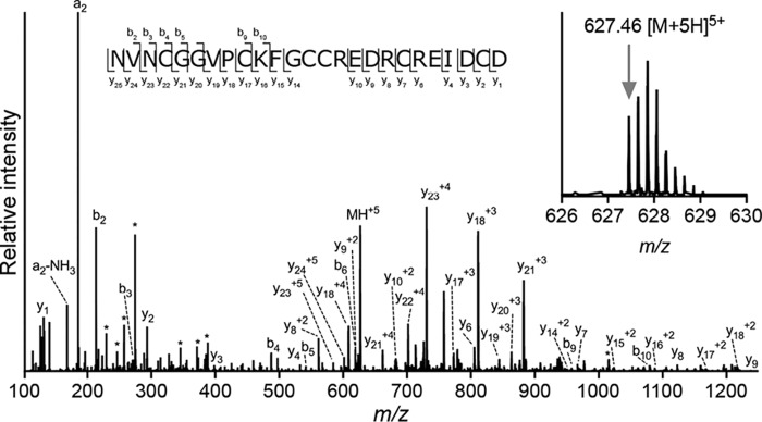
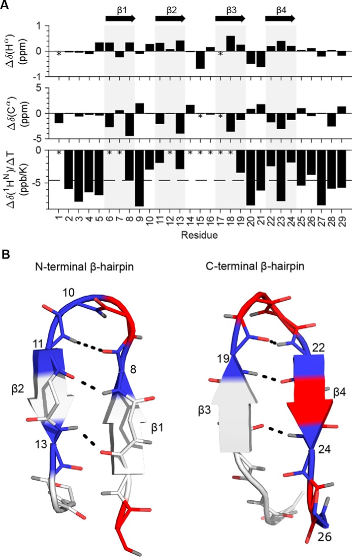
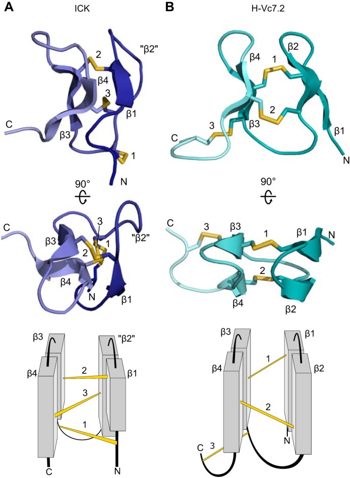
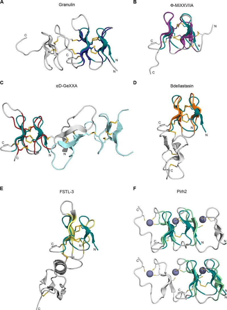
Venomous marine cone snails produce peptide toxins (conotoxins) that bind ion channels and receptors with high specificity and therefore are important pharmacological tools. Conotoxins contain conserved cysteine residues that form disulfide bonds that stabilize their structures. To gain structural insight into the large, yet poorly characterized conotoxin H-superfamily, we used NMR and CD spectroscopy along with MS-based analyses to investigate H-Vc7.2 from Conus victoriae, a peptide with a VI/VII cysteine framework. This framework has CysI-CysIV/CysII-CysV/CysIII-CysVI connectivities, which have invariably been associated with the inhibitor cystine knot (ICK) fold. However, the solution structure of recombinantly expressed and purified H-Vc7.2 revealed that although it displays the expected cysteine connectivities, H-Vc7.2 adopts a different fold consisting of two stacked β-hairpins with opposing β-strands connected by two parallel disulfide bonds, a structure homologous to the N-terminal region of the human granulin protein. Using structural comparisons, we subsequently identified several toxins and nontoxin proteins with this "mini-granulin" fold. These findings raise fundamental questions concerning sequence-structure relationships within peptides and proteins and the key determinants that specify a given fold.

Keywords: CD spectroscopy; NMR spectroscopy; antistasin; conotoxin; disulfide; disulfide bond; granulin; inhibitor cystine knot; protein conformation; protein evolution; protein expression; protein structure; protein-disulfide isomerase; toxin; β-hairpin.


Conflict of interest statement

The authors declare that they have no conflicts of interest with the contents of this article

Figures

Figure 1.
Figure 1.
H-Vc7.2 is a 25-residue conotoxin devoid of post-translational modifications. Top, b/y ladder diagram summarizing observed b- and y-ions within the amino acid sequence of H-Vc7.2. Bottom, MS/MS spectrum of H-Vc7.2 in the reduced and alkylated venom of C. victoriae. *, internal fragment ions. Inset, H-Vc7.2 [M + 5H]5+ precursor ion selected for MS/MS had monoisotopic m/z of 627.46 (z = 5, predicted m/z 627.46).
Figure 2.
Figure 2.
NextH-Vc7. 2 expressed in E. coli was purified as a single major species. A, overview of the two plasmids used in this study. pLE577 encodes Erv1p, hPDI, and csPDI under control of a tac promoter and confers chloramphenicol resistance. pLE566 encodes Ub–His10–NextH-Vc7.2 under the control of a T7 promoter and confers kanamycin resistance. This vector contains the lacI gene, which encodes the lac repressor. B, overview of the Ub–His10–NextH-Vc7.2 fusion protein. C, nonreducing SDS-polyacrylamide gel of E. coli extracts from untreated cells (lane 1) and cells treated with 1 mm IPTG overnight at 30 °C (lanes 2–4). Total, soluble, and pellet fractions were loaded as indicated. The mobility of molecular mass marker bands in kDa is shown on the left-hand side of the gel. The expression of both PDI proteins was verified by MALDI MS (see “Experimental procedures”). D, preparative RP-HPLC indicating A215 (black line), A280 (gray line), and percent of solvent B (broken line). The material eluting over the volume indicated by the black line labeled C was reanalyzed by analytical RP-HPLC as shown in E. A single main peak eluted at 25 ml.
Figure 3.
Figure 3.
rH-Vc7.2 elutes with the same retention time as the native venom peptide and disulfide bonds are critical for stability. A, RP-HPLC profiles of rH-Vc7.2 peptide (gray line) and C. victoriae venom (black line). rH-Vc7.2 and venom fractions with the same retention time as rH-Vc7.2 were collected and analyzed by MALDI-TOF MS. B, MALDI-TOF spectra of rH-Vc7.2 showing a monoisotopic mass of [M + H]1+ = 2785.21 (inset, calculated [M + H]1+ = 2785.12). C, venom fraction eluting at the same retention time as rH-Vc7.2 contained a peptide of identical mass ([M + H]1+ = 2785.15) suggesting that the recombinant and native venom peptides have identical physicochemical properties. D, CD spectra of oxidized and reduced rH-Vc7.2 recorded at 25, 90, and again at 25 °C (after heating to 90 °C), and color-coded as indicated above the spectra.
Figure 4.
Figure 4.
NextH-Vc7. 2 structure is characterized by two short, stacked β-hairpins. A, amino acid sequence of NextH-Vc7.2. The four N-terminal non-native residues are underlined; disulfide bonds are numbered, and β-strands are indicated with red arrows. B, 15N HSQC spectrum of NextH-Vc7.2 at different temperatures ranging from 5 to 45 °C and color-coded from pink to dark red as indicated by the color bar. The peaks from assigned backbone amides are labeled with one-letter code and residue number. Unassigned peaks are not labeled. Peaks with temperature coefficients higher than −4.6 ppb/K are labeled in bold. C, lowest energy NMR structure showing the three disulfide bonds connecting Cys-8 and Cys-18 (1), Cys-13 and Cys-23 (2), and Cys-17 and Cys-28 (3), and the two β-hairpins. Structural elements are colored with β-strands in red, sulfur atoms of the six cysteine residues in yellow, and loops in teal. Positions of the N and C termini are indicated. D, superimposition of the 20 lowest energy structures. The structures are shown in the same overall orientation as the conformer in C and with the same color-coding (see also Table 1).
Figure 5.
Figure 5.
Secondary chemical shifts, amide hydrogen temperature coefficients, and β-sheet hydrogen bonds. A, NextH-Vc7.2 Hα and Cα secondary chemical shifts and temperature coefficients plotted per residue. Asterisks mark unassigned residues, and gray bars indicate the position of the four β-strands as indicated with arrows at the top. The dashed line at −4.6 ppb/K in the bottom plot indicates the cutoff for hydrogen bonding of amide hydrogens. B, residues of the β-hairpins formed by β-strands 1 and 2 (left) and β-strands 3 and 4 (right) are colored blue (with residue numbers labeled) if the temperature coefficient is higher than −4.6 ppb/K, red if lower than −4.6 ppb/K, and light gray if unassigned. Backbone atoms are shown with oxygens in red and hydrogens in gray. Black dashed lines indicate the hydrogen bonds between the β-strands.
Figure 6.
Figure 6.
Structure topology of NextH-Vc7. 2 is unlike the ICK fold. Top and middle, 3D structures with β-strands, disulfides, and N and C termini labeled in two different orientations. Bottom, schematic topology representation of the structures shown above. A, a representative ICK (the SHL-I lectin (PDB code 1QK7) (64)), where “β2” indicates the position of the corresponding secondary structural element (β2) in NextH-Vc7.2, which is not present in the ICK. B, NextH-Vc7.2.
Figure 7.
Figure 7.
NextH-Vc7. 2 adopts a fold similar to the N-terminal domain of granulin. NextH-Vc7.2 (dark green) is aligned to different structural homologs (displayed in gray outside the region of structural homology). A, N-terminal domain of the human progranulin A module (dark blue, PDB code 2JYE) (33). B, Φ-MIXXVIIA conotoxin (violet) (31). C, C-terminal domain of the αD-GeXXa conotoxin (red, PDB code 4X9Z) (40). The second molecule of the αD-GeXXa dimer is depicted in light blue. D, N-terminal domain of the leech serine protease inhibitor bdellastasin (orange, PDB code 1C9T) (41). E, N-terminal domain of the follistatin-related protein 3 (FSTL-3) (yellow, PDB code 2KCX). F, zinc-binding β-hairpin repeat from the C-lobe of the N-terminal domain of Pirh2 (light green, PDB code 2K2C) (44). Top, alignment with the second and third β-hairpins; bottom, alignment with the first two β-hairpins. Zinc atoms are shown as purple spheres.

References

    1. Pennington M. W., Czerwinski A., and Norton R. S. (2018) Peptide therapeutics from venom: Current status and potential. Bioorg. Med. Chem. 26, 2738–2758 10.1016/j.bmc.2017.09.029 - DOI - PubMed
    1. Miljanich G. P. (2004) Ziconotide: neuronal calcium channel blocker for treating severe chronic pain. Curr. Med. Chem. 11, 3029–3040 10.2174/0929867043363884 - DOI - PubMed
    1. Robinson S. D., and Safavi-Hemami H. (2017) Venom peptides as pharmacological tools and therapeutics for diabetes. Neuropharmacology 127, 79–86 10.1016/j.neuropharm.2017.07.001 - DOI - PubMed
    1. Safavi-Hemami H., Brogan S. E., and Olivera B. M. (2019) Pain therapeutics from cone snail venoms: from Ziconotide to novel non-opioid pathways. J. Proteomics 190, 12–20 10.1016/j.jprot.2018.05.009 - DOI - PMC - PubMed
    1. Balsara R., Dang A., Donahue D. L., Snow T., and Castellino F. J. (2015) Conantokin-G attenuates detrimental effects of NMDAR hyperactivity in an ischemic rat model of stroke. PLoS One 10, e0122840 10.1371/journal.pone.0122840 - DOI - PMC - PubMed

Publication types

LinkOut - more resources