Review
. 2019 Jul;20(7):436-450.
doi: 10.1038/s41580-019-0123-5.

Metabolic regulation of cell growth and proliferation

Affiliations
Review

Metabolic regulation of cell growth and proliferation

Jiajun Zhu et al. Nat Rev Mol Cell Biol. 2019 Jul.

Abstract

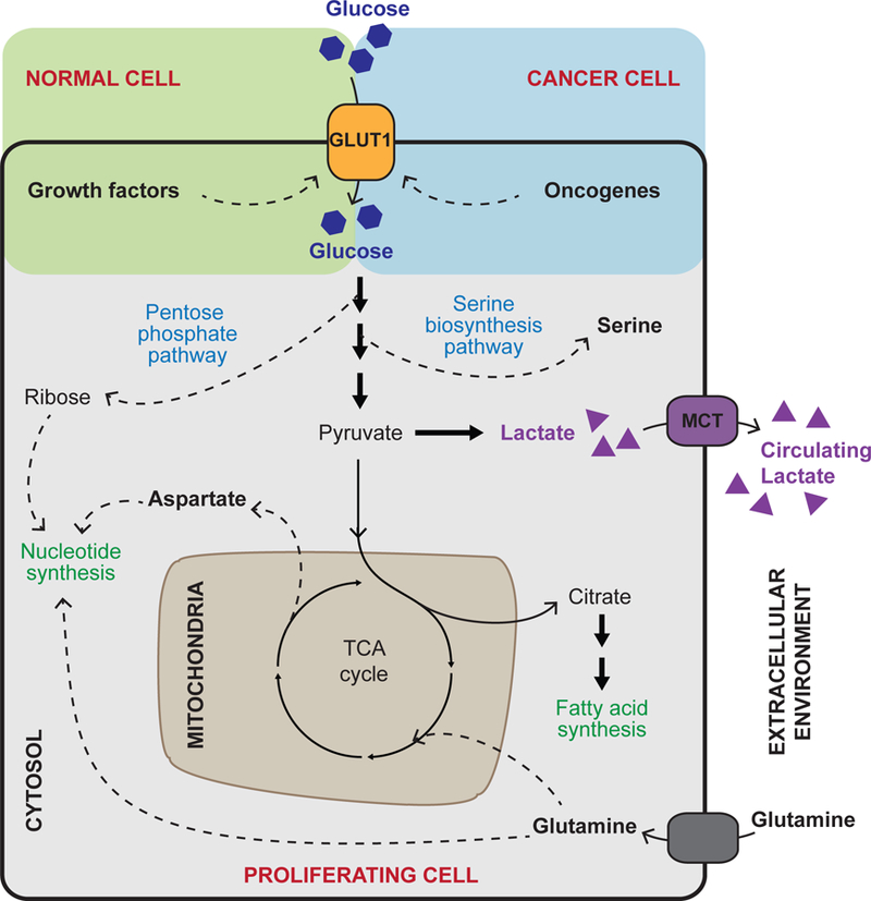
Cellular metabolism is at the foundation of all biological activities. The catabolic processes that support cellular bioenergetics and survival have been well studied. By contrast, how cells alter their metabolism to support anabolic biomass accumulation is less well understood. During the commitment to cell proliferation, extensive metabolic rewiring must occur in order for cells to acquire sufficient nutrients such as glucose, amino acids, lipids and nucleotides, which are necessary to support cell growth and to deal with the redox challenges that arise from the increased metabolic activity associated with anabolic processes. Defining the mechanisms of this metabolic adaptation for cell growth and proliferation is now a major focus of research. Understanding the principles that guide anabolic metabolism may ultimately enhance ways to treat diseases that involve deregulated cell growth and proliferation, such as cancer.


Conflict of interest statement

Competing interests

C.B.T. is a founder of Agios Pharmaceuticals and a member of its scientific advisory board. He is also a former member of the Board of Directors and stockholder of Merck and Charles River Laboratories. He has patents related to cellular metabolism.

Figures

Figure 1
Figure 1. Glucose uptake and utilization
In order to support cell growth and proliferation, glucose uptake is promoted by the growth factor signalling pathways in normal cells or by oncogenic mutations in cancer cells, which may render growth signalling pathways constantly active. Glucose is an important carbon source in energy-generating pathways of oxidative metabolism, fuelling the tricarboxylic acid (TCA) cycle and subsequent oxidative phosphorylation in mitochondria (not shown). In proliferating cells, intermediates of glucose metabolism are often diverted from glycolysis and the TCA cycle (dashed arrows) and used for biosynthetic purposes such as the production of nucleotides, amino acids and fatty acids. The TCA cycle is kept active by anaplerotic reactions involving glutamine, which — beyond supporting anabolic reactions — replenishes TCA cycle intermediates that are diverted from the cycle to support anabolism. As a result of aerobic glycolysis, a large portion of glucose carbon is also converted to lactate and secreted. Cancer associated adaptations are highlighted in red. GLUT1, glucose transporter 1; MCT, monocarboxylate transporter.
Figure 2
Figure 2. Amino acid sensing and acquisition
Cellular levels of amino acids can be increased by increased expression and cell membrane retention of amino acid transporters, which is downstream of receptor tyrosine kinase signalling mediated by growth factors. In addition, mechanistic target of rapamycin complex 1 (mTORC1) senses growth factor signalling and the presence of amino acids to promote the utilization of amino acids by activating protein translation. CASTOR1, Sestrin2, and SAMTOR are amino acid sensor proteins for arginine, leucine, and methionine (through S-adenosylmethionine (SAM)), respectively. These sensors control the activity of the GATOR2–GATOR1 complexes, negatively regulating the activity of RAG GTPases and hence inhibiting mTORC1 localization to the lysosome and activation. The suppressive functions of these sensors are diminished in the presence of the amino acids that they sense, leading to the activation of mTORC1. When amino acid levels are low, general control nonderepressible (GCN2) is activated by the increase of uncharged tRNAs and results in the inhibition of global protein translation (not shown). Paradoxically, GCN2 activation leads to increased expression of activating transcription factor 4 (ATF4). ATF4 mediates integrated stress response that includes the upregulation of genes that mediate amino acid uptake, synthesis and utilization (translation) to eventually support cell adaptation and recovery from stress. Note that ATF4 increases the expression of a variety of tRNA synthetases that allows the cell to better capture the limited amount of cellular amino acid for protein translation, which facilitates the production of proteins that are critical in stress adaptation and recovery. Cells can also acquire amino acids through macropinocytosis of extracellular proteins followed by their lysosomal degradation. Macropinocytic utilization is inhibited by mTORC1 and is promoted in cancer via oncogenic RAS and phosphoinositide 3-kinase (PI3K) pathway. Certain cancers have also been associated with upregulation of phosphoglycerate dehydrogenase (PHGDH), leading to increased serine synthesis. These cancers depend on PHGDH for growth and interference with de novo serine biosynthesis could be explored as a potential strategy in cancer treatment. Cancer associated adaptations are highlighted in red. ASNS, asparagine synthetase; PSAT1, phosphoserine aminotransferase 1; RHEB, Ras homolog enriched in brain; TSC, tuberous sclerosis complex; x(c)(−), cystine–glutamine antiporter.
Figure 3
Figure 3. Fatty acid synthesis
Fatty acids can be taken up from the environment (for example, from the circulation) through the activity of lipoprotein lipase (LPL) and fatty acid translocase. In addition, fatty acid synthesis can occur de novo. Growth factor signalling promotes utilization of glucose for fatty acid synthesis via redirecting citrate (dashed arrow) away from the tricarboxylic acid (TCA) cycle. AKT activation results in the increase in fatty acid synthesis, partly by promoting the activity of ATP citrate lyase (ACLY). Acetyl-CoA carboxylase (ACC) and fatty acid synthase (FASN) are both involved in the early steps of fatty acid synthesis leading to the formation of palmitate. These enzymes have been shown to be elevated in cancers and hence are potential targets in cancer treatment (highlighted in red boxes). The production of saturated fatty acids needs to be balanced with that of unsaturated fatty acids to avoid endoplasmic reticulum (ER) stress, which occurs in response to accumulation of saturated fatty acids in cellular (including ER) membranes and consequent disruption of membrane homeostasis. This balance is partly achieved through the regulation of oxygen-dependent and iron-dependent enzyme, stearoyl-CoA desaturase (SCD). Note that acetyl-CoA acts as the substrate for acetylation of histones as well as a variety of non-histone proteins. This link has been demonstrated to be an important mechanism by which cells coordinate metabolic status with gene expression and protein activities. GLUT1, glucose transporter 1.
Figure 4
Figure 4. Synthesis of pyrimidine and purine nucleotides
Nucleotide synthesis is key for cell proliferation as it is required for DNA replication, gene transcription and ribosome biogenesis, and the uptake of nucleotides from extracellular sources is negligible. Nucleotide biosynthesis interrogates multiple metabolic pathways and requires the utilization of various substrates as the carbon and nitrogen sources, including amino acids and metabolites of the folate cycle (the colours of carbon and nitrogen in the chemical structure of pyrimidines and purines correspond to the contributing source). Pyrimidine synthesis intersects with amino acid metabolism via positive regulation of carbamoyl-phosphate synthetase 2, aspartate transcarbamylase, and dihydroorotase (CAD) downstream of mTROC1 as well as with ATP production and energy metabolism (electron transport chain (ETC) via the activity of dihydroorotate dehydrogenase (DHODH); DHODH, which is highlighted in red, has been implicated as a potential target for cancer treatment). Purine synthesis is indirectly regulated by mechanistic target of rapamycin complex 1 (mTORC1) via its stimulation of the mitochondrial folate cycle. In addition, the key energy sensor AMP-activated protein kinase (AMPK) negatively regulates purine biosynthesis by inhibiting the activity of phosphoribosyl pyrophosphate synthetase (PRPS). This allows fine tuning of nucleotide synthesis in accordance with cellular energy metabolism. IMPDH, inosine monophosphate dehydrogenase; PRPP, phosphoribosyl pyrophosphate; QH2, dihydroxyquinone; Ribose-5-P, Ribose-5-phosphate; RPA, phosphoribosyl amine; THF, tetrahydrofolate.
Figure 5
Figure 5. Metabolic interactions with the extracellular environment
Extensive metabolic interactions exist between proliferating cells and the microenvironment. In the case of cancer cells and the tumour microenvironment, cancer cells receive metabolic signalling cues such as growth factors, which can be provided by infiltrating immune cells. These cues rewire their metabolic programmes to support biosynthesis and growth (see Figs. 1 to 4). Hypoxia (low O2 levels), which is a hallmark of solid tumours further promotes metabolic rewiring by activating hypoxia-inducible factor-1α (HIF-1α), which, among other functions, promotes aerobic glycolysis and lactate production. Lactate secreted by cancer cells can have effects on the surrounding environment. For example, cancer cell-derived lactate has been shown to induce secretion of vascular endothelial growth factor (VEGF), thereby promoting angiogenesis and increasing the potential of cancer dissemination. Cancer cells also acquire nutrients directly from the environment, including glucose, amino acids and macromolecules, such as components of the extracellular matrix (ECM) via integrin receptor-mediated endocytosis as well as other extracellular proteins and lysophospholipids via macropinocytosis. Specialized fibroblasts, known as cancer-associated fibroblasts (CAFs), have a particularly important role in cancer. CAFs have the ability to support cancer cell growth by mechanisms involving ECM deposition and autophagy. Autophagy in CAFs and other stromal cells present in cancer environment can result in the release of nutrients, such as amino acids, which can be taken up and utilized by cancer cells (for example, as building blocks for protein synthesis or for bioenergetics). GLUT1, glucose transporter 1; MCT, monocarboxylate transporter; RTK, receptor tyrosine kinase; TGFβ, transforming growth factor beta.
Figure 6
Figure 6. Maintaining redox homeostasis in proliferating cells
Owing to high glycolytic rate and associated biosynthetic reactions, proliferating cells face the problem of an elevated NADH to NAD+ ratio and thus, maintaining redox homeostasis is critical during cell proliferation. Mechanisms to maintain NADH to NAD+ ratio include the malate–aspartate shuttle, which regenerates cytosolic NAD+ to sustain constant flow through biosynthetic pathways. Also, an increase in lactate production supports regeneration of NAD+. At the same time, proliferating cells face the problem of increased generation of reactive oxygen species (ROS), primarily derived from electron transport chain in mitochondria and NADPH oxidases (NOXs). Excessive ROS cause oxidative stress, which curbs cell proliferation and viability. Thus, proliferating cells require efficient antioxidant systems to counteract the destructive activity of ROS. The two key cellular antioxidant defense systems include the thioredoxin system (not shown) and the glutathione system. Glutathione is synthesized from glutamate, cysteine and glutamine. Glutamine importantly regulates this process by supplying glutamate and promoting cystine uptake. Additionally, NADPH is required for efficient ROS detoxification as it is involved in generating reduced glutathione — which is active against ROS — from its oxidized form (not shown). NADPH is supplied by the conversion of glycolysis intermediates in the pentose phosphate pathway as well as by mitochondrial serine metabolism in the folate pathway. Antioxidant defense has been shown to be important for cancer cell survival and tumour growth. For example, cancer cells were demonstrated to rely on glutathione synthesis and to upregulate mitochondrial folate pathway (highlighted in red). MCT, monocarboxylate transporter; THF, tetrahydrofolate; x(c)(−), cystine–glutamine antiporter.
None

References

    1. Galdieri L, Mehrotra S, Yu S & Vancura A Transcriptional regulation in yeast during diauxic shift and stationary phase. OMICS 14, 629–638, doi:10.1089/omi.2010.0069 (2010). - DOI - PMC - PubMed
    1. Marijuan PC, Navarro J & del Moral R On prokaryotic intelligence: strategies for sensing the environment. Biosystems 99, 94–103, doi:10.1016/j.biosystems.2009.09.004 (2010). - DOI - PubMed
    1. Schaller GE, Shiu SH & Armitage JP Two-component systems and their co-option for eukaryotic signal transduction. Curr Biol 21, R320–330, doi:10.1016/j.cub.2011.02.045 (2011). - DOI - PubMed
    1. DeBerardinis RJ et al. Beyond aerobic glycolysis: transformed cells can engage in glutamine metabolism that exceeds the requirement for protein and nucleotide synthesis. Proc Natl Acad Sci U S A 104, 19345–19350, doi:10.1073/pnas.0709747104 (2007). - DOI - PMC - PubMed
    1. Rheinwald JG & Green H Epidermal growth factor and the multiplication of cultured human epidermal keratinocytes. Nature 265, 421–424 (1977). - PubMed

Publication types