Case Reports
. 2019 Aug;27(8):1225-1234.
doi: 10.1038/s41431-019-0397-2. Epub 2019 Apr 11.

Variants in DOCK3 cause developmental delay and hypotonia

Affiliations
Case Reports

Variants in DOCK3 cause developmental delay and hypotonia

Kimberly Wiltrout et al. Eur J Hum Genet. 2019 Aug.

Abstract

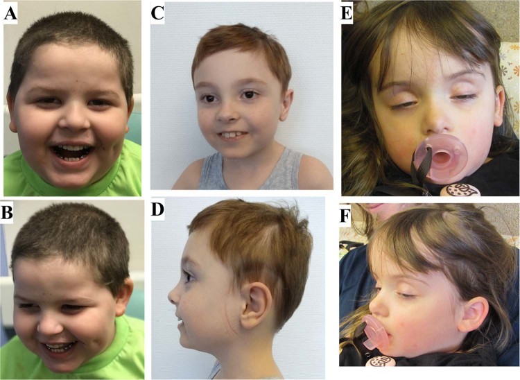
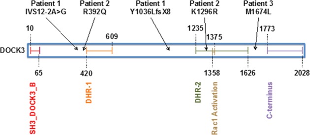
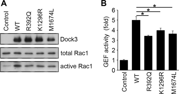
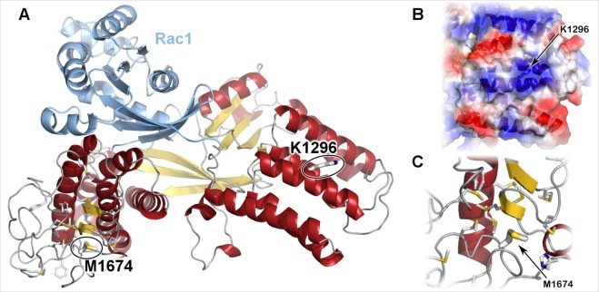
The DOCK3 gene encodes the Dedicator of cytokinesis 3 (DOCK3) protein, which belongs to the family of guanine nucleotide exchange factors and is expressed almost exclusively in the brain and spinal cord. We used whole exome sequencing (WES) to investigate the molecular cause of developmental delay and hypotonia in three unrelated probands. WES identified truncating and splice site variants in Patient 1 and compound heterozygous and homozygous missense variants in Patients 2 and 3, respectively. We studied the effect of the three missense variants in vitro by using site-directed mutagenesis and pull-down assay and show that the induction of Rac1 activation was significantly lower in DOCK3 mutant cells compared with wild type human DOCK3 (P < 0.05). We generated a protein model to further examine the effect of the two missense variants within or adjacent to the DHR-2 domain in DOCK3 and this model supports pathogenicity. Our results support a loss of function mechanism but the data on the patients with missense variants should be cautiously interpreted because of the variability of the phenotypes and limited number of cases. Prior studies have described DOCK3 bi-allelic loss of function variants in two families with ataxia, hypotonia, and developmental delay. Here, we report on three patients with DOCK3-related developmental delay, wide-based or uncoordinated gait, and hypotonia, further supporting DOCK3's role in a neurodevelopmental syndrome and expanding the spectrum of phenotypic and genotypic variability.


Conflict of interest statement

RES discloses her employment with GeneDx. The other authors declare that they have no conflict of interest.

Figures

Fig. 1
Fig. 1
Facial images of 3 patients with DOCK3 variants. From left to right: Patient 1 (a, b), Patient 2 (c, d), and Patient 3 (e, f)
Fig. 2
Fig. 2
DOCK3 protein domains (UniProt: Q8IZD9) and location of variants in Patients 1, 2, and 3. Numbers refer to amino acids. The SH3_DOCK3_B domain (amino acids 10–65) is shown in red. The DHR-1 domain (amino acids 420–609) is orange. The DHR-2 domain (amino acids 1235–1626) is green and contains the catalytic activity for Rho/Rac GTPases. Amino acids 1358–1375 are conserved amino acids required for Rac1 activation, which is outlined in brown. The last conserved domain is the proline-rich C terminus (amino acids 1773–2028) in purple, which is involved in the internalization and degradation of the NMDA receptor. The variants described in this paper are shown above the protein diagram
Fig. 3
Fig. 3
Dock3 GEF activity. Lysates from Cos-7 cells transfected with the indicated plasmids were subjected to the pull-down assay for measurement of Rac1 activity using the GST-CRIB fusion protein. Expression of Dock3, total Rac1, and activated Rac1 were detected by immunoblot analysis (left panel). The level of activated Rac1 was quantified and shown in the histogram (right panel). The data are presented as means ± S.E.M. of four samples. *P < 0.05
Fig. 4
Fig. 4
The DOCK3 DHR-2 domain structure is shown in cartoon representation colored by protein and secondary structure. The sites altered by genetic variation are shown in thick sticks. a Our protein model shows that neither of the missense variant positions, p.(Lys1296) and p.(Met1674), are at the Rac1 interface. b Focusing on the surface around p.(Lys1296), it is within a large positively charged patch (blue semi-transparent surface) surrounded by a triad of negative patches (red semi-transparent surface). c p.(Met1674) is within the hydrophobic core of the methionine-rich region of the catalytic domain. Additional methionine and aromatic residues within the region are shown in sticks representation

References

    1. Michelson DJ, Shevell MJ, Sherr EH, Moeschler JB, Gropman AL, Ashwal S. Evidence report: genetic and metabolic testing on children with global developmental delay. Neurology. 2011;77:1629–35. doi: 10.1212/WNL.0b013e3182345896. - DOI - PubMed
    1. Soden SE, Saunders CJ, Willig LK, Farrow EG, Smith LD, Petrikin JE, et al. Effectiveness of exome and genome sequencing guided by acuity of illness for diagnosis of neurodevelopmental disorders. Sci Transl Med. 2014;6:265ra168. doi: 10.1126/scitranslmed.3010076. - DOI - PMC - PubMed
    1. De Silva MG, Elliott K, Dahl HH, Fitzpatrick E, Wilcox S, Delatycki M, et al. Disruption of a novel member of a sodium/hydrogen exchanger family and DOCK3 is associated with an attention deficit hyperactivity disorder-like phenotype. J Med Genet. 2003;40:733–40. doi: 10.1136/jmg.40.10.733. - DOI - PMC - PubMed
    1. Namekata K, Enokido Y, Iwasawa K, Kimura H. MOCA induces membrane spreading by activating Rac1. J Biol Chem. 2004;279:14331–7. doi: 10.1074/jbc.M311275200. - DOI - PubMed
    1. Côté JF, Vuori K. Identification of an evolutionarily conserved superfamily of DOCK180-related proteins with guanine nucleotide exchange activity. J Cell Sci. 2002;115:4901–13. doi: 10.1242/jcs.00219. - DOI - PubMed

Publication types

Substances