Review
. 2019 Feb;10(1-2):83-97.
doi: 10.1159/000495739. Epub 2019 Jan 16.

Syndromic Craniosynostosis: Complexities of Clinical Care

Affiliations
Review

Syndromic Craniosynostosis: Complexities of Clinical Care

Justine O'Hara et al. Mol Syndromol. 2019 Feb.

Abstract

Patients with syndromic craniosynostosis have a molecularly identified genetic cause for the premature closure of their cranial sutures and associated facial and extra-cranial features. Their clinical complexity demands comprehensive management by an extensive multidisciplinary team. This review aims to marry genotypic and phenotypic knowledge with clinical presentation and management of the craniofacial syndromes presenting most frequently to the craniofacial unit at Great Ormond Street Hospital for Children NHS Foundation Trust.

Keywords: Craniofrontonasal dysplasia; Fronto-orbital remodelling; Intracranial pressure; Syndromic craniosynostosis.


Figures

Fig. 1
Fig. 1
Algorithm for the treatment of raised intracranial pressure.
Fig. 2
Fig. 2
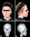
Apert syndrome patient, unoperated, with plump ventricles, and a pre-shunt at age 10 weeks. Clinical images. A Front view. B Side view. C Hand. D Foot. Radiological images. E Front view. F Side view.
Fig. 3
Fig. 3
Apert syndrome patient before midface surgery with clinical features of hypertelorism, downslanting palpebral fissures, AOB, and midface hypoplasia at 10 years of age. A Front view. B Side view.
Fig. 4
Fig. 4
Apert syndrome patient after Le Fort II/III osteotomies with zygomatic repositionings with Le Fort 2 segment distraction and dorsal bone graft. Ventriculoperitoneal shunting due to persistent symptomatic hydrocephalus; age 12 years. Clinical and radiological images, respectively. A, C Front view. B, D Side view.
Fig. 5
Fig. 5
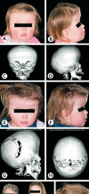
A, B Unoperated Crouzon patient with exorbitism, midface hypoplasia, Class III occlusion pattern at age 27 weeks. C, D CT images demonstrating patent sutures. E, F Older unoperated patient with developing clinical features at age 75 weeks. G, H CT images showing the development of pansynostosis. I, J After posterior vault expansion at 2 years of age.
Fig. 6
Fig. 6
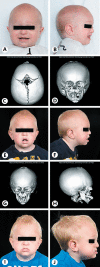
Pfeiffer syndrome. A-D Unoperated child with brachyturricephaly, exorbitism, midface hypoplasia, short nose, and broad thumbs and toes with deviation at age 54 days. E, F CT images showing pansynostosis. G, H Post-ventriculoperitoneal shunt and nasopharyngeal airway and globe subluxations (age 20 weeks). I, J CT images showing pansynostosis with ventriculoperitoneal shunt and turribrachycephaly. K, L Immediate post monobloc with rigid external distractor (RED) frame with resolution of exorbitism (age 46 weeks). M, N Post-monobloc CT images with RED frame in situ demonstrating osteotomies. O, P Changes after monobloc with resolution of exorbitism threatening ocular surface protection and improvement in midface hypoplasia (age 79 weeks). Q, R CT images showing post-monobloc bone consolidation and improvement in position. S, T After posterior vault expansion (PVE) due to signs of increased intracranial pressure, not decannulated because of multi-level airway obstruction and tracheal sleeve (age 4 years).
Fig. 7
Fig. 7
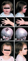
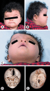
Muenke syndrome. A-D Phenotype with right unicoronal synostosis demonstrating orbital and facial scoliosis (age 83 weeks). E, F Skull reconstruction image showing right unicoronal synostosis. G, H After fronto-orbital remodelling (age 2 years).
Fig. 8
Fig. 8
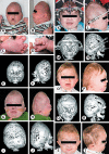
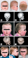
Saethre-Chotzen syndrome. A, B Phenotypic features including brachycephaly and a tall flat forehead (age 75 weeks). C, D Bicoronal synostosis and typical orbital morphology. E, F After PVE with springs (age 83 weeks). G, H CT images showing PVE with springs demonstrating the expansion achieved. I Three generations of Saethre-Chotzen syndrome phenotype with a strong family history. J After fronto-orbital remodelling completing a 2-stage reconstruction (age 2.5 years).
Fig. 9
Fig. 9
Craniofrontonasal dysplasia. A-C Clinical images showing left unicoronal synostosis, asymmetric hypertelorism, and a broad nasal bridge and tip (age 65 weeks). D, E CT images showing left unicoronal synostosis, asymmetric hypertelorism, and an open metopic suture typical for craniofrontonasal dysplasia.
Fig. 10
Fig. 10
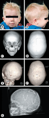
A child with a TCF12 mutation (c.1916del; p.Thr639Lysfs*16). A-C Brachycephaly with a tall, broad forehead. D, E CT images showing bicoronal synostosis. F-I Early post-PVE and CT images of PVE with springs demonstrating the expansion achieved. J, K Late after PVE. L Fronto-orbital remodelling results showing an acceptable appearance and function (age 3.5 years).
Fig. 11
Fig. 11
A boy with an ERF mutation (mutation c.247C>T; p.Arg83Trp) at 2 years of age. A, B Mild hypertelorism, mild scaphocephaly. The clinical features are within normal range. C, D Patent sutures. E, F Late closure of sagittal and bilambdoid sutures associated with increased intracranial pressure (ICP). G After PVE for raised ICP.

References

    1. Abu-Sittah GS, Jeelani O, Dunaway D, Hayward R. Raised intracranial pressure in Crouzon syndrome: incidence, causes, and management. J Neurosurg Pediatr. 2016;17:469–475. - PubMed
    1. Agochukwu NB, Solomon BD, Muenke M. Impact of genetics on the diagnosis and clinical management of syndromic craniosynostoses. Childs Nerv Syst. 2012a;28:1447–1463. - PMC - PubMed
    1. Agochukwu NB, Solomon BD, Gropman AL, Muenke M. Epilepsy in Muenke syndrome: FGFR3-related craniosynostosis. Pediatr Neurol. 2012b;47:355–361. - PMC - PubMed
    1. Ahmad F, Cobb AR, Mills C, Jones BM, Hayward RD, Dunaway DJ. Frontofacial monobloc distraction in the very young: a review of 12 consecutive cases. Plast Reconstr Surg. 2012;129:488e–497e. - PubMed
    1. Anderson PJ, Hall C, Evans RD, Harkness WJ, Hayward RD, Jones BM. The cervical spine in Crouzon syndrome. Spine (Phila Pa 1976) 1997a;22:402–405. - PubMed