Review
. 2019 Mar 1;3(4):456-470.
doi: 10.1002/hep4.1331. eCollection 2019 Apr.

Role of the Gut-Liver Axis in Liver Inflammation, Fibrosis, and Cancer: A Special Focus on the Gut Microbiota Relationship

Affiliations
Review

Role of the Gut-Liver Axis in Liver Inflammation, Fibrosis, and Cancer: A Special Focus on the Gut Microbiota Relationship

Naoko Ohtani et al. Hepatol Commun. .

Abstract

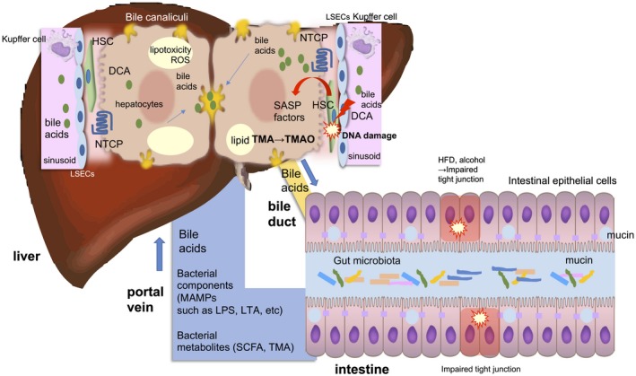
The gut and the liver are anatomically and physiologically connected, and this "gut-liver axis" exerts various influences on liver pathology. The gut microbiota consists of various microorganisms that normally coexist in the human gut and have a role of maintaining the homeostasis of the host. However, once homeostasis is disturbed, metabolites and components derived from the gut microbiota translocate to the liver and induce pathologic effects in the liver. In this review, we introduce and discuss the mechanisms of liver inflammation, fibrosis, and cancer that are influenced by gut microbial components and metabolites; we include recent advances in molecular-based therapeutics and novel mechanistic findings associated with the gut-liver axis and gut microbiota.


Figures

Figure 1
Figure 1
The gut–liver axis. The intestinal tract and the liver are anatomically and physiologically connected. This relationship between the intestine and liver has been called the “gut–liver axis.” Impaired tight junction results in the breakage of the gut barrier function and renders large amounts of MAMPs and bacterial metabolites or even the gut microbiota itself susceptible to transfer to the liver. BAs are actively absorbed by the BA transporter in terminal ileum and enter the colon epithelium through passive diffusion. Secondary BAs, such as DCA, are known to be toxic and elicit DNA damage and thereby producing SASP factors in the HSCs. The gut microbiota is also involved in choline metabolism by converting it into choline metabolites, such as TMA. It is transferred to liver and converted into TMAO, which causes liver inflammation and damage. Abbreviation: SCFA, short‐chain fatty acid.
Figure 2
Figure 2
Pathways of HSC activation and their fate in the resolution stage. In intact liver, HSCs localize in the Disse space and contain lipid droplets consisting of mainly vitamin A. They show a nonproliferative phenotype and express quiescent markers, such as LRAT and Lhx2. HSCs take part in the regulation of sinusoidal microcirculation through their contractility. When liver injury occurs by, e.g., HBV/HCV infection, alcohol abuse, or obesity (NASH), HSCs are exposed to oxidative stress signals (reactive oxygen intermediates), DAMPs, PAMPs, LPS, and paracrine stimuli, including cytokines, chemokines, and growth factors (PDGF, VEGF, TGFβ1, FGF2, CTGF, ANG II) secreted from neighboring cells, such as hepatic Kupffer cells/bone marrow‐derived macrophages, sinusoidal endothelial cells, hepatocytes, and platelets, and undergo the process of “activation.” Activated HSCs exhibit a number of specific phenotypes, including proliferation, contractility (mediated by ET‐1, ANG‐II, NO/CO, and ECM production), altered matrix degradation, chemotaxis, immune modulation, inflammatory signaling, and contribution to the cancer microenvironment. In the resolution stage of the underling liver disease, activated HSCs undergo apoptosis through Fas (CD95), TNFR1, p75NTR, and TRAIL; senescence showing p16, p21, γH2AX, and SASP; and inactivation exhibiting low TGFβ and collagen production and high expression of PPARγ and CYGB. Restorative macrophages take part in fibrolysis basically by producing MMP‐9, MMP‐12, and MMP‐13. Abbreviations: ANG‐II, angiotensin II; CTGF, connective tissue growth factor; ET‐1, endothelin 1; γH2AX, gamma‐histone family member X; HBV, hepatitis B virus; HCV, hepatitis C virus; Lhx2, LIM homeobox 2; LRAT, lecithin:retinol acyltransferase; MT‐1‐MMP, membrane‐type matrix metalloproteinase 1; N‐CAM, neural cell adhesion molecule; p75NTR, p75neurotrophin receptor; TIMP‐1, tissue inhibitor of metalloproteinase 1; TNFR1, tumor necrosis factor receptor 1; VEGF, vascular endothelial growth factor.
Figure 3
Figure 3
The role of the gut microbiota in liver cancer. In the leaky gut situation, a large amount of gut microbial components and metabolites are transferred to the liver and are likely to promote liver cancer progression. HCCs in the cirrhotic liver were associated with continuous exposure of LPS from gram‐negative bacteria of the gut microbiota. The development of NASH/NAFLD is also associated with LPS exposure. In addition, LTA from gram‐positive bacteria of the gut microbiota is likely to be involved in noncirrhotic NASH‐associated HCC. The HSCs in this type of HCC tumor region undergo cellular senescence and SASP. These HSCs do not appear to be proliferating or producing collagens.
Figure 4
Figure 4
BA‐mediated signaling pathways and related therapeutic drugs for NASH. In the leaky gut situation, a large amount of BAs as well as MAMPs, such as LPS and LTA, transfer to the liver through the portal vein. FXR agonists reduce the expression of CYP7A1, a rate limiting enzyme for BA synthesis, and NTCP, one of the key transporters in hepatocytes that uptake BAs from the sinusoids but up‐regulate the expression of BSEP, the major transporter for the excretion of BAs from hepatocytes into bile canaliculi. FXR activation reduces the excess BA pool in the hepatocyte, thereby preventing cholestasis and/or accumulation of toxic secondary BAs, such as DCA (drawn in the hepatocyte on the left). The secondary BA, DCA, the level of which is known to be increased in individuals with NASH, provokes DNA damage as well as stress response signals, such as JNK and p38‐mediated signaling pathways. Lipotoxicity derived from lipid storage in hepatocytes in NASH also activate these signals. ASK‐1 inhibitors suppress these stress response signals and improve NASH‐associated inflammation (drawn in the hepatocyte on the right). Abbreviation: BSEP, bile salt export pump.

References

    1. Tripathi A, Debelius J, Brenner DA, Karin M, Loomba R, Schnabl B, et al. The gut‐liver axis and the intersection with the microbiome. Nat Rev Gastroenterol Hepatol 2018;15:397‐411. - PMC - PubMed
    1. Yu LX, Schwabe RF. The gut microbiome and liver cancer: mechanisms and clinical translation. Nat Rev Gastroenterol Hepatol 2017;14:527‐539. - PMC - PubMed
    1. Seo YS, Shah VH. The role of gut‐liver axis in the pathogenesis of liver cirrhosis and portal hypertension. Clin Mol Hepatol 2012;18:337‐346. - PMC - PubMed
    1. Wiest R, Albillos A, Trauner M, Bajaj JS, Jalan R. Targeting the gut‐liver axis in liver disease. J Hepatol 2017;67:1084‐1103. Erratum. J Hepatol 2018;68:1336. - PubMed
    1. Kasubuchi M, Hasegawa S, Hiramatsu T, Ichimura A, Kimura I. Dietary gut microbial metabolites, short‐chain fatty acids, and host metabolic regulation. Nutrients 2015;7:2839‐2849. - PMC - PubMed