. 2019 Jul 1;29(7):530-542.
doi: 10.1093/glycob/cwz029.

Characterizing the selectivity of ER α-glucosidase inhibitors

Affiliations

Characterizing the selectivity of ER α-glucosidase inhibitors

Sarah O'Keefe et al. Glycobiology. .

Erratum in

Abstract

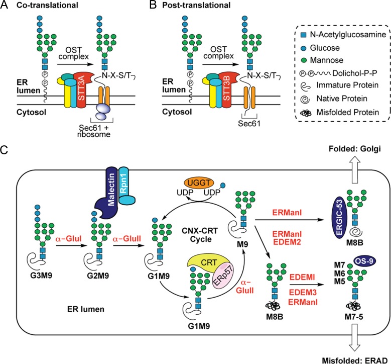
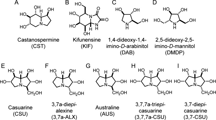
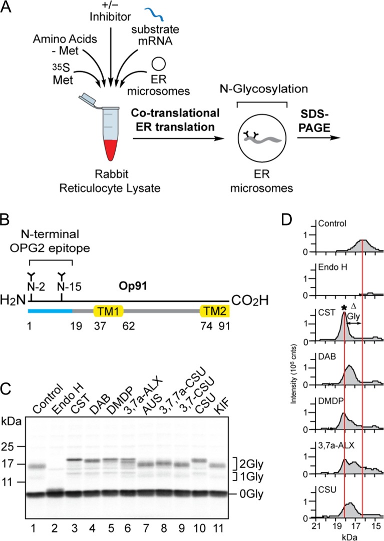
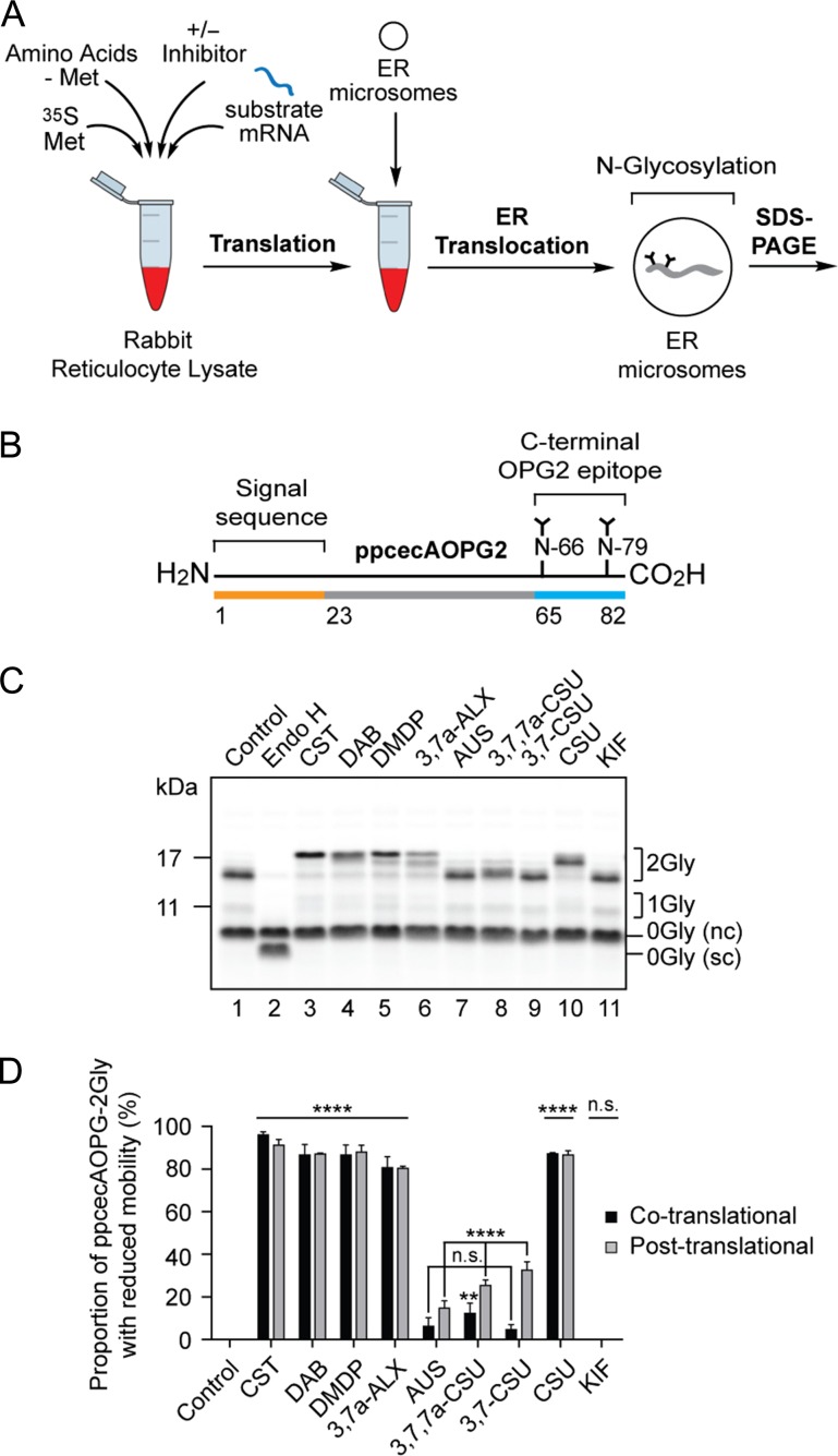
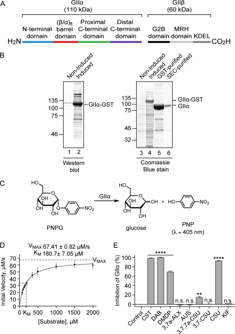
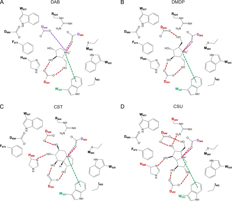
The endoplasmic reticulum (ER) contains both α-glucosidases and α-mannosidases which process the N-linked oligosaccharides of newly synthesized glycoproteins and thereby facilitate polypeptide folding and glycoprotein quality control. By acting as structural mimetics, iminosugars can selectively inhibit these ER localized α-glycosidases, preventing N-glycan trimming and providing a molecular basis for their therapeutic applications. In this study, we investigate the effects of a panel of nine iminosugars on the actions of ER luminal α-glucosidase I and α-glucosidase II. Using ER microsomes to recapitulate authentic protein N-glycosylation and oligosaccharide processing, we identify five iminosugars that selectively inhibit N-glycan trimming. Comparison of their inhibitory activities in ER microsomes against their effects on purified ER α-glucosidase II, suggests that 3,7a-diepi-alexine acts as a selective inhibitor of ER α-glucosidase I. The other active iminosugars all inhibit α-glucosidase II and, having identified 1,4-dideoxy-1,4-imino-D-arabinitol (DAB) as the most effective of these compounds, we use in silico modeling to understand the molecular basis for this enhanced activity. Taken together, our work identifies the C-3 substituted pyrrolizidines casuarine and 3,7a-diepi-alexine as promising "second-generation" iminosugar inhibitors.

Keywords: N-linked glycosylation; endoplasmic reticulum; glucose trimming; iminosugar inhibitors.


Figures

Fig. 1.
Fig. 1.
N-linked glycosylation in the ER. The multimeric oligosaccharyltransferase (OST) complex facilitates transfer of a lipid-linked glycan chain (G3M9GlcNAc2-) to suitable asparagine residues (N-X-S/T) of newly synthesized polypeptides via the STT3A/B catalytic subunit. (A) In the co-translational pathway, preassembled glycan chains are covalently attached to the growing nascent chain (via STT3A) as it emerges from the luminal side of the ER translocon (cf. Wild et al. 2018). (B) In the strictly post-translational pathway completed precursor proteins transit the Sec61 translocon and are N-glycosylated during or after complete translocation into the ER lumen (via STT3B). Once attached to the polypeptide chain, N-glycans (G3M9) undergo a series of trimming reactions in the ER lumen (C) catalyzed by α-glycosidase enzymes which sequentially remove glucose and mannose residues. In the first instance, cleavage of the terminal α-1,2-glucose residue by α-glucosidase I (α-Glu I) liberates a di-glucosylated N-glycan (G2M9) which associates with a membrane-bound lectin called malectin whilst the polypeptide associates with the OST subunit ribophorin I (Rpn1) (Qin et al. 2012). Following malectin-association, α-glucosidase II (α-Glu II) sequentially removes the two inner α-1,3-glucose residues. After the first cleavage by α-Glu II, resultant mono-glucosylated N-glycans (G1M9) are recognized by the ER luminal chaperones calreticulin (CRT) and calnexin (CNX) (not shown for simplicity), each in complex with the co-chaperone ERp57 (Oliver et al. 1999) which helps prevent aggregation and aids in polypeptide folding. The second cleavage by α-Glu II removes the innermost glucose residue generating an N-glycan comprised only of mannose residues (M9). Removal of this final glucose residue precludes further N-glycan-mediated binding to the CNX/CRT complexes but selective re-glucosylation by UDP-Glc:glycoprotein glucosyltransferase (UGGT) regenerates a G1M9 glycoform capable of rebinding CNX/CRT. Whilst exit from the CNX-CRT cycle is not fully understood, ER α-mannosidase I (ER Man I) removes a mannose residue (M8B) and, if the polypeptide has reached its native conformation, glycoproteins bearing M8B N-glycans are exported from the ER (which may be assisted by the lectin ERGIC-53) and progress through the secretory pathway. Terminally misfolded proteins, however, remain in the ER and are processed further by ER Man I and ER degradation-enhancing mannosidase-like proteins (EDEMs) (Słomińska-Wojewódzka and Sandvig 2015); i) ER Man I and/or EDEM2 recognize terminally misfolded proteins and trim a mannose residue to yield a M8B glycoform, ii) further mannose trimming of M8B by EDEMI together with EDEM3 and/or ER Man I generates M7, M6 and M5 N-glycans. Glycoproteins bearing these extensively trimmed N-glycans (assisted by the lectin OS9) are then targeted for ER-associated degradation (ERAD) (Vembar and Brodsky 2008). Monosaccharide symbols follow the SNFG (Symbol Nomenclature for Glycans) system (Varki et al. 2015).
Fig. 2.
Fig. 2.
Structures of iminosugars evaluated as inhibitors of ER glycoprotein processing. The commercially available indolizidines (A) castanospermine (CST) and (B) kifunensine (KIF), and the pyrrolidines (C) 1,4-dideoxy-1,4-imino-D-arabinitol (DAB) and (D) 2,5-dideoxy-2,5-imino-D-mannitol (DMDP) provided well defined control inhibitors of ER α-glycosidases (see main text). These compounds were compared against the effects of a subset of pyrrolizidines bearing a hydroxymethyl substituent at the C-3 position; (E) casuarine (CSU), (F) 3,7a-diepi-alexine (3,7a-ALX), (G) australine (AUS) and the synthetic analogs (H) 3,7,7a-triepi-casuarine (3,7,7a-CSU) and (I) 3,7-diepi-casuarine (3,7-CSU).
Fig. 3.
Fig. 3.
A subset of compounds alter N-glycan processing of the model glycoprotein Op91. (A) A schematic of the in vitro assay for N-glycan trimming; radiolabelled precursor proteins synthesized in the presence of ER microsomes undergo co-translational translocation, N-glycosylation and ER dependent N-glycan trimming events which can be studied by recovering the ER membrane fraction and analyzing the radiolabelled products by SDS-PAGE and phosphorimaging. (B) Op91 is an N-terminal fragment of bovine rhodopsin that includes the first transmembrane domain (TM1), part of the second transmembrane domain (TM2) and two endogenous sites for N-glycosylation (N2 and N15) that is efficiently inserted into ER microsomes (Crawshaw et al. 2004). (C) The effects of nine compounds (cf. Figure 2), each at 5 mM, on the processing of the N-glycans attached to Op91 during its synthesis (co-translationally) were assessed via a gel shift assay. Reduced migration of the major N-glycosylated species (2Gly) when compared to the non-inhibitor control (lane 1) was used to assess changes in N-glycan trimming. Treatment with Endoglycosidase H (Endo H) confirmed the identity of the N-glycosylated Op91 products (lane 2). (D) Gel shifts present in C were analyzed using AIDA software with peaks corresponding to the migration and signal intensity of bands. Migration profiles of the doubly N-glycosylated Op91 species generated in the presence of CST, DAB, DMDP, 3,7a-ALX and CSU were aligned with the control (C, lanes 3, 4, 5, 6 and 10 versus lane 1). Alterations in N-glycan trimming (ΔGly) as judged by changes in glycoprotein mobility are depicted between the center of the control peak and the center of the peak generated in the presence of CST which was benchmarked as the G3M9 N-glycan form and denoted by an asterisk (*).
Fig. 4.
Fig. 4.
Inhibition of N-glycan trimming with a post-translationally modified glycoprotein substrate. (A) The radiolabelled precursor protein was synthesized as before (cf. Figure 3), but in the absence of ER microsomes. Following puromycin-induced termination of protein synthesis, completed polypeptides were incubated with ER microsomes ensuring ER translocation and subsequent N-glycosylation were strictly post-translational. (B) ppcecAOPG2 is a modified form of preprocecropin A containing residues 1 to 18 of bovine rhodopsin, with two sites for N-glycosylation (cf. Figure 3B), added to its C-terminus (Johnson et al. 2012). (C) The effect of nine compounds, each at 5 mM, on the processing of the N-glycans attached to ppcecAOPG2 post-synthesis was assessed as described in the legend to Figure 3. nc, non-cleaved signal sequence form of ppcecAOPG2; sc, signal cleaved form. (D) The efficiency of inhibition of N-glycan trimming in the post-translational system (light gray bars) was estimated by quantifying the signal intensity of the distinct 2Gly species with reduced mobility, as compared to the control sample, that were observed in the presence of tested compounds (e.g., Figure 4C, cf. lanes 1 and 3). The proportion of 2Gly species with reduced mobility was then expressed as percentage of the total signal for all 2Gly species. Similar calculations were performed for ppcecAOPG2-2Gly synthesized in the presence of ER microsomes where the co-translational pathway is also available (see Supplementary Figure S1Biv), and these results are included for comparison (black bars). In each case the experiments were performed in triplicate (n = 3). Statistical significance of compound-induced inhibition relative to the control (one-way ANOVA) was determined using Tukey’s multiple comparisons test and shown in the figure. Statistical significance comparing the levels of compound-induced inhibition between co- and post-translational mechanisms of translocation (two-way ANOVA) was determined using Sidak’s multiple comparisons test and are as follows: n.s. CST, DAB, DMDP, 3,7a-ALX, CSU; **AUS; **** 3,7,7a-CSU, 3,7-CSU. Statistical significance is given as n.s., non-significant; **, P < 0.01 and ****, P < 0.0001.
Figure 5.
Figure 5.
The effect of compounds on α-glucosidase II enzyme activity. (A) ER α-Glu II is a heterodimeric enzyme consisting of the catalytic GIIα subunit (110 kDa) and a non-catalytic regulatory GIIβ subunit (60 kDa). The active site of GIIα is located in its (β/α)8 barrel domain (Caputo et al. 2016; Satoh et al. 2016). The GIIβ subunit contains an N-terminal GIIα-binding domain (G2B), a mannose-6-phosphate receptor homology (MRH) domain involved in N-glycan recognition and a KDEL ER-retention signal (Olson et al. 2013; Caputo et al. 2016). (B) A C-terminally GST-tagged version of Chaetomium thermophilum GIIα lacking the signal sequence (Satoh et al. 2016) was expressed in E. coli (western blot using anti-GST, lanes 1–2; Coomassie Blue staining, lanes 3–4; see GIIα-GST). Following cell lysis, recombinant protein was purified using a Glutathione-Sepharose Column and GIIα released by on-column cleavage with tobacco etch virus (TEV) protease (lane 5), followed by size exclusion chromatography (SEC) (lane 6). (C) A schematic depicting the calorimetric reaction used to measure GIIα inhibition. GIIα-catalyzed cleavage of p-nitrophenyl-α-D-glucopyranoside (PNPG) produces glucose and yellow p-nitrophenol (PNP). The amount of PNP liberated during the course of the reaction was monitored by absorbance measurements. (D) Different concentrations of PNPG (ranging from 75 μM to 2 mM) were incubated with GIIα (6 μg/mL) at 37°C and absorbance measurements (λ = 410 nm, 1 min intervals, 90 min) used to generate a substrate-velocity curve. Using the Michaelis-Menten model, values for VMAX (67.41 ± 0.82 μM/s) and KM (180.7 ± 7.05 μM) were estimated (n = 12, R2 0.9534). (E) Compounds at 100 μM were incubated with 125 μM PNPG and 6 μg/mL GIIα at 37°C and absorbance measurements (λ = 410 nm, 1 min intervals, 90 min) used to calculate the % inhibition of GIIα relative to control reactions. Assays were performed in triplicate (n = 3) and statistical significance (one-way ANOVA) determined using Tukey’s multiple comparisons test. Statistical significance is given as n.s., non-significant; **, P < 0.01 and ****, P < 0.0001.
Figure 6.
Figure 6.
Inhibitors of ER α-Glu II exhibit similar binding modes but form different bonding interactions with GIIα when docked in the substrate binding site. (A) DAB, (B) DMDP, (C) CST and (D) CSU share common hydrogen bonding interactions (red dashed line) with D451 and D564, cation-π interactions (green dashed line) with W423 and an ionic interaction (purple dashed line) with D564. DAB may form a second ionic interaction with D640 not present in the other three compounds whose variable bonding network involves additional hydrogen bonding interactions with residues D460 and/or H698 and/or R624.

Similar articles

Cited by

References

    1. Alonzi DS, Scott KA, Dwek RA, Zitzmann N. 2017. Iminosugar antivirals: the therapeutic sweet spot. Biochem Soc Trans. 45:571–582. - PMC - PubMed
    1. Araki K, Nagata K. 2011. Protein folding and quality control in the ER. Cold Spring Harb Perspect Biol. 4:a015438. - PMC - PubMed
    1. Asano N. 2008. Glycosidase-inhibiting alkaloids: isolation, structure and application In: Fattorusso E, Taglialatela-Scafati O, editors. Modern Alkaloids: Structure, Isolation, Synthesis and Biology. Weinheim: Wiley-VCH Verlag; p. 111–138.
    1. Asano N, Nash RJ, Molyneux RJ, Fleet GWJ. 2000. Sugar-mimic glycosidase inhibitors: natural occurrence, biological activity and prospects for therapeutic application. Tetrahedron: Asymmetry. 11:1645–1680.
    1. Asano N, Oseki K, Kizu H, Matsui K. 1994. Nitrogen-in-the-ring pyranoses and furanoses: structural basis of inhibition of mammalian glycosidases. J Med Chem. 37:3701–3706. - PubMed

Publication types