. 2019 Sep;76(18):3657-3665.
doi: 10.1007/s00018-019-03090-9. Epub 2019 Apr 11.

Mechanisms of neurodegeneration in a preclinical autosomal dominant retinitis pigmentosa knock-in model with a RhoD190N mutation

Affiliations

Mechanisms of neurodegeneration in a preclinical autosomal dominant retinitis pigmentosa knock-in model with a RhoD190N mutation

Javier Sancho-Pelluz et al. Cell Mol Life Sci. 2019 Sep.

Abstract

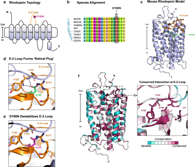
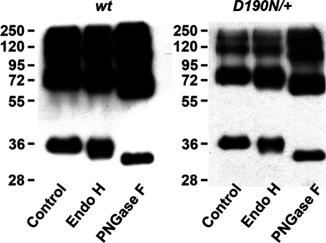
D190N, a missense mutation in rhodopsin, causes photoreceptor degeneration in patients with autosomal dominant retinitis pigmentosa (adRP). Two competing hypotheses have been developed to explain why D190N rod photoreceptors degenerate: (a) defective rhodopsin trafficking prevents proteins from correctly exiting the endoplasmic reticulum, leading to their accumulation, with deleterious effects or (b) elevated mutant rhodopsin expression and unabated signaling causes excitotoxicity. A knock-in D190N mouse model was engineered to delineate the mechanism of pathogenesis. Wild type (wt) and mutant rhodopsin appeared correctly localized in rod outer segments of D190N heterozygotes. Moreover, the rhodopsin glycosylation state in the mutants appeared similar to that in wt mice. Thus, it seems plausible that the injurious effect of the heterozygous mutation is not related to mistrafficking of the protein, but rather from constitutive rhodopsin activity and a greater propensity for chromophore isomerization even in the absence of light.

Keywords: D190N; Excitotoxicity; GPCR; Mouse model; Retina; Retinitis pigmentosa; Rhodopsin.


Figures

Fig. 1
Fig. 1
Mechanism of rhodopsin D190N mutation. a Membrane topological structure of rhodopsin. The E-2 loop is located on the extracellular side of the protein between helices TM4 and TM5. b Multiple sequence alignment highlighting the location and conservation of the D190N mutation. This residue is conserved multiple species. c Homology model of mouse rhodopsin based off the bovine rhodopsin crystal structure (PDB: 1U19). d Ion pair between D190 and R177 that stabilizes the E-2 loop. e D190N mutation disrupts the ion pair formed by D190/R177. f Conservation analysis in ConSurf reveals that R177/D190 ion pair is 100% conserved in 150 opsin sequences
Fig. 2
Fig. 2
Photoreceptor degeneration in retinas of control and D190N animals. a Hematoxylin–eosin-stained paraffin sections of wild type (wt, at P30), heterozygous (D190N/+, at P30), and homozygous (D190N/D190N) mice at P12, 21, 30, and 210. At P12, D190N/D190N mice have 5–6 rows of photoreceptors. By P21, only 2 rows worth of cells remained in the ONL. At P210, only a single row of photoreceptors was observed. b Anti-rhodopsin-stained cryosections (red) showed that in D190N/+ mice, rhodopsin appears to localize correctly in the OS of the retina. In the D190N homozygotes, rhodopsin appears mislocalized within the photoreceptor nuclei, and its expression continues to decline with time. c Anti-Glyphos antibody (green) was used to stain for cones. At P12, cones resembled that of wt mice, although the IS/OS were not completely formed. The INL and the outer plexiform layer are partially stained. At P21, staining was decreased, although some remained in the ONL. At P210, only a few cells stained positively for glyphos in the ONL. Outer segments, OS; inner segments, IS; outer nuclear layer, ONL; inner nuclear layer, INL; ganglion cell layer, GCL. DAPI (blue) was used to counterstain nuclei. Scale bar: 50 μm. n = 3 for each genotype and each time point
Fig. 3
Fig. 3
D190N/+ mice show relatively normal outer segment discs compared to D190N/D190N mice, which degenerate sooner. OS of wt, D190N/+, and D190N/D190N were observed with electron microscopy. Compared to wt (a), shortened OS were observed in D190N/+ mice at P21 (b) and in D190N/D190N mice at P12, for which the OS and IS were indistinguishable (c). D190N/+ OS were not only shortened, but also presented an irregular morphology (e) compared to wt (d). Photoreceptors undergoing apoptosis were observed in D190N/D190N at P12 (f). Regular elongated mitochondria in wt animals (arrows in g) were observed, while swollen mitochondria were seen in D190N/+ (arrows in h) at P21. Accumulation of dysmorphic mitochondria was observed also in the D190/D190N background (arrows in i), and disrupted cilia epithelium was observed (arrowheads in i). RPE retinal pigment epithelium, ONL outer nuclear layer, OS outer segments, IS inner segments, N nucleus
Fig. 4
Fig. 4
Decreased visual function in homozygous D190N animals. a Scotopic dim light rod-specific ERG traces for a B6 control mouse (blue), a D190N/+ (orange) and a D190N/D190N mouse (red) at 3–4 weeks of age. Quantification of dim light scotopic a-waves did not show any difference among the groups, whereas the b-wave of the homozygous mice was significantly decreased when compared to the wt and the heterozygous mice (b). Scotopic maximum ERG traces were recorded. Heterozygous and wt a- and b-waves were similar, but the homozygous trace was significantly flatter. c Photopic cone-specific traces were recorded. Again, wt and heterozygous signals were similar, while the homozygous trace was significantly diminished. (Error bars show SEM for each time point. Significance was calculated using a ratio paired t test analysis. n ≥ 3 mice. *p < 0.05; **p < 0.01; ***p < 0.001)
Fig. 5
Fig. 5
Glycosylation state of rhodopsin. Retinas from wt and D190N/+ animals at P30 were collected and homogenized. After treating with Endo-H or PNGase, proteins were separated by electrophoresis. Blots from both wt and D190N/+ were found at the same level

References

    1. Boughman JA, Conneally PM, Nance WE. Population genetic studies of retinitis pigmentosa. Am J Hum Genet. 1980;32:223–235. - PMC - PubMed
    1. Berson EL. Retinitis pigmentosa: the friedenwald lecture. Invest Opthal Vis Sci. 1993;34:1655–1676. - PubMed
    1. Hartong DT, Berson EL, Dryja TP. Retinitis pigmentosa. Lancet. 2006;368:1795–1809. doi: 10.1016/S0140-6736(06)69740-7. - DOI - PubMed
    1. Rivolta C, Sharon D, De Angelis MM, Dryja TP. Retinitis pigmentosa and allied diseases: numerous diseases, genes, and inheritance patterns. Hum Mol Genet. 2002;11:1219–1227. doi: 10.1093/hmg/11.10.1219. - DOI - PubMed
    1. Wilson JH, Wensel TG. The nature of dominant mutations of rhodopsin and implications for gene therapy. Mol Neurobiol. 2003;28:149–158. doi: 10.1385/MN:28:2:149. - DOI - PubMed