. 2019 Apr 11;5(1):58.
doi: 10.1186/s40792-019-0621-x.

Laparoscopy-assisted extended right hepatectomy for giant hemorrhagic hepatic cyst mimicking biliary cystadenocarcinoma: a case report

Affiliations

Laparoscopy-assisted extended right hepatectomy for giant hemorrhagic hepatic cyst mimicking biliary cystadenocarcinoma: a case report

Yasunari Fukuda et al. Surg Case Rep. .

Abstract

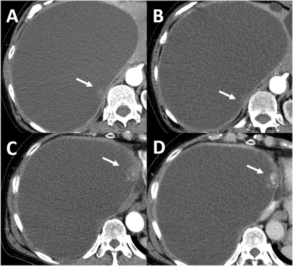
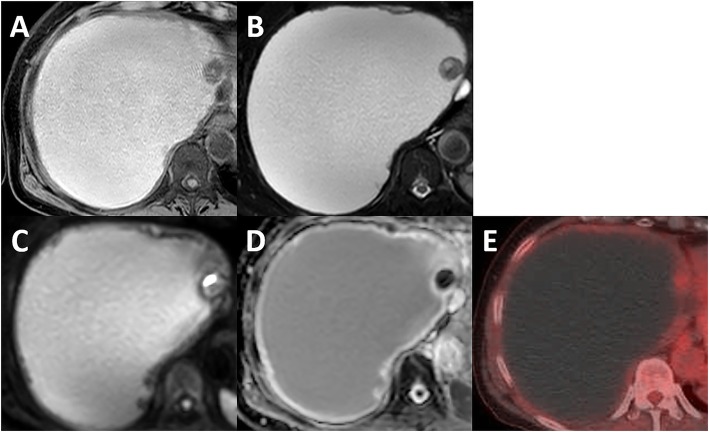
Background: Hemorrhagic hepatic cysts infrequently involve several iconographic changes requiring a differential diagnosis, primarily with a cystic malignancy. We herein report a case of laparoscopy-assisted extended right hepatectomy for a giant hemorrhagic hepatic cyst with an enhancing mural nodule that was clinically suspected of being biliary cystadenocarcinoma.

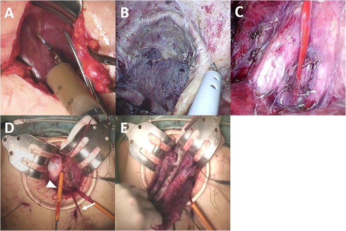
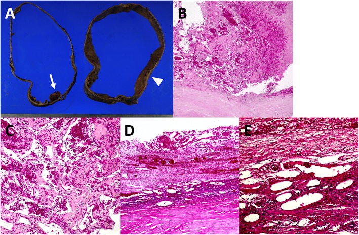
Case presentation: A 73-year-old woman was followed up for giant hepatic cyst occupying the right lobe of the liver. During the follow-up, an enhancing mural nodule was newly noted on computed tomography in 2016. Based on additional clinical examinations, biliary cystadenocarcinoma was undeniable, and laparoscopy-assisted extended right hepatectomy was performed for diagnostic and therapeutic purposes. She had no perioperative complications and was discharged on postoperative day 13. A histological examination of the mural nodule showed neovascularization within an organized hematoma.

Conclusion: We herein report a rare case of giant hemorrhagic hepatic cyst mimicking biliary cystadenocarcinoma that was successfully treated with laparoscopy-assisted extended right hepatectomy. Laparoscopic surgery in our case was an effective procedure performed with the utmost care.

Keywords: Hemorrhagic hepatic cyst; Laparoscopy-assisted surgery; Neovascularization; Organized hematoma.


Conflict of interest statement

Ethics approval and consent to participate

Not applicable.

Consent for publication

Written informed consent was obtained from the patient for publication of this case report and any accompanying images. A copy of the written consent document is available for review by the Editor-in-Chief of this journal.

Competing interests

The authors declare that they have no competing interests.

Publisher’s Note

Springer Nature remains neutral with regard to jurisdictional claims in published maps and institutional affiliations.

Figures

Fig. 1
Fig. 1
CT images of mural nodules in giant cyst. a Plain CT showed a dorsal unenhanced mural nodule within the cyst wall (arrow), b which did not change thereafter (arrow). c Enhanced CT showed a ventral mural nodule with weak enhancement in the early phase (arrow) and d with centripetal prolonged enhancement in delayed phase (arrow)
Fig. 2
Fig. 2
MRI and FDG-PET images of the ventral mural nodule. a MRI showed that the cyst content was a high-signal and the mural nodule was a low-signal on T1 W1. b MRI showed the cyst content was a high-signal and the mural nodule was partially high-signal on T2WI. c MRI showed that the mural nodule was partially strong high-signal on DWI, d with low ADC value (ADCmin 0.00 mm2/s, ADCmax 0.91 mm2/s, ADCmean 0.6 × 10−3 mm2/s). e FDG-PET showed an abnormal uptake in the mural nodule with SUV-max 2.3
Fig. 3
Fig. 3
Surgical procedures. a The cyst content with chocolate color was trans-hepatically aspirated from a 7-cm upper midline incision. b The adhesiotomy between the cyst wall and right diaphragm was laparoscopically performed without rupturing the cyst wall. c Preparation for a hanging maneuver was performed. d Parenchymal division was performed under direct vision through the small laparotomy wound using a hanging maneuver (arrow) and the Pringle’s maneuver (arrowhead). e The specimen was carefully excised from the mini-laparotomy
Fig. 4
Fig. 4
Macroscopic and microscopic findings of the resected specimen. a (left-hand): Macroscopically, a dark-red solid protuberance was found in the cyst wall (arrow). a (right-hand): Macroscopically, a thickened portion of the cyst wall where the dorsal mural nodule seemed to have originally existed was detected (arrowhead). b, c Hematoxylin and eosin staining showed that the cyst wall mainly consisted of thick fibrous stroma and the ventral mural nodule consisted of several sizes of blood-filled vascular cavities surrounded by endothelial cells within the organized hematoma (original magnification: b × 40, c × 100). d, e Hematoxylin and eosin staining showed that the thickened portion of the wall consisted of malformed veins of various sizes within the calcified hematoma (original magnification: d × 40, e × 200)
Fig. 5
Fig. 5
Immunohistochemical staining. Immunohistochemical staining of ventral mural nodule was a positive for CD31 (original magnification × 40) and b negative for αSMA and c MDM2 (original magnification × 200). d The Ki-67 labeling index was 19.0% (original magnification × 200). e Immunohistochemical staining of the thickened portion of the cyst wall was positive for αSMA (original magnification × 200)

References

    1. Hagiwara A, Inoue Y, Shutoh T, Kinoshita H, Wakasa K. Haemorrhagic hepatic cyst: a differential diagnosis of cystic tumour. Br J Radiol. 2001;74:270–272. doi: 10.1259/bjr.74.879.740270. - DOI - PubMed
    1. Kitajima Y, Okayama Y, Hirai M, Hayashi K, Imai H, Okamoto T, et al. Intracystic hemorrhage of a simple liver cyst mimicking a biliary cystadenocarcinoma. J Gastroenterol. 2003;38:190–193. doi: 10.1007/s005350300032. - DOI - PubMed
    1. Zhang YL, Yuan L, Shen F, Wang Y. Hemorrhagic hepatic cysts mimicking biliary cystadenoma. World J Gastroenterol. 2009;15:4601–4603. doi: 10.3748/wjg.15.4601. - DOI - PMC - PubMed
    1. Tanaka T, Gobara H, Tomita K, Hiraki T, Tanaka T, Kanazawa S. Hepatic intracystic organizing hematoma mimicking biliary cystadenocarcinoma in a patient with polycystic liver disease. Intern Med. 2015;54:2001–2005. doi: 10.2169/internalmedicine.54.4218. - DOI - PubMed
    1. Kohno Shigeshi, Arizono Shigeki, Isoda Hiroyoshi, Yoshizawa Akihiko, Togashi Kaori. Imaging findings of hemorrhagic hepatic cysts with enhancing mural nodules. Abdominal Radiology. 2019;44(4):1205–1212. doi: 10.1007/s00261-019-01898-4. - DOI - PubMed

LinkOut - more resources