. 2019 Oct;35(5):815-825.
doi: 10.1007/s12264-019-00371-y. Epub 2019 Apr 11.

MicroRNA-365 Knockdown Prevents Ischemic Neuronal Injury by Activating Oxidation Resistance 1-Mediated Antioxidant Signals

Affiliations

MicroRNA-365 Knockdown Prevents Ischemic Neuronal Injury by Activating Oxidation Resistance 1-Mediated Antioxidant Signals

Jia-Lin Mo et al. Neurosci Bull. 2019 Oct.

Abstract

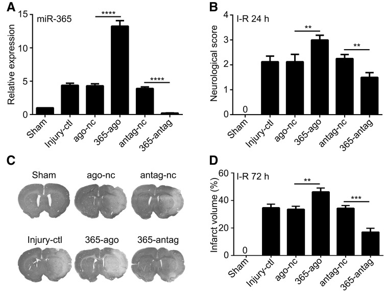
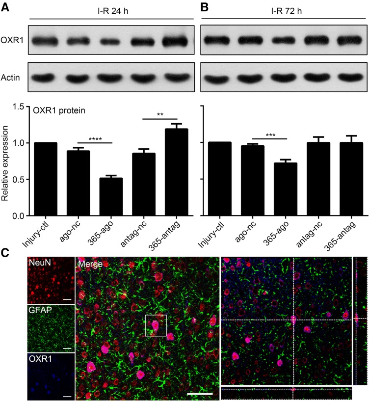
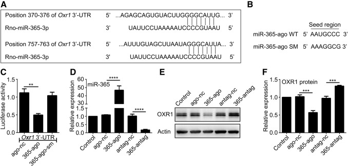
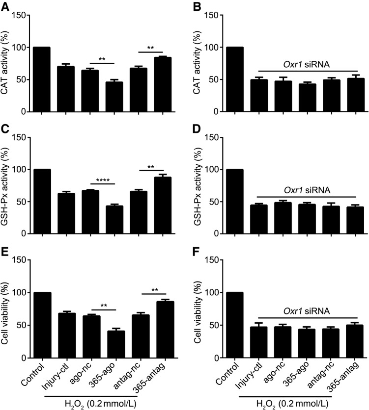
MicroRNA-365 (miR-365) is upregulated in the ischemic brain and is involved in oxidative damage in the diabetic rat. However, it is unclear whether miR-365 regulates oxidative stress (OS)-mediated neuronal damage after ischemia. Here, we used a transient middle cerebral artery occlusion model in rats and the hydrogen peroxide-induced OS model in primary cultured neurons to assess the roles of miR-365 in neuronal damage. We found that miR-365 exacerbated ischemic brain injury and OS-induced neuronal damage and was associated with a reduced expression of OXR1 (Oxidation Resistance 1). In contrast, miR-365 antagomir alleviated both the brain injury and OXR1 reduction. Luciferase assays indicated that miR-365 inhibited OXR1 expression by directly targeting the 3'-untranslated region of Oxr1. Furthermore, knockdown of OXR1 abolished the neuroprotective and antioxidant effects of the miR-365 antagomir. Our results suggest that miR-365 upregulation increases oxidative injury by inhibiting OXR1 expression, while its downregulation protects neurons from oxidative death by enhancing OXR1-mediated antioxidant signals.

Keywords: Ischemic stroke; MicroRNA; Neuronal damage; Neuroprotection; Oxidative stress.


Conflict of interest statement

The authors claim that there are no conflicts of interest.

Figures

Fig. 1
Fig. 1
MiR-365 antagomir reduces brain injury after ischemic stroke. A qRT-PCR analysis of miR-365 expression in the ischemic striatum 24 h after injection of miR-365 agomir (365-ago), agomir negative control (ago-nc), miR-365 antagomir (365-antag), or antagomir negative control (antag-nc) (n = 3). Injury control rats (Injury-ctl) were subjected to ischemia-reperfusion (I-R) without other interventions. B Neurological scores according to Longa’s method 24 h after I-R (n = 3 in the sham group; n = 8 in the other groups). C Representative images of cresyl violet stained brain sections from rats 3 days after I-R (white indicates infarct area). D Infarct volume as a percentage of contralateral hemisphere volume (n = 3 in the sham group; n = 6 in the other groups). **P < 0.01, ***P < 0.001, ****P < 0.0001, one-way ANOVA with Tukey’s post-hoc test. Data are presented as the mean ± SEM.
Fig. 2
Fig. 2
MiR-365 antagomir reduces γH2AX expression in the ischemic striatum. A Representative images of immunolabeling of γH2AX in sections from rats 3 days after I-R (scale bars, 100 μm). B Schematic showing the areas in which immunoreactive cells were counted (five fields of view). C Numbers of γH2AX-positive cells (average of total positive cells/mm2 in the five fields; n = 3 in the sham group; n = 5 in the other groups). D Representative images of immunofluorescent triple labeling of NeuN, γH2AX, and GFAP in sections from rats 3 days after MCAO (scale bars, 50 μm).**P < 0.01, ***P < 0.001, one-way ANOVA with Tukey’s post-hoc test. Data are presented as the mean ± SEM.
Fig. 3
Fig. 3
OXR1 expression is reduced in the ischemia-induced striatum and primary neurons. A, B Expression levels of OXR1 protein and miR-365 in the ischemic striatum 24 and 72 h after I-R analyzed by Western blotting (WB) and qRT-PCR, respectively (n = 3 for qRT-PCR, n = 4 for WB). C, D Expression levels of OXR1 protein and miR-365 in primary neurons after 12, 24, and 36 h exposure to ischemia-like conditions analyzed by WB and qRT-PCR, respectively (n = 3). **P < 0.01, ***P < 0.001, ****P < 0.0001 versus sham or control, unpaired two-tailed Student’s t-test.
Fig. 4
Fig. 4
MiR-365 antagomir increases OXR1 expression in the ischemic striatum. A, B Expression levels of OXR1 protein in the ischemic striatum 24 and 72 h after injection of 365-ago, ago-nc, 365-antag, or antag-nc analyzed by WB (n = 4 in the injury-control groups; n = 5 in the other groups). C Representative images of immunofluorescent triple labeling of NeuN, OXR1, and GFAP in brain sections from rats 3 days after MCAO (scale bars, 50 μm).**P < 0.01, ***P < 0.001, ****P < 0.0001, one-way ANOVA with Tukey’s post-hoc test. Data are presented as the mean ± SEM.
Fig. 5
Fig. 5
MiR-365 directly targets OXR1 expression in primary neurons. A Nucleotide sequences of the predicted miR-365 binding sites in the 3′-UTR of Oxr1. Shown are the seed sequence (CCCGUAA) and wild-type miR-365 binding site (GGGCAUU). B The mutant seed sequence from mutant miR-365 agomir (miR-365-ago SM). Shown are the wild-type (CCCGUAA) and mutant (GCGGAAA) seed sequences. C Luciferase assays in PC12 cells 48 h after transfection of luciferase reporter plasmid containing the wild-type Oxr1 3′-UTR, together with ago-nc, 365-ago, or 365-ago-sm. Luciferase activity was calculated as the Renilla: firefly ratio (n = 4). D qRT-PCR analysis of miR-365 expression in primary neurons 48 h after transfection of 365-ago, ago-nc, 365-antag, or antag-nc (n = 3). E, F Expression levels of OXR1 protein in primary neurons 48 h after transfection of 365-ago, ago-nc, 365-antag, or antag-nc analyzed by Western blotting (n = 4). **P < 0.01, unpaired two-tailed Student’s t-test in C,***P < 0.001, ****P < 0.0001, one-way ANOVA with Tukey’s post-hoc test in D and F. Data are presented as the mean ± SEM.
Fig. 6
Fig. 6
Oxr1 siRNA abolishes the protective effect of miR-365 antagomir on primary neurons. A CAT activity in primary neurons after transfection with 365-ago, ago-nc, 365-antag, or antag-nc and exposure to H2O2 (n = 6). B CAT activity in primary neurons after co-transfection of 365-ago, ago-nc, 365-antag, or antag-nc with Oxr1 siRNA and exposure to H2O2 (n = 6). C GSH-Px activity in primary neurons after transfection with 365-ago, ago-nc, 365-antag, or antag-nc and exposure to H2O2 (n = 6). D GSH-Px activity in primary neurons after co-transfection of 365-ago, ago-nc, 365-antag, or antag-nc with Oxr1 siRNA and exposure to H2O2 (n = 6). E, F Cell viability of primary neurons after co-transfection of 365-ago, ago-nc, 365-antag, or antag-nc with (F) or without (E) Oxr1 siRNA and exposure to H2O2 (n = 5). **P < 0.01, ****P < 0.0001, one-way ANOVA with Tukey’s post-hoc test. Data are presented as the mean ± SEM.

References

    1. Lipton P. Ischemic cell death in brain neurons. Physiol Rev. 1999;79:1431–1568. doi: 10.1152/physrev.1999.79.4.1431. - DOI - PubMed
    1. Zhao H, Han Z, Ji X, Luo Y. Epigenetic regulation of oxidative stress in ischemic stroke. Aging Dis. 2016;7:295–306. doi: 10.14336/AD.2015.1009. - DOI - PMC - PubMed
    1. Li XJ, Zhang LM, Gu J, Zhang AZ, Sun FY. Melatonin decreases production of hydroxyl radical during cerebral ischemia-reperfusion. Acta Pharmacol Sin. 1997;18:394–396. - PubMed
    1. Davis SM, Pennypacker KR. Targeting antioxidant enzyme expression as a therapeutic strategy for ischemic stroke. Neurochem Int. 2017;107:23–32. doi: 10.1016/j.neuint.2016.12.007. - DOI - PMC - PubMed
    1. Ramos E, Patino P, Reiter RJ, Gil-Martin E, Marco-Contelles J, Parada E, et al. Ischemic brain injury: new insights on the protective role of melatonin. Free Radic Biol Med. 2017;104:32–53. doi: 10.1016/j.freeradbiomed.2017.01.005. - DOI - PubMed