Review
. 2019 May;289(1):158-172.
doi: 10.1111/imr.12743.

G-protein coupled receptors and ligands that organize humoral immune responses

Affiliations
Review

G-protein coupled receptors and ligands that organize humoral immune responses

Erick Lu et al. Immunol Rev. 2019 May.

Abstract

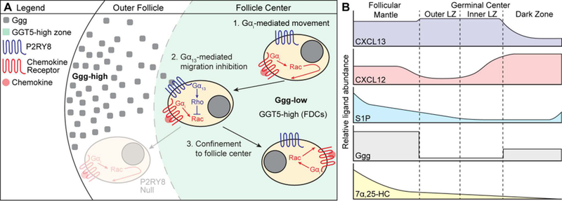
B-cell responses are dynamic processes that depend on multiple types of interactions. Rare antigen-specific B cells must encounter antigen and specialized systems are needed-unique to each lymphoid tissue type-to ensure this happens efficiently. Lymphoid tissue barrier cells act to ensure that pathogens, while being permitted entry for B-cell recognition, are blocked from replication or dissemination. T follicular helper (Tfh) cells often need to be primed by dendritic cells before supporting B-cell responses. For most responses, antigen-specific helper T cells and B cells need to interact, first to initiate clonal expansion and the plasmablast response, and later to support the germinal center (GC) response. Newly formed plasma cells need to travel to supportive niches. GC B cells must become confined to the follicle center, organize into dark and light zones, and interact with Tfh cells. Memory B cells need to be positioned for rapid responses following reinfection. Each of these events requires the actions of multiple G-protein coupled receptors (GPCRs) and their ligands, including chemokines and lipid mediators. This review will focus on the guidance cue code underlying B-cell immunity, with an emphasis on findings from our laboratory and on newer advances in related areas. We will discuss our recent identification of geranylgeranyl-glutathione as a ligand for P2RY8. Our goal is to provide the reader with a focused knowledge about the GPCRs guiding B-cell responses and how they might be therapeutic targets, while also providing examples of how multiple types of GPCRs can cooperate or act iteratively to control cell behavior.

Keywords: CXCR4; CXCR5; EBI2; P2RY8; S1PR2; chemoattractant; migration-inhibition.


Figures

Figure 1.
Figure 1.. In situ hybridization for CXCL13 defines B cell follicles in the spleen.
CXCL13 mRNA expression (left panel, purple signal) was identified in spleen sections using in situ hybridization. Shown is a single white pulp cord. CXCL13 is abundantly expressed by follicular stromal cells. The right panel shows a diagram corresponding to the splenic architecture of the section shown on the left. B cell follicles (B, purple) can be distinguished from the T cell zone (T, white) at the center of the white pulp cord. The marginal zone (MZ, light blue) and marginal sinus (yellow) surrounds the outer areas of the white pulp cord. Bridging channels (BC) are areas in which the T cell zone directly contacts the red pulp.
Figure 2.
Figure 2.. Model of EBI2-mediated B and T cell co-localization.
(A) RNAscope in situ hybridization for Ch25h and Cyp7b1 mRNA expression (red signal) relative to endogenous B cells (IgD, brown) in lymph node sections. Ch25h signal is enriched in outer- and inter-follicular regions and at the follicle-T zone interface. Cyp7b1 signal is also detected in these regions. (B) Model for cognate B cell and CD4+ T cell movement after immunization. Early after immunization (Day 0–1), B cells upregulate EBI2 and CCR7 to promote positioning along the follicle-T zone interface. Concurrently, cognate CD4+ T cells also upregulate EBI2 to position at this interface, where they interact with activated cDC2s as well as the activated B cells. A subset of T zone fibroblastic reticular cells (TRCs) expresses Ch25h, supporting the 7α,25-HC gradient that guides cells to this location. During days 2–3, B cells downregulate CCR7 and maintain upregulated EBI2 expression, promoting their positioning at outer-follicular and inter-follicular locations. In these locations, Ch25h-expressing marginal reticular cells (MRCs) support the 7α,25-HC gradient. The cognate CD4+ T cells continue to receive signals from activated cDC2s to promote their differentiation into T follicular helper (Tfh) cells. During days 4–7, the cognate B cells downregulate EBI2 to promote their positioning at the center of the follicle to form GCs. The cognate CD4+ T cells that have developed into Tfh cells utilize CXCR5 to move into the follicle, where they support the GC response. These cells have been shown to express lower levels of EBI2, supporting their positioning in GCs (144).
Figure 3.
Figure 3.. Plasma cell subsets, egress and tissue tropism.
Plasma cells (PCs) in the spleen utilize CXCR4 to position in the red pulp, and those in the LN use CXCR4 to position in the medulla. PCs egress into circulation using S1PR1 (blue cells), while a subset remains resident in spleen and LNs (green cells). Depending on the type of immune response, different subsets of PCs develop. These subsets express distinct chemokine receptors that govern their tissue tropism, with CXCR4hi PCs predominantly homing to BM sinusoids, CXCR3+ PCs homing to inflamed tissue, and CCR9 / CCR10+ PCs from Peyer’s or cecal patches homing to gut tissues.
Figure 4.
Figure 4.. Model of P2RY8-mediated B cell confinement and proposed distribution of B cell guidance cues.
(A) A gradient of Ggg is formed through GGT5-mediated degradation of Ggg at the center of the follicle, resulting in higher levels of Ggg in the outer follicle and lower levels at the center. When P2RY8-expressing B cells experience movement (either random or chemokine-directed) towards outer areas of the follicle, Ggg engages P2RY8 to induce Gα13-mediated activation of Rho, which inhibits Rac-based movement. This confines a P2RY8-expressing cell to the center of the follicle. Movement towards the center of the follicle is not inhibited by Ggg, promoting central clustering. The distribution of chemokine (CXCL13, CXCL12 – red dot) is not shown. (B) Proposed model of B cell guidance cue distribution within GC-containing follicles. We speculate that the high density of GGT5 expression within GCs (being higher on LZ than DZ FDCs) results in a sharp, stepwise drop in Ggg levels. In contrast, degradation of S1P does not depend on FDCs, but may be mediated by many cell types (including B cells), suggesting that the S1P gradient may decay in a more linear fashion. The interplay between these two migration-inhibitory ligands may help support the formation of inner and outer zones in the GC LZ that have been reported in tonsil GCs (117, 145). CXCL13 is present throughout the follicular mantle (region around the GC) and is abundant within the LZ, while CXCL12 is enriched in the DZ. As suggested by Figure 2, the 7α,25-HC gradient is high in the follicular mantle and low at the follicle center. These cues together support GC confinement and zonal organization.

References

    1. Cyster JG, Ansel KM, Reif K, et al. Follicular stromal cells and lymphocyte homing to follicles. Immunol Rev.2000;176:181–193. - PubMed
    1. Mueller SN, Germain RN. Stromal cell contributions to the homeostasis and functionality of the immune system. Nat Rev Immunol.2009;9:618–629. - PMC - PubMed
    1. Rodda LB, Lu E, Bennett ML, et al. Single-Cell RNA Sequencing of Lymph Node Stromal Cells Reveals Niche-Associated Heterogeneity. Immunity.2018;48:1014–1028 e1016. - PMC - PubMed
    1. Griffith JW, Sokol CL, Luster AD. Chemokines and chemokine receptors: positioning cells for host defense and immunity. Annu Rev Immunol.2014;32:659–702. - PubMed
    1. Schulz O, Hammerschmidt SI, Moschovakis GL, Forster R. Chemokines and Chemokine Receptors in Lymphoid Tissue Dynamics. Annu Rev Immunol.2016;34:203–242. - PubMed

Publication types

Substances