Review
. 2019 Jun 1;316(6):H1426-H1438.
doi: 10.1152/ajpheart.00772.2018. Epub 2019 Apr 12.

Mitochondrial angiotensin receptors and cardioprotective pathways

Affiliations
Review

Mitochondrial angiotensin receptors and cardioprotective pathways

Nelson Escobales et al. Am J Physiol Heart Circ Physiol. .

Abstract

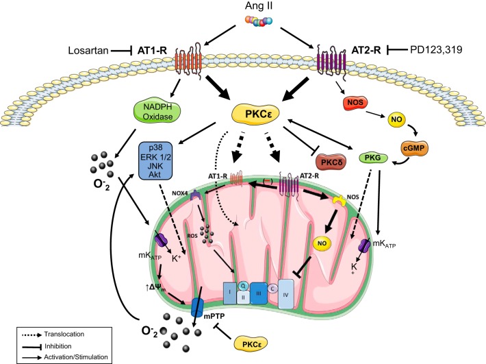
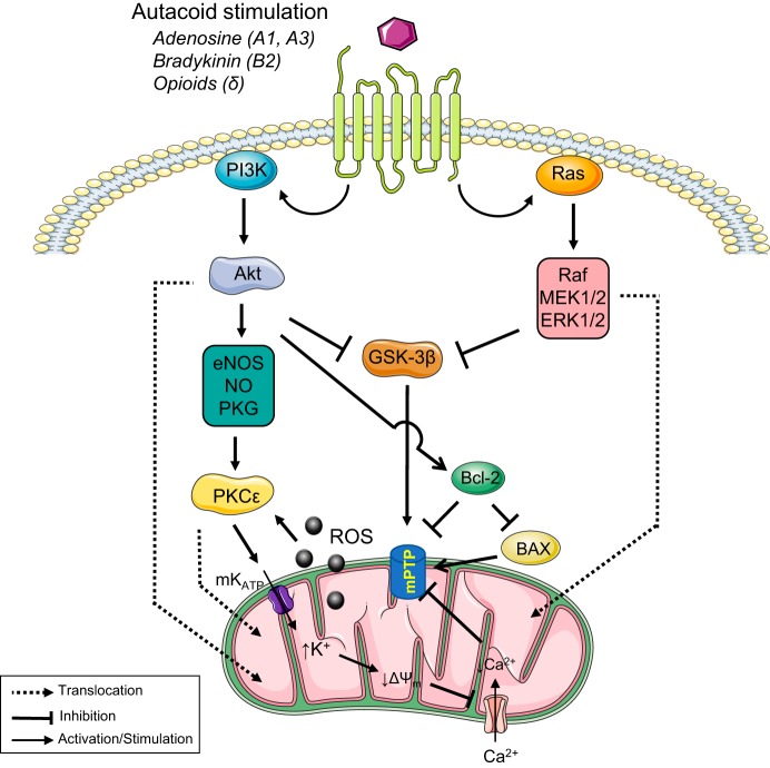
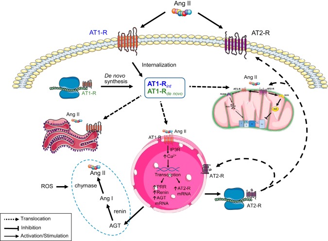
A growing body of data provides strong evidence that intracellular angiotensin II (ANG II) plays an important role in mammalian cell function and is involved in the pathogenesis of human diseases such as hypertension, diabetes, inflammation, fibrosis, arrhythmias, and kidney disease, among others. Recent studies also suggest that intracellular ANG II exerts protective effects in cells during high extracellular levels of the hormone or during chronic stimulation of the local tissue renin-angiotensin system (RAS). Notably, the intracellular RAS (iRAS) described in neurons, fibroblasts, renal cells, and cardiomyocytes provided new insights into regulatory mechanisms mediated by intracellular ANG II type 1 (AT1Rs) and 2 (AT2Rs) receptors, particularly, in mitochondria and nucleus. For instance, ANG II through nuclear AT1Rs promotes protective mechanisms by stimulating the AT2R signaling cascade, which involves mitochondrial AT2Rs and Mas receptors. The stimulation of nuclear ANG II receptors enhances mitochondrial biogenesis through peroxisome proliferator-activated receptor-γ coactivator-1α and increases sirtuins activity, thus protecting the cell against oxidative stress. Recent studies in ANG II-induced preconditioning suggest that plasma membrane AT2R stimulation exerts protective effects against cardiac ischemia-reperfusion by modulating mitochondrial AT1R and AT2R signaling. These studies indicate that iRAS promotes the protection of cells through nuclear AT1R signaling, which, in turn, promotes AT2R-dependent processes in mitochondria. Thus, despite abundant data on the deleterious effects of intracellular ANG II, a growing body of studies also supports a protective role for iRAS that could be of relevance to developing new therapeutic strategies. This review summarizes and discusses previous studies on the role of iRAS, particularly emphasizing the protective and counterbalancing actions of iRAS, mitochondrial ANG II receptors, and their implications for organ protection.

Keywords: angiotensin II receptors; cardioprotection; heart; intracellular renin-angiotensin system; mitochondria.


Conflict of interest statement

No conflicts of interest, financial or otherwise, are declared by the authors.

Figures

Fig. 1.
Fig. 1.
Proposed mechanisms of mitochondria-mediated cardioprotection by ANG II-preconditioning (APC). APC operates through plasma membrane ANG II type 1 (AT1Rs) and 2 (AT2Rs) receptors that are blocked by losartan and PD123319, respectively. Stimulation of these receptors leads to PKCε activation and translocation of AT1R, AT2R, Akt, ERK1/2, JNK, p38, and PKG to the mitochondria where they exert their protective role. AT1Rs also stimulate NAD(P)H oxidase (NOX) activity leading to superoxide generation, mitoKATP channel activity, membrane depolarization, and mitochondrial permeability transition pores (mPTP) opening. This process enhances the efflux of superoxide into the cytoplasm and promotes the activation of prosurvival kinases. By contrast, AT2R stimulation leads to nitric oxide synthase (NOS)-nitric oxide (NO)-PKG stimulation that further stimulates PKCε. In the mitochondria, the upregulation of AT2R/AT1R protein levels controls the respiratory chain activity through a stimulatory mechanism composed of NOX4-reactive oxygen species (ROS) and an inhibitory pathway involving NOS-NO. The latter is proposed to exert a master control on respiratory function as it modulates AT1Rs while tonically suppressing electron transport chain activity.
Fig. 2.
Fig. 2.
Main signaling pathways involved in mitochondria-mediated cardioprotection by ischemic preconditioning (IPC). IPC is triggered by ligands (i.e., adenosine, bradykinin, and opioids, among others) that through receptor-mediated processes stimulate signaling pathways involved in cardioprotection. Prosurvival kinases involved in these pathways [phosphatidylinositol 3-kinase (PI3K)-Akt and Ras-Raf-MEK1/2-ERK1/2] are activated and translocated to the mitochondria to exert protective action. Both pathways lead to inhibition of GSK-3β, which blocks the mitochondrial permeability transition pores (mPTP). Also, the activation of the PI3K-Akt signaling mechanism, promote activation of PKCє through the endothelial nitric oxide synthase (eNOS)-nitric oxide (NO)-PKG pathway. Stimulation of mitoKATP channel leads to reduced mitochondrial potential and inhibition of mPTP opening. Reactive oxygen species (ROS) generation by stimulation of respiratory chain activity promotes the continuous stimulation of PKCε and cardioprotection.
Fig. 3.
Fig. 3.
Proposed mechanism of regulation of mitochondrial function by extracellular ANG II receptors. Activation of plasma membrane receptors of ANG II leads to endocytic internalization and trafficking of the ANG II-ANG II type 1 receptor (AT1R) complex to an intracellular (“processing”) pool that also contains the synthesized AT1Rs. ANG II from intracellular synthesis binds to translocated AT1Rs in the nuclear membrane and the mitochondria. At the nucleus, the complex stimulates transcription and synthesis of ANG II type 2 receptors (AT2Rs) that are also translocated to the mitochondria, the plasma membrane and the nucleus. Nuclear AT1Rs also lead to the synthesis of intracellular renin-angiotensin system (RAS) components and ANG II generation. The process can also be stimulated by oxidative stress. ANG II receptors localized at the plasma membrane, nucleus, endoplasmic reticulum, and mitochondria regulate cell metabolism through signaling pathways including reactive oxygen species (ROS), nitric oxide (NO), and PKCε-dependent cascades, among others (see Fig. 1). AGT, angiotensinogen.

References

    1. Abadir PM, Foster DB, Crow M, Cooke CA, Rucker JJ, Jain A, Smith BJ, Burks TN, Cohn RD, Fedarko NS, Carey RM, O’Rourke B, Walston JD. Identification and characterization of a functional mitochondrial angiotensin system. Proc Natl Acad Sci USA 108: 14849–14854, 2011. doi: 10.1073/pnas.1101507108. - DOI - PMC - PubMed
    1. Abadir PM, Walston JD, Carey RM. Subcellular characteristics of functional intracellular renin-angiotensin systems. Peptides 38: 437–445, 2012. doi: 10.1016/j.peptides.2012.09.016. - DOI - PMC - PubMed
    1. Arnaud C, Joyeux-Faure M, Bottari S, Godin-Ribuot D, Ribuot C. New insight into the signalling pathways of heat stress-induced myocardial preconditioning: protein kinase Cepsilon translocation and heat shock protein 27 phosphorylation. Clin Exp Pharmacol Physiol 31: 129–133, 2004. doi: 10.1111/j.1440-1681.2004.03966.x. - DOI - PMC - PubMed
    1. Baker KM, Chernin MI, Schreiber T, Sanghi S, Haiderzaidi S, Booz GW, Dostal DE, Kumar R. Evidence of a novel intracrine mechanism in angiotensin II-induced cardiac hypertrophy. Regul Pept 120: 5–13, 2004. doi: 10.1016/j.regpep.2004.04.004. - DOI - PubMed
    1. Benigni A, Cassis P, Remuzzi G. Angiotensin II revisited: new roles in inflammation, immunology and aging. EMBO Mol Med 2: 247–257, 2010. doi: 10.1002/emmm.201000080. - DOI - PMC - PubMed

Publication types

MeSH terms

LinkOut - more resources