. 2019 Jun 10:303:42-54.
doi: 10.1016/j.jconrel.2019.04.008. Epub 2019 Apr 9.

Combining activatable nanodelivery with immunotherapy in a murine breast cancer model

Affiliations

Combining activatable nanodelivery with immunotherapy in a murine breast cancer model

Azadeh Kheirolomoom et al. J Control Release. .

Abstract

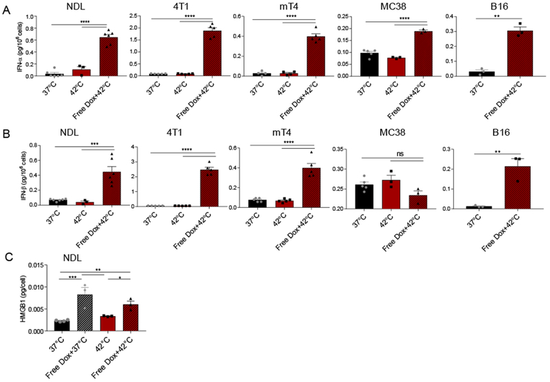
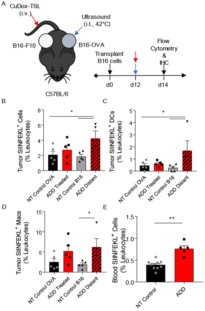
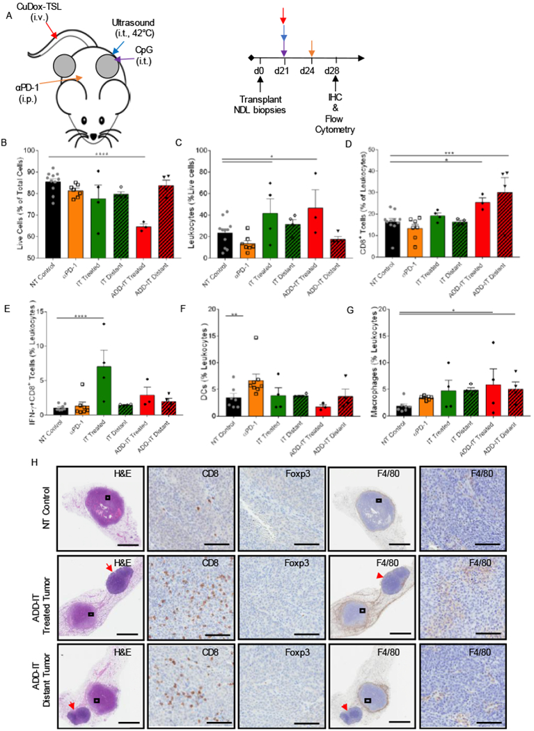
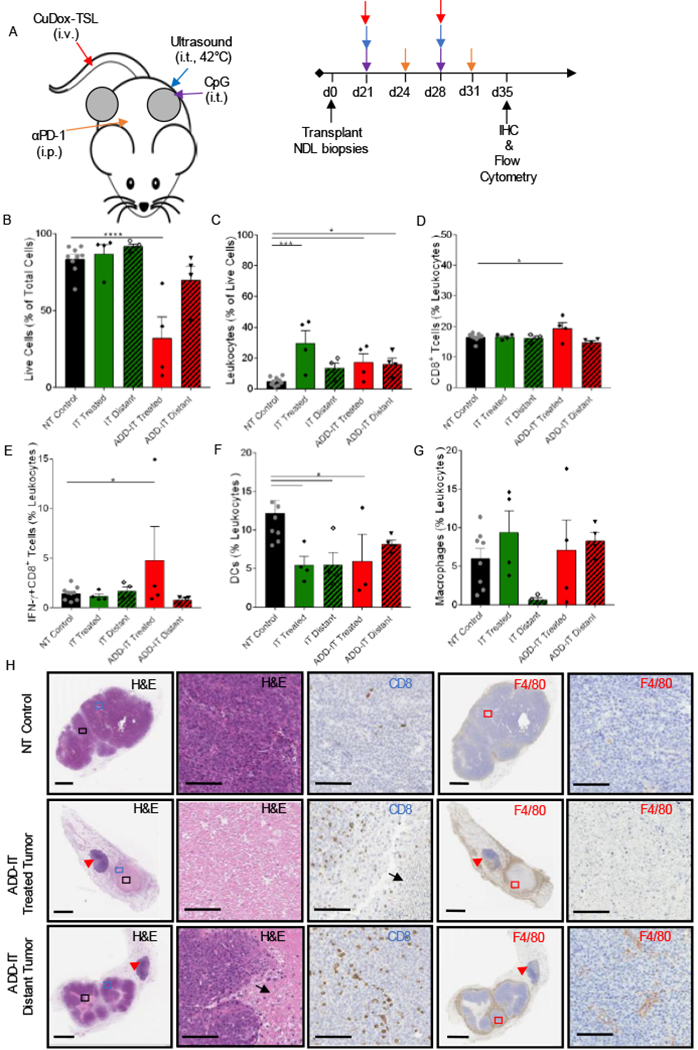
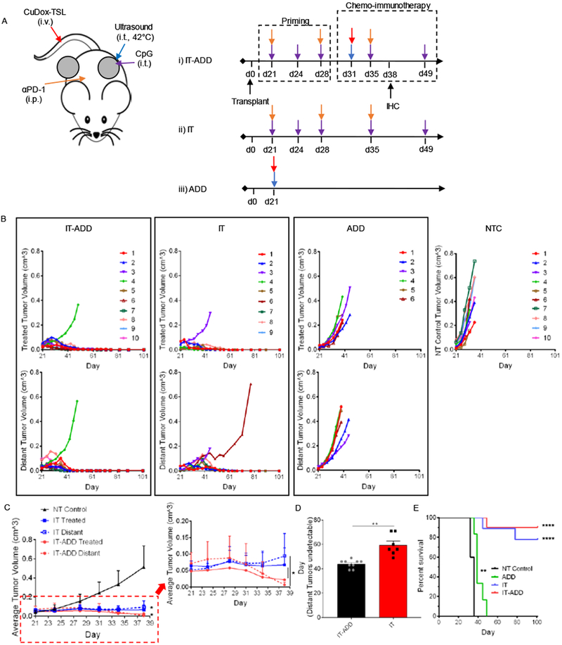
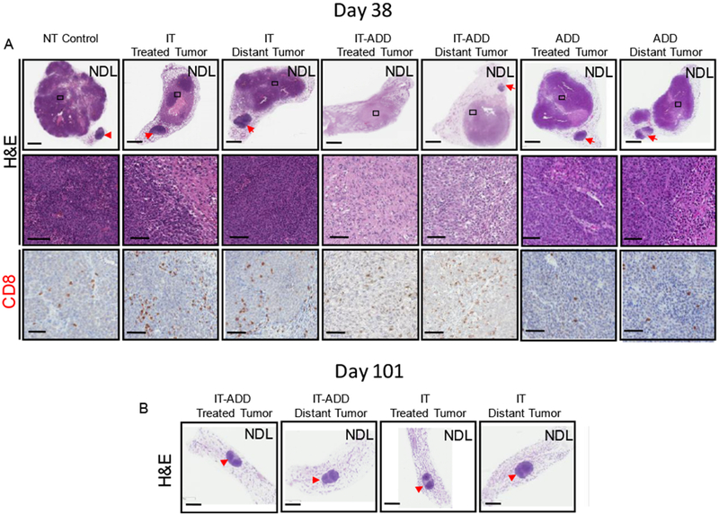
A successful chemotherapy-immunotherapy solid-tumor protocol should accomplish the following goals: debulk large tumors, release tumor antigen for cross-presentation and cross-priming, release cancer-suppressive cytokines and enhance anti-tumor immune cell populations. Thermally-activated drug delivery particles have the potential to synergize with immunotherapeutics to accomplish these goals; activation can release chemotherapy within bulky solid tumors and can enhance response when combined with immunotherapy. We set out to determine whether a single protocol, combining locally-activated chemotherapy and agonist immunotherapy, could accomplish these goals and yield a potentially translational therapy. For effective delivery of free doxorubicin to tumors with minimal toxicity, we stabilized doxorubicin with copper in temperature-sensitive liposomes that rapidly release free drug in the vasculature of cancer lesions upon exposure to ultrasound-mediated hyperthermia. We found that in vitro exposure of tumor cells to hyperthermia and doxorubicin resulted in immunogenic cell death and the local release of type I interferons across murine cancer cell lines. Following intravenous injection, local activation of the liposomes within a single tumor released doxorubicin and enhanced cross-presentation of a model antigen at distant tumor sites. While a variety of protocols achieved a complete response in >50% of treated mice, the complete response rate was greatest (90%) when 1 week of immunotherapy priming preceded a single activatable chemotherapeutic administration. While repeated chemotherapeutic delivery reduced local viable tumor, the complete response rate and a subset of tumor immune cells were also reduced. Taken together, the results suggest that activatable chemotherapy can enhance adjuvant immunotherapy; however, in a murine model the systemic adaptive immune response was greatest with a single administration of chemotherapy.

Keywords: Breast cancer; CpG; Doxorubicin; Immunotherapy; Temperature-sensitive liposome; Ultrasound; αPD-1.


Conflict of interest statement

Conflict of interest

The authors declare no completing financial of interest.

Figures

Figure 1.
Figure 1.. Doxorubicin increased type I interferon production in murine cancer cell lines and immunogenic cell death in NDL cells.
In vitro release of IFN-α (A), IFN-β (B) in NDL and 4T1 murine mammary carcinoma, mT4 murine pancreatic cancer, MC-38 murine colon cancer cells, B16 murine melanoma, and HMGB 1 in NDL cell culture 24 h post treatment, respectively. Cells were preincubated for 5 min (type I IFN) or 1 min (HMGB 1) at 42°C prior to addition of media only (42°C) or a solution of 5 µg/mL Dox in media (Free Dox+42°C) at 42°C for another 5 min. * p < 0.05, ** p< 0.01, *** p< 0.001, **** p< 0.0001.
Figure 2.
Figure 2.. Tumor-specific antigen presentation increased in tumor and blood after treatment.
A) Schematic diagram illustrating the treatment protocol of CuDox+US (ADD) in C57BL/6 mice (n = 13) bearing one B16-OVA tumor (locally treated tumor) and one control B16-F10 tumor on the opposite side (distant tumor). B-D) Frequency of SIINFEKL-labeled immune cells, and specifically DCs, and macrophages as a percentage of leukocytes in tumors directly treated with ADD or in the distant tumor of the treated mice (n = 5) compared to B16-OVA and B16-F10 tumors of untreated control mice (n = 5), respectively. E) Frequency of SIINFEKL-labeled blood immune cells as a percentage of leukocytes in blood of mice treated with ADD compared to no-treatment (NT) control mice (n = 3). * p < 0.05, ** p< 0.01.
Figure 3.
Figure 3.. Combining ADD with CpG and αPD-1 increased cytotoxic T lymphocytes in local and distant NDL tumors.
A) Representative treatment protocol and timeline for treatment of NDL tumor-bearing mice. One tumor of bilateral tumors of NDL orthotopically transplanted into FVB/n mice was treated with one complete administration of each component of CuDox+US+CpG (ADD-CpG) followed by αPD-1 post three days. B-G) On day 28 after one complete treatment of ADD-IT (n = 4) or IT (n = 4) or a single administration of αPD-1 (n = 4), tumors/inguinal lymph nodes of treated mice were stained with the antibody cocktail as listed below and analyzed via flow cytometry and compared with untreated control tumors. Fraction of live cells given as a percentage of total isolated cells (B), frequency of leukocytes (CD45+ cells) as a percentage of live cells (C) and frequency of cytotoxic T lymphocytes (CD3+CD8+ cells) as a percentage of total leukocytes (D), populations of IFN-γ producing CD8+ (CD3+CD8+IFN-γ+ cells) T-cells as a percentage of total leukocytes (E), fraction of DCs (CD11c+MHCII+ F4/80 cells) as a percentage of leukocytes (F), fraction of macrophages (CD11b+F4/80+Gr-1 cells) as a percentage of leukocytes (G) across various treatments. H) Histological sections of the no-treatment control, the tumor locally treated with CuDox+US+CpG+αPD-1 (ADD-IT) and the distant tumor on day 28 were stained with H&E (the left first column), CD8 (second column), Foxp3 (third column), F4/80 (fourth column) and the magnified images (fifth column), respectively. CD8, F4/80, and Foxp3 staining (brown) identify CD8+ T-cells, macrophages, and T regs, respectively. Red arrowheads denote the inguinal lymph nodes. Scale bars are 3 mm for whole tumor image and 100 µm for the magnified image. * p < 0.05, ** p< 0.01, *** p< 0.001, **** p< 0.0001.
Figure 4.
Figure 4.. Two treatments of ADD-IT increased IFN-ɣ-producing T-cells in the treated NDL tumor.
A) Representative treatment protocol and timeline for two treatments of ADD-IT administered to NDL tumor-bearing mice. B-G) After two sequential treatments of CuDox+US+CpG+ αPD-1 (ADD-IT, n = 4), the entire inguinal fat pads containing tumor and lymph node were harvested from treated mice and stained for CD45, CD3, CD4, CD8, F4/80, CD11b, CD11c, MHCII, Gr-1, and IFN-γ on day 35 and compared to untreated control mice (n = 4) via flow cytometry analysis. Number of live cells given as a percentage of total isolated cells (B), frequency of leukocytes (CD45+ cells) as a percentage of live cells (C), frequency of cytotoxic T lymphocytes (CD3+ CD8+ cells) as a percentage of total leukocytes (D), fraction of IFN-γ producing CD8+ (CD3+CD8+IFN-γ+) T-cells given as a percentage of total leukocytes (E), fraction of DCs (CD11c+ MHCII+ F4/80 cells) as a percentage of leukocytes (F), and fraction of macrophages (CD11b+ F4/80+ Gr-1 cells) as a percentage of leukocytes (G). H) Histological sections of no-treatment control, local and distant tumors of mice treated with two administrations of ADD-IT on day 35 and stained for H&E (the left first column, whole tumor view) and (second column, magnified view), CD8 (third column), F4/80 (fourth column, whole tumor) and the magnified panels (fifth column), respectively. CD8 and F4/80 staining (brown) identify CD8+ T-cells and macrophages, respectively. Black arrows and red arrowheads denote the necrotic cells and inguinal lymph nodes, respectively. Scale bars are 3 mm for whole tumor image and 100 µm for the magnified image. * p < 0.05, *** p< 0.001, **** p< 0.0001
Figure 5.
Figure 5.. Administration of CpG and αPD-1 prior to a combined ADD and immunotherapy protocol reduced tumor viability in both local and distant tumors of NDL-tumor bearing mice.
A) Treatment protocols of an immunopriming sequence with and without chemotherapy and chemotherapy alone. One tumor was treated directly with either CpG intratumorally in CpG+αPD-1 Prime (IT) or with CuDox+US (ADD) in CuDox+US+CpG+αPD-1 Prime (IT-ADD) and in CuDox+US (ADD) treatment groups. B) Growth of directly treated and distant tumors in bilateral NDL-tumor bearing mice treated with IT-ADD (n = 10), IT (n = 9), and ADD (n = 6) (B) compared to no-treatment control tumors (n = 10). C) Average growth of NDL tumors treated with either IT-ADD (n = 3) or IT (n = 5) treatment protocols compared to no-treatment control group over 38 days. The inset shows a representative magnified area. D) Temporal response of distant tumors to IT-ADD and IT treatments. E) The overall survival over the course of 101 days. In D, the survival curves for both primed & non-primed ADD-IT (p<0.0001) and ADD (p<0.01) were found statistically significant compared to NT Control group as evaluated by Log-rank (Martel-Cox) test. * p< 0.05, ** p< 0.01, **** p<0.0001).
Figure 6.
Figure 6.. Histological assessment of local and distant tumors confirmed antitumor efficacy of the ADD-IT treatment protocol.
A) On day 38, a subset of mice was euthanized and tumors were isolated for histology and IHC. Histological sections stained for H&E (upper row, whole tumor view) and (middle row, magnified view), CD8 (lower row, magnified view). Whole tumor sections and the magnified views enclosed by black boxes are shown. B) H&E stained histological sections of mice treated with either IT-ADD or IT and survived 101 days. Red arrowheads denote the inguinal lymph nodes. Scale bars correspond to 3 mm (whole tumor panels) and 100 µm (magnified panels).

References

    1. Topalian SL, Drake CG, Pardoll DM, Immune checkpoint blockade: a common denominator approach to cancer therapy, Cancer Cell. 27 (2015) 450–461. doi:10.1016/j.ccell.2015.03.001. - DOI - PMC - PubMed
    1. Rizvi NA, Hellmann MD, Snyder A, Kvistborg P, Makarov V, Havel JJ, Lee W, Yuan J, Wong P, Ho TS, Miller ML, Rekhtman N, Moreira AL, Ibrahim F, Bruggeman C, Gasmi B, Zappasodi R, Maeda Y, Sander C, Garon EB, Merghoub T, Wolchok JD, Schumacher TN, Chan TA, Cancer immunology. Mutational landscape determines sensitivity to PD-1 blockade in non-small cell lung cancer, Science. 348 (2015) 124–128. doi:10.1126/science.aaa1348. - DOI - PMC - PubMed
    1. Van Allen EM, Miao D, Schilling B, Shukla SA, Blank C, Zimmer L, Sucker A, Hillen U, Foppen MHG, Goldinger SM, Utikal J, Hassel JC, Weide B, Kaehler KC, Loquai C, Mohr P, Gutzmer R, Dummer R, Gabriel S, Wu CJ, Schadendorf D, Garraway LA, Genomic correlates of response to CTLA-4 blockade in metastatic melanoma, Science. 350 (2015) 207–211. doi:10.1126/science.aad0095. - DOI - PMC - PubMed
    1. McGranahan N, Furness AJ, Rosenthal R, Ramskov S, Lyngaa R, Saini SK, Jamal-Hanjani M, Wilson GA, Birkbak NJ, Hiley CT, Watkins TB, Shafi S, Murugaesu N, Mitter R, Akarca AU, Linares J, Marafioti T, Henry JY, Van Allen EM, Miao D, Schilling B, Schadendorf D, Garraway LA, Makarov V, Rizvi NA, Snyder A, Hellmann MD, Merghoub T, Wolchok JD, Shukla SA, Wu CJ, Peggs KS, Chan TA, Hadrup SR, Quezada SA, Swanton C, Clonal neoantigens elicit T cell immunoreactivity and sensitivity to immune checkpoint blockade, Science. 351 (2016) 1463–1469. doi:10.1126/science.aaf1490. - DOI - PMC - PubMed
    1. Gajewski TF, The Next Hurdle in Cancer Immunotherapy: Overcoming the Non-T-Cell-Inflamed Tumor Microenvironment, Semin Oncol. 42 (2015) 663–671. doi:10.1053/j.seminoncol.2015.05.011. - DOI - PMC - PubMed

Publication types